. 2020 May 22;3(1):254.
doi: 10.1038/s42003-020-0972-7.

The strategic combination of trastuzumab emtansine with oncolytic rhabdoviruses leads to therapeutic synergy

Affiliations

The strategic combination of trastuzumab emtansine with oncolytic rhabdoviruses leads to therapeutic synergy

Rozanne Arulanandam et al. Commun Biol. .

Abstract

We have demonstrated that microtubule destabilizing agents (MDAs) can sensitize tumors to oncolytic vesicular stomatitis virus (VSVΔ51) in various preclinical models of cancer. The clinically approved T-DM1 (Kadcyla®) is an antibody-drug conjugate consisting of HER2-targeting trastuzumab linked to the potent MDA and maytansine derivative DM1. We reveal that combining T-DM1 with VSVΔ51 leads to increased viral spread and tumor killing in trastuzumab-binding, VSVΔ51-resistant cancer cells. In vivo, co-treatment of VSVΔ51 and T-DM1 increased overall survival in HER2-overexpressing, but trastuzumab-refractory, JIMT1 human breast cancer xenografts compared to monotherapies. Furthermore, viral spread in cultured HER2+ human ovarian cancer patient-derived ascites samples was enhanced by the combination of VSVΔ51 and T-DM1. Our data using the clinically approved Kadcyla® in combination with VSVΔ51 demonstrates proof of concept that targeted delivery of a viral-sensitizing molecule using an antibody-drug conjugate can enhance oncolytic virus activity and provides rationale for translation of this approach.


Conflict of interest statement

The authors declare no competing interests.

Figures
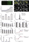
Fig. 1
Fig. 1. Conjugation of DM1 to trastuzumab increases VSVΔ51 spread and bystander killing in VSVΔ51-resistant cancer cells.
a 786-0 cells were pretreated with T-DM1 or trastuzumab for 4 h followed by infection with VSVΔ51-GFP at MOI 0.01 and fluorescent images taken 24 h later, n = 3. b A high-throughput method was used to quantify viral titer from 786-0 pretreated with T-DM1 and infected with VSVΔ51-Fluc at MOI 0.1 (n = 9; mean ± SEM; one-way ANOVA compared to 0 µg/ml). c 786-0 were pretreated with either 100 μg/ml trastuzumab or T-DM1, 100 nM colchicine or Mock-treated, infected with VSVΔ51-GFP at MOI 0.01 and plaque assay performed after 48 h (n = 3; mean ± SEM; one-way ANOVA compared to Mock). d 786-0 were plated on glass coverslips, treated with 100 µg/ml T-DM1 or trastuzumab or mock, and then stained for β-tubulin and DAPI 24 h later. Objective = ×63, scale bar = 50 µm. White arrows indicate polynuclear cells. e Quantification of polynuclear 786-0 or GM38 cells from d. Images were taken 24 h post infection and 6 frames per experimental condition from n = 3 were scored for % polynuclear cells with SEM. Two-way ANOVA with Sidak’s multiple comparisons test was performed over mock or trastuzumab, or between groups indicated with bars, for each cell line. f, g 786-0 were treated as in a, and viability assessed 48 h post infection (n = 3; mean ± SEM; two-way ANOVA with Sidak’s multiple comparisons test across concentrations). h 786-0 treated as in c were administered 0 or 100 ng/ml TNFα and viability assessed after 72 h (n = 3; mean ± SEM); two-way ANOVA with Sidak’s multiple comparisons test across pretreatment conditions. i 786-0 were pretreated as in a, infected with VSVΔ51-GFP at MOI 0.1 or Mock. and supernatants assayed for IFNβ production 20 h later (n = 3; mean ± SEM; one-way ANOVA with Tukey’s multiple comparisons test across either Mock or VSVΔ51). j, k 786-0 were treated indicated and infected with VSVΔ51-Fluc at MOI 0.01 (j, n = 6) or MOI 1 (k, n = 3) and titered as in b (mean ± SEM; two-way ANOVA with Dunnett’s multiple comparison test compared to Mock for each time point).
Fig. 2
Fig. 2. T-DM1 increases VSVΔ51 activity in a panel of HER2-expressing breast and ovarian cancer lines.
a A panel of human breast and ovarian carcinoma lines were lysed and subject to western blotting for human HER2. b Mouse breast carcinoma 4T1 cells, and a panel of human cancer cell lines were subject to extracellular staining with Herceptin®, followed by goat anti-human IgG-PE. PE signal (y axis) was analyzed by flow cytometry, median MFI are shown; n = 3 ± SD. c 786-0 and JIMT1 cells were pretreated with T-DM1 for 4 h, washed and infected with VSVΔ51 (MOI 0.05 and 0.01, respectively) and plaque assay performed 45 h later (n = 3; mean ± SEM; one-way ANOVA with Dunnett’s multiple comparisons test compared to 0 µg/ml T-DM1 for each cell line). d Indicated cell lines were pretreated with either 100 nM colchicine, 100 μg/ml T-DM1 or trastuzumab, or mock, for 3 h, then washed and infected with VSVΔ51 (MOI 0.01 for JIMT1 and MOI 0.001 for others). 1 h later, wells were overlayed with agarose, and plaques visualized with Coomassie blue 48 h later. Diameter of 30 plaques per well were graphed (mean ± SEM; one-way ANOVA with Dunnett’s multiple comparisons test compared to Mock for each cell line). e JIMT1 cells were plated on glass coverslips, treated with 100 µg/ml T-DM1 or trastuzumab, or mock-treated, then stained for β-tubulin and DAPI 24 h later. Objective = ×63, scale bar = 50 µm, n = 3. White arrow indicates polynuclear cells. f Cell lines in d were pretreated as indicated then administered 0 or 100 ng/ml TNFα. Viability was determined 72 h later (n = 3; mean ± SEM; two-way ANOVA with Sidak’s multiple comparisons test for each pretreatment condition). g 786-0 or JIMT1 cells were either Mock-treated or treated with 1000 µg/ml T-DM1 (786-0) or 250 µg/ml T-DM1 (JIMT1) for 2 h, followed by an equivalent concentration of trastuzumab or IVIG. 2 h later, cells were infected with VSVΔ51 at MOI 0.01 and plaque assay performed 45 h later (n = 4; mean ± SEM; two-way ANOVA with Sidak’s multiple comparisons test compared to Mock for each cell line).
Fig. 3
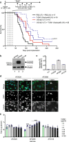
Fig. 3. T-DM1 increases oncolytic activity of VSVΔ51 in vivo and in human patient samples.
a BALB/c nude mice were implanted with 1 × 10⁷ JIMT1 cells. Once tumors were ~100 mm3, mice were treated weekly four times (dotted lines) with the regimen of VSVΔ51-Fluc (1E8 pfu) or PBS (intratumorally) followed by 10 mg/kg T-DM1 (Kadcyla®) or PBS 4 h later (intravenously). Survival was monitored over time (n = 16–20 mice per group as indicated, pooled over three independent experiments) and was observed to be significantly reduced with the combination of VSVΔ51 + T-DM1 compared to either monotherapies (p = 0.039 compared to Kadcyla; p = 0.004 compared to VSV; p < 0.0001 compared to PBS, Gehan–Breslow–Wilcoxon test). b Ascites fluid cells derived from three different human ovarian cancer patients were each lysed and blotted for HER2 or actin as a loading control. c Mouse breast carcinoma 4T1 cells, and the ascites fluid-derived cell lines were subject to extracellular staining and flow cytometry. Cells were stained with Herceptin® (trastuzumab, 1:1000), followed by goat anti-human IgG-PE (1:100). PE signal (y axis) was analyzed by flow cytometry, median MFI are shown; n = 3 ± SD. d AF2028 and AF2068 were plated on glass coverslips, treated with 100 µg/ml T-DM1 or trastuzumab, then fixed and stained for β-tubulin and DAPI 24 h later. Objective = ×63, scale bar = 50 µm, n = 3. White arrow indicates polynuclear cells. e The ascites fluid-derived cell lines were each pretreated with 0–100 µg/ml of T-DM1 for 4 h, washed and infected with VSVΔ51 MOI 0.01 and supernatants tittered by plaque assay after 45 h (n = 3 mean ± SEM; one-way ANOVA with Dunnett’s multiple comparisons test was performed and compared to 0 µg/ml condition for each cell line).

References

    1. Verri E, et al. HER2/neu oncoprotein overexpression in epithelial ovarian cancer: evaluation of its prevalence and prognostic significance. Clin. Study Oncol. 2005;68:154–161. - PubMed
    1. Chung CH, et al. Increased epidermal growth factor receptor gene copy number is associated with poor prognosis in head and neck squamous cell carcinomas. J. Clin. Oncol. 2006;24:4170–4176. doi: 10.1200/JCO.2006.07.2587. - DOI - PubMed
    1. Palle, J. et al. Human epidermal growth factor receptor 2 (HER2) in advanced gastric cancer: current knowledge and future perspectives. Drugs80, 401–415 (2020). - PubMed
    1. Iqbal N, Iqbal N. Human epidermal growth factor receptor 2 (HER2) in cancers: overexpression and therapeutic implications. Mol. Biol. Int. 2014;2014:852748. doi: 10.1155/2014/852748. - DOI - PMC - PubMed
    1. Slamon DJ, et al. Human breast cancer: correlation of relapse and survival with amplification of the HER-2/neu oncogene. Science. 1987;235:177–182. doi: 10.1126/science.3798106. - DOI - PubMed

Publication types