Review
. 2021 Dec;336(8):666-679.
doi: 10.1002/jez.b.22952. Epub 2020 May 23.

Somatic cell conversion to a germ cell lineage: A violation or a revelation?

Affiliations
Review

Somatic cell conversion to a germ cell lineage: A violation or a revelation?

Gary M Wessel et al. J Exp Zool B Mol Dev Evol. 2021 Dec.

Abstract

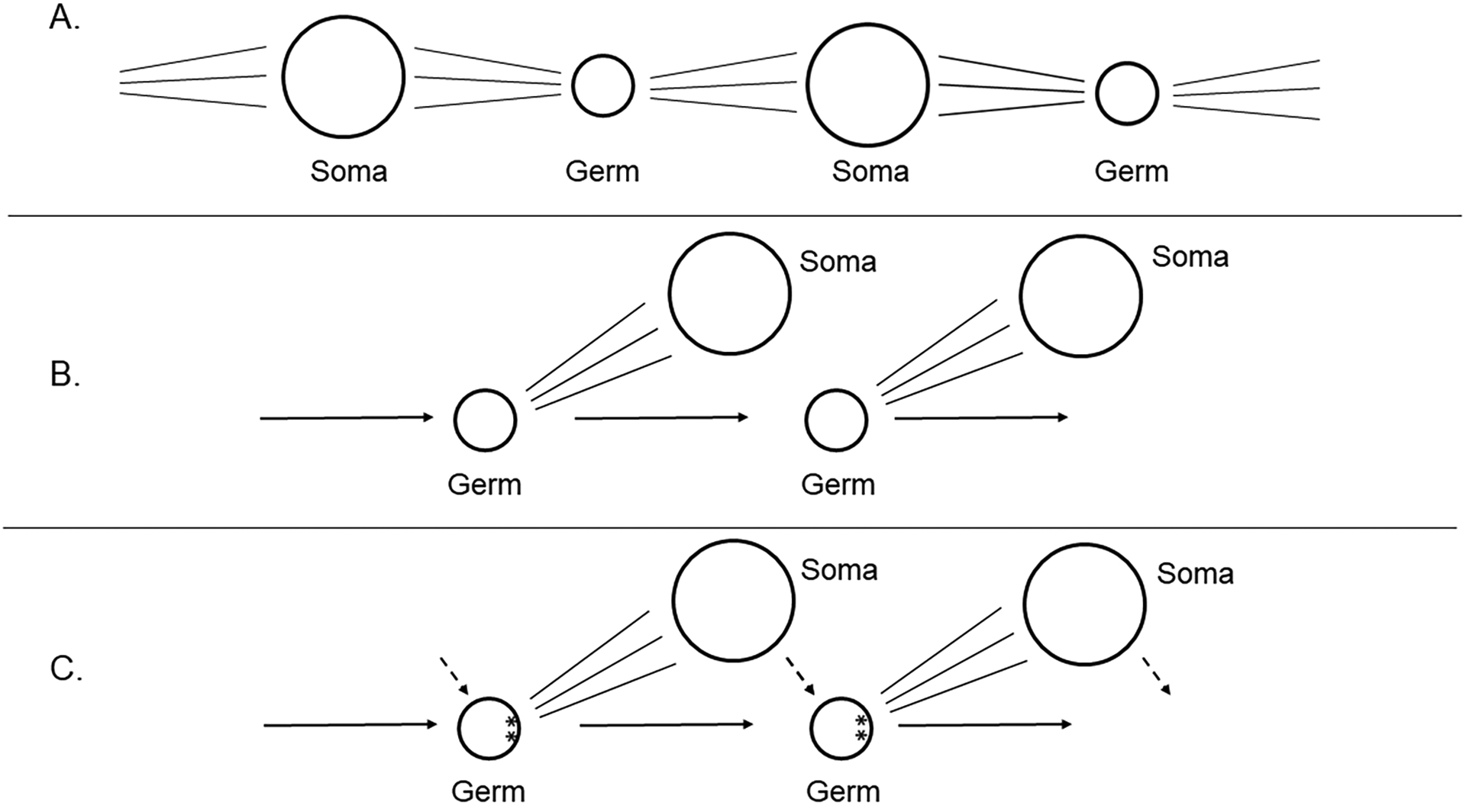
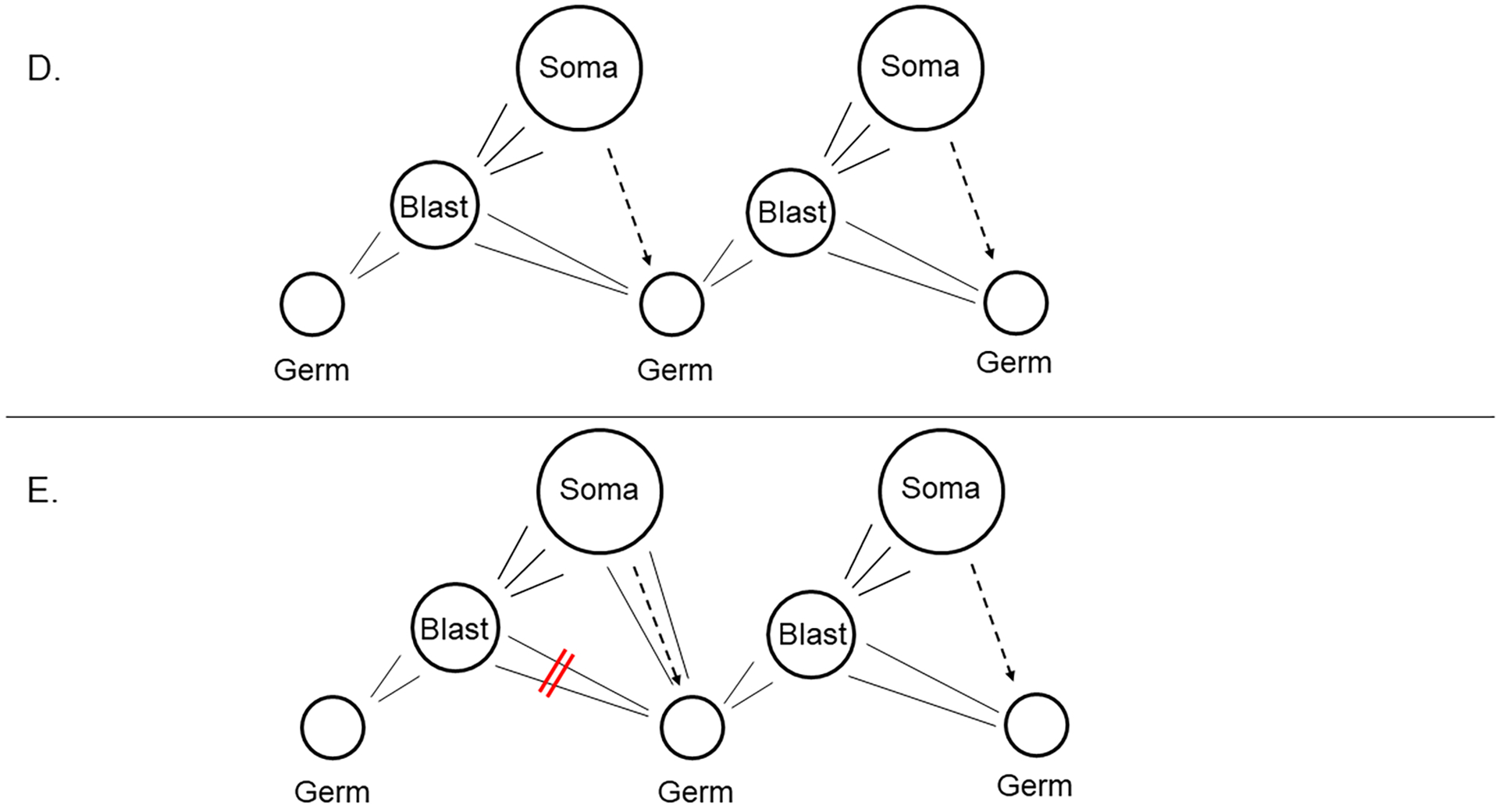
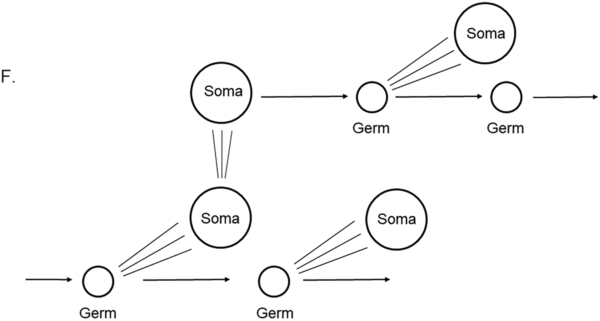
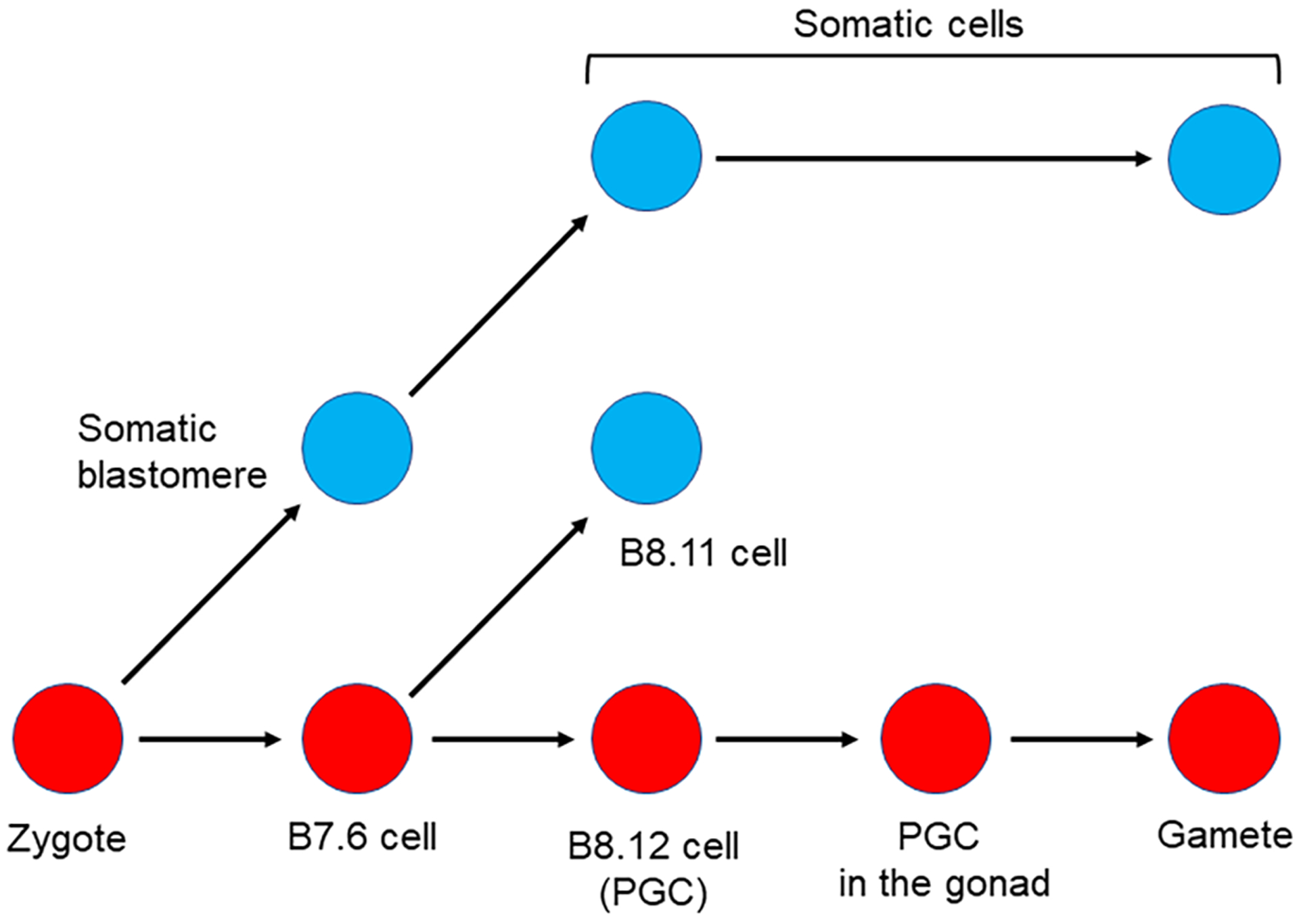
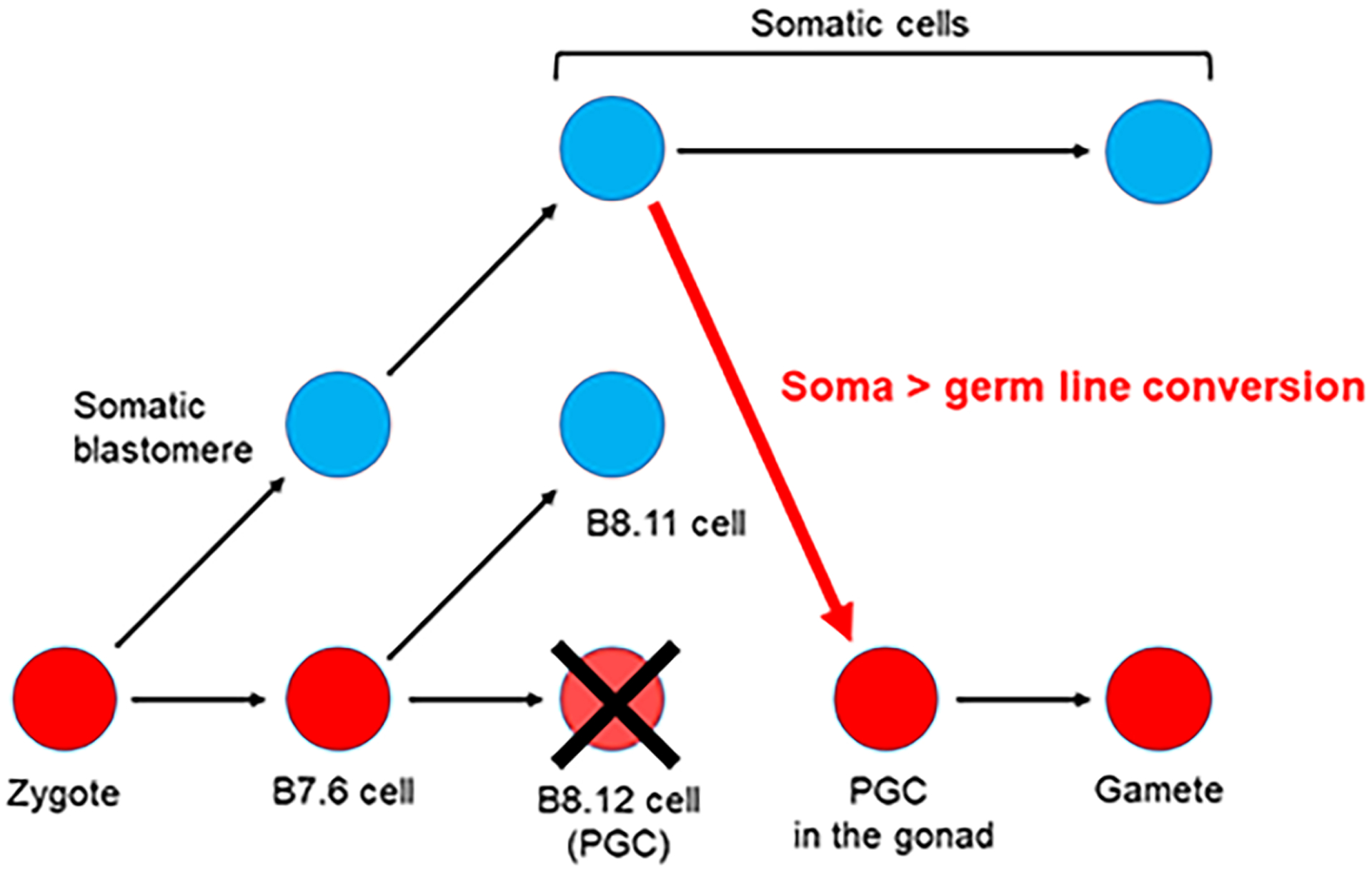
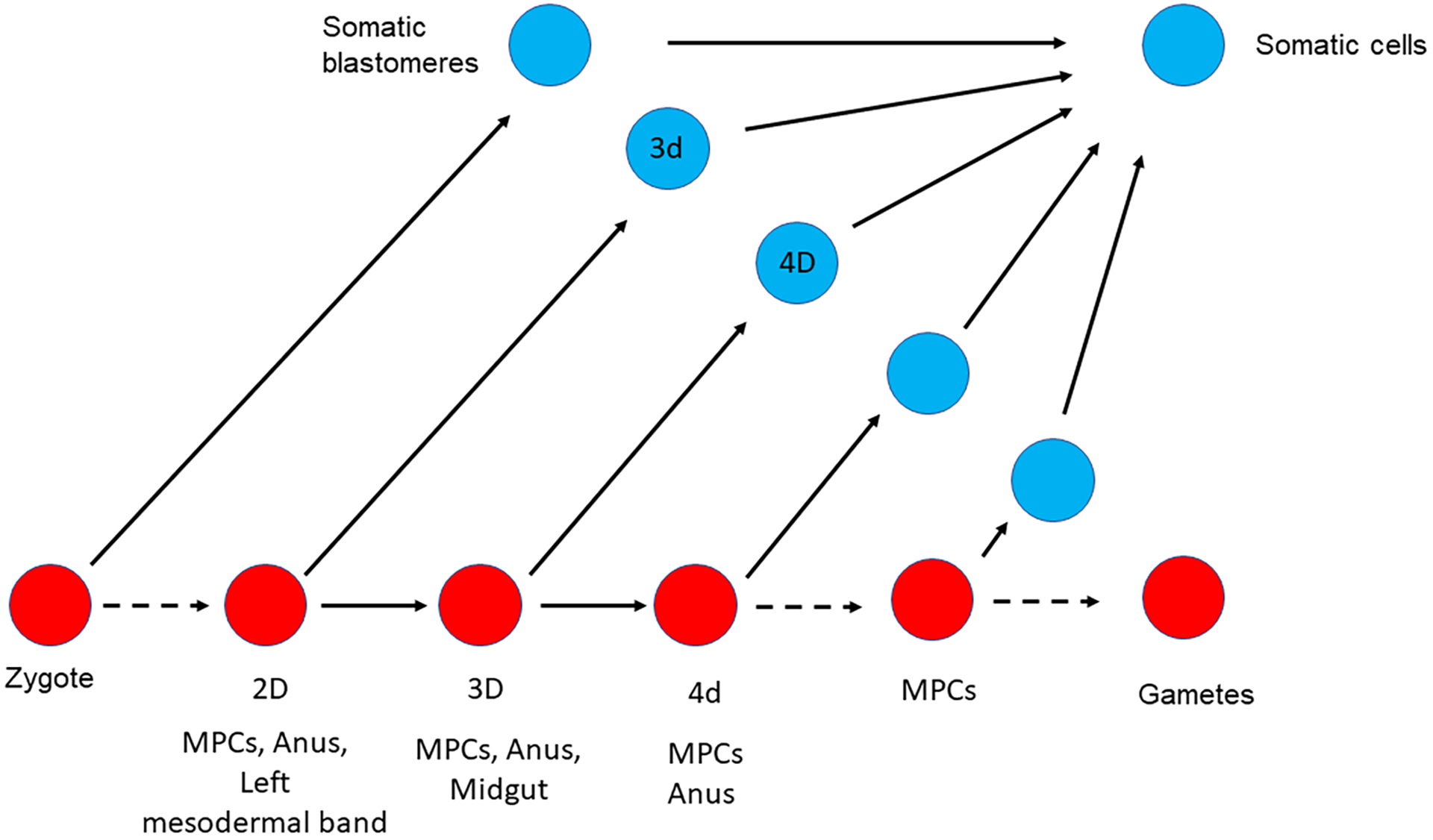
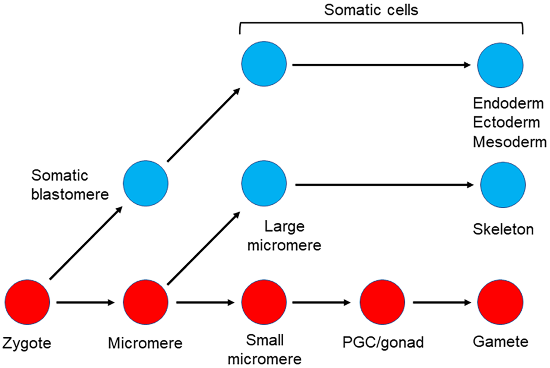
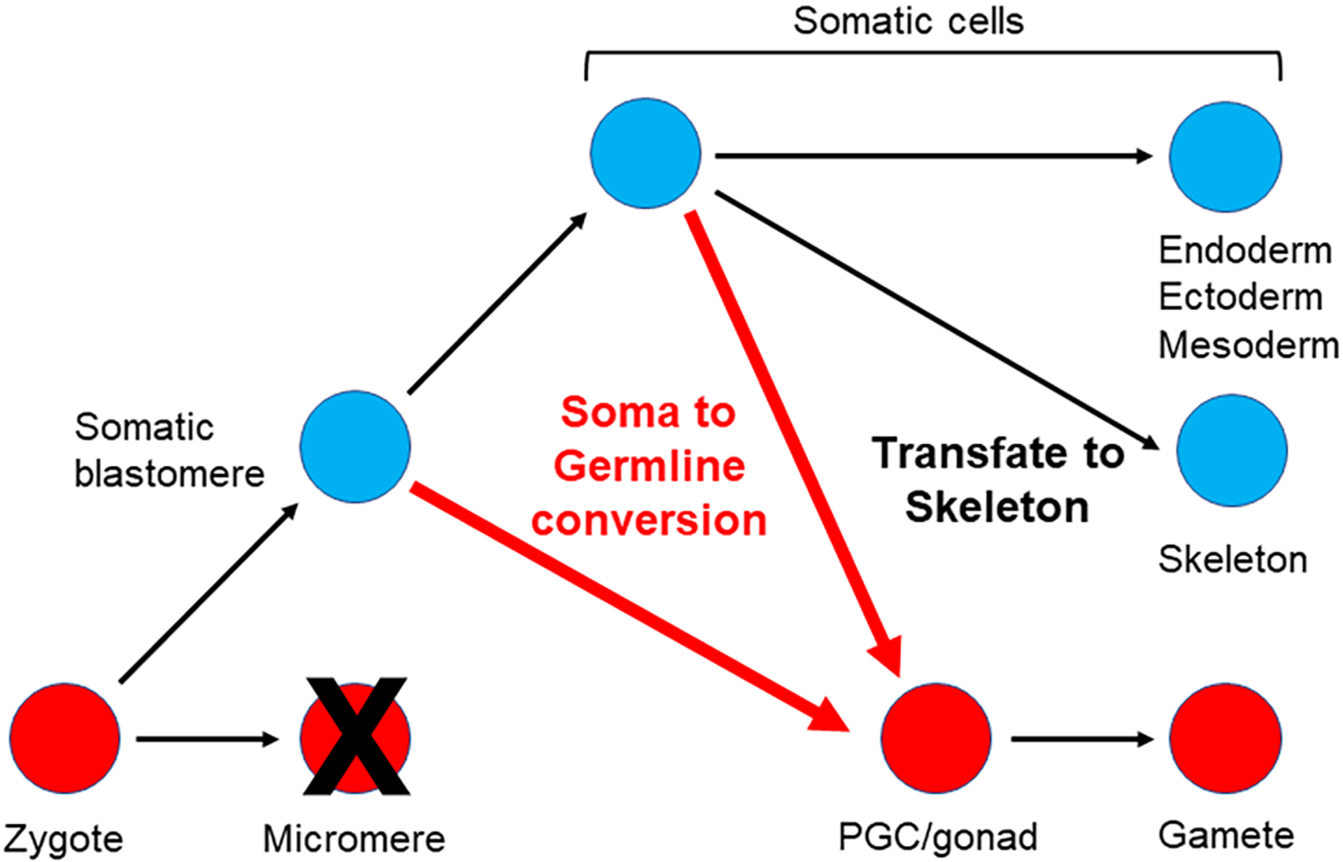
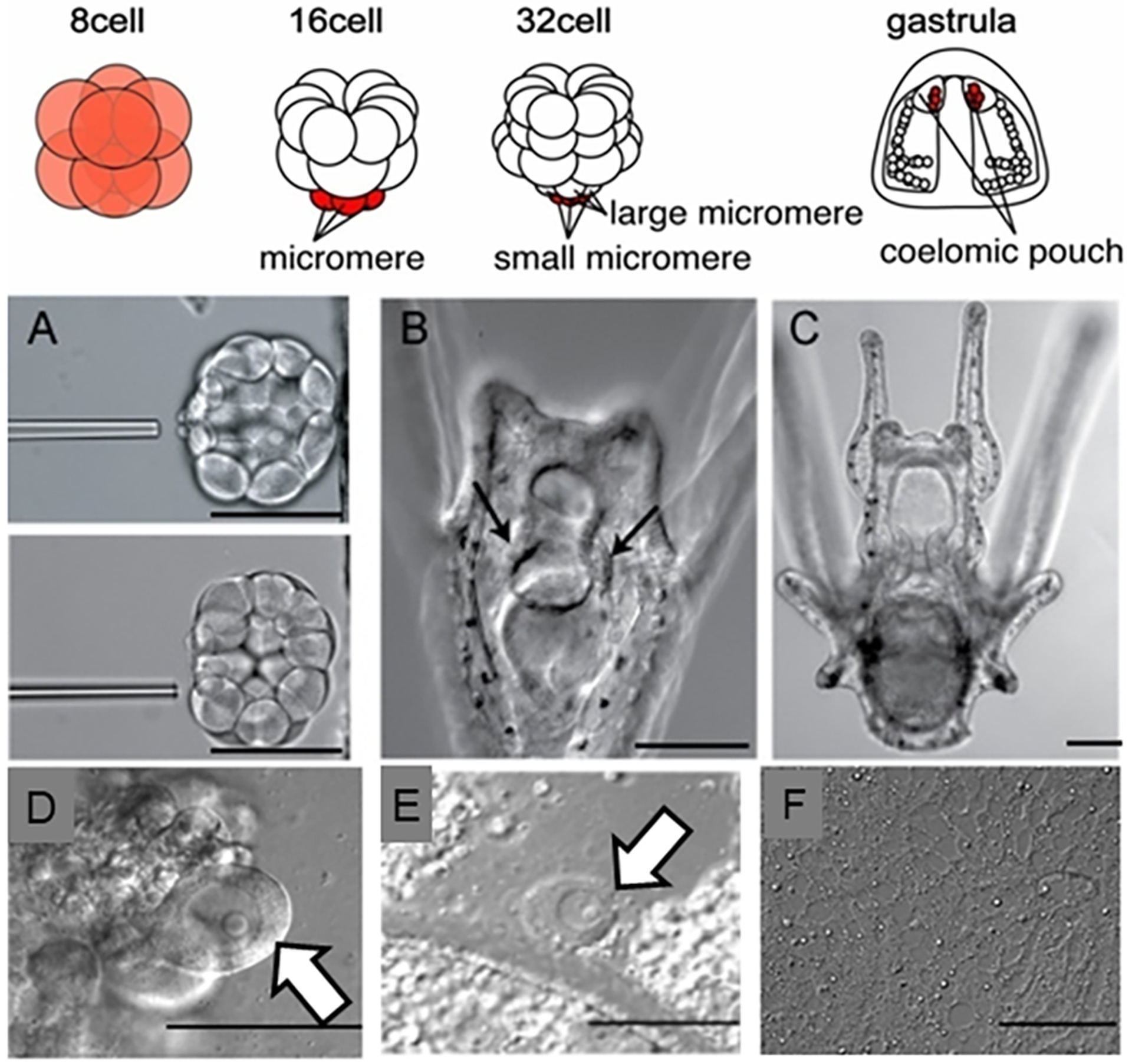
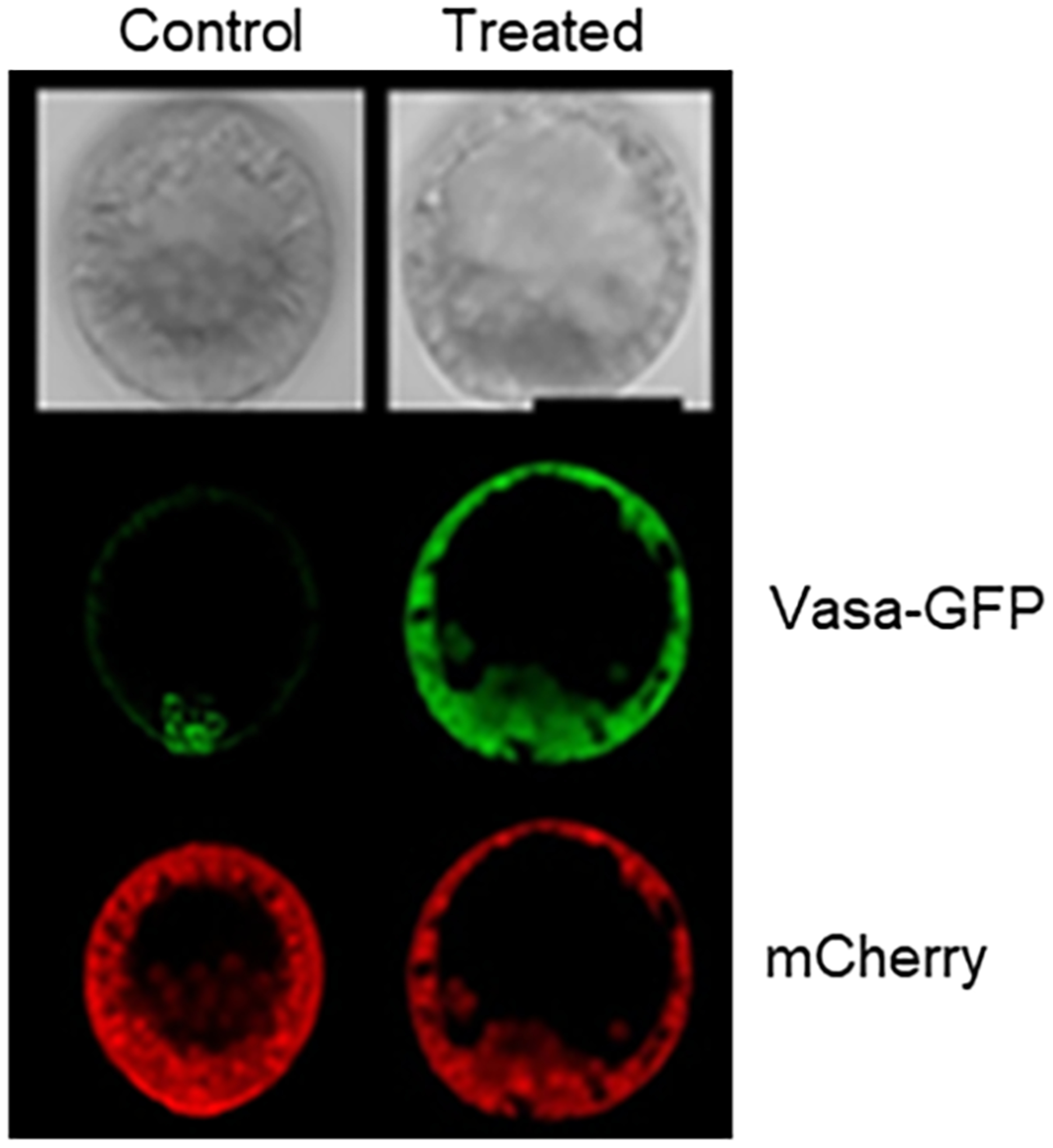
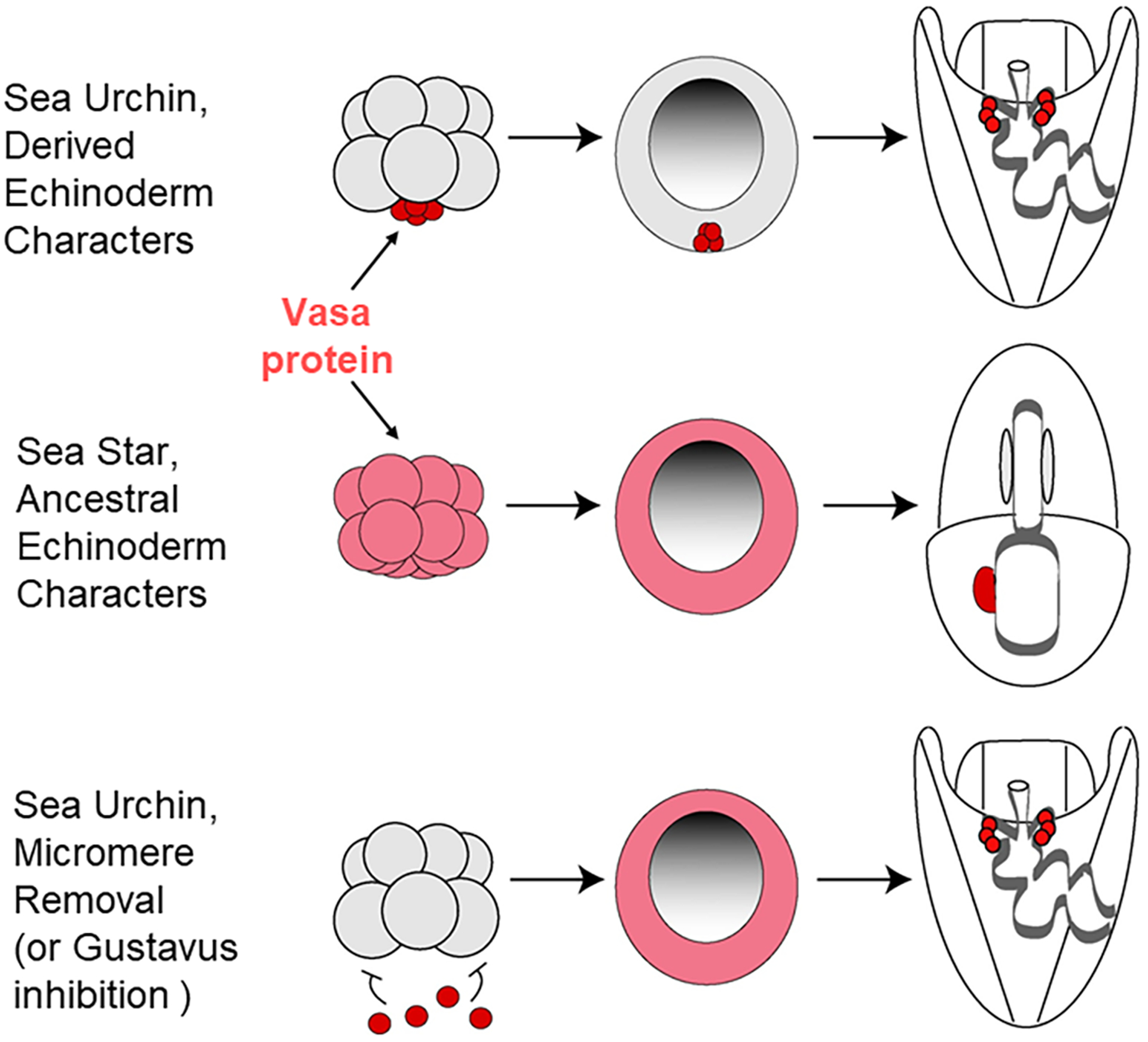
The germline is unique and immortal (or at least its genome is). It is able to perform unique jobs (meiosis) and is selected for genetic changes. Part of being this special also means that entry into the germline club is restricted and cells of the soma are always left out. However, the recent evidence from multiple animals now suggests that somatic cells may join the club and become germline cells in an animal when the original germline is removed. This "violation" may have garnered acceptance by the observation that iPScells, originating experimentally from somatic cells of an adult, can form reproductively successful eggs and sperm, all in vitro. Each of the genes and their functions used to induce pluripotentiality are found normally in the cell and the in vitro conditions to direct germline commitment replicate conditions in vivo. Here, we discuss evidence from three different animals: an ascidian, a segmented worm, and a sea urchin; and that the cells of a somatic cell lineage can convert into the germline in vivo. We discuss the consequences of such transitions and provide thoughts as how this process may have equal precision to the original germline formation of an embryo.

Keywords: cell fate conversion; germ cell; germline; soma.


Figures

Figure 1:
Figure 1:
A. Darwin’s concept of transgenerational germ cells B. Weismann’s concept of soma divergence from the germline to eliminate somatic cell contribution to heritable cells (Weismann, 1893). C. Some germlines lead to localized information in the egg that is asymmetric and that directs germline formation in those cells that acquire it. The soma however does impact the epigenetics of the germline (dotted line), and thereby has an effect on the germline, even though it is not a direct DNA sequence contribution. D. McLaren (McLaren, 1981) posited the epiblast cell concept in which, in contrast to C, the germline comes from one of any cells within a pluripotential cell population, all of which are capable of becoming soma or germline. McLaren further emphasized the strong influence of the soma on the germline through epigenetic mechanisms (dotted line). E. Were blast cells blocked from becoming germ cells, or if the precursor cells to the germ cells were lost, a replacement phenomenon from soma to germline is seen in some animals. F. Buss (L. W. Buss, 1983) emphasized consideration of somatic mutations, which if the soma did give rise to a germ cell normally, or to germ cells in a replacement/conversion phenomena, the germ cells and subsequent offspring would be genetically distinct.
Figure 1:
Figure 1:
A. Darwin’s concept of transgenerational germ cells B. Weismann’s concept of soma divergence from the germline to eliminate somatic cell contribution to heritable cells (Weismann, 1893). C. Some germlines lead to localized information in the egg that is asymmetric and that directs germline formation in those cells that acquire it. The soma however does impact the epigenetics of the germline (dotted line), and thereby has an effect on the germline, even though it is not a direct DNA sequence contribution. D. McLaren (McLaren, 1981) posited the epiblast cell concept in which, in contrast to C, the germline comes from one of any cells within a pluripotential cell population, all of which are capable of becoming soma or germline. McLaren further emphasized the strong influence of the soma on the germline through epigenetic mechanisms (dotted line). E. Were blast cells blocked from becoming germ cells, or if the precursor cells to the germ cells were lost, a replacement phenomenon from soma to germline is seen in some animals. F. Buss (L. W. Buss, 1983) emphasized consideration of somatic mutations, which if the soma did give rise to a germ cell normally, or to germ cells in a replacement/conversion phenomena, the germ cells and subsequent offspring would be genetically distinct.
Figure 2:
Figure 2:
Somatic cell to Germ cell conversion in the ascidian Ciona robusta. Germline cells (red circles) and somatic cells (blue circles) are segregated during the gastrula stage when B7.6 cells undergo asymmetric division to produce B8.12 and B8.11 cells. The B8.12 cells migrate into the gonad and differentiate into gametes. If tissues including the B8.12 descendants are surgically removed during larval stages, some somatic cells presumably convert (red arrow), thereby enabling recovery from loss of the original germline cells, B8.12 cells (Dannenberg & Seaver, 2018; Maki Shirae-Kurabayashi & Nakamura, 2018).
Figure 2:
Figure 2:
Somatic cell to Germ cell conversion in the ascidian Ciona robusta. Germline cells (red circles) and somatic cells (blue circles) are segregated during the gastrula stage when B7.6 cells undergo asymmetric division to produce B8.12 and B8.11 cells. The B8.12 cells migrate into the gonad and differentiate into gametes. If tissues including the B8.12 descendants are surgically removed during larval stages, some somatic cells presumably convert (red arrow), thereby enabling recovery from loss of the original germline cells, B8.12 cells (Dannenberg & Seaver, 2018; Maki Shirae-Kurabayashi & Nakamura, 2018).
Figure 3:
Figure 3:
Somatic cell to Germ cell conversion in Capitella teleta. Germline cells are represented with red circles, somatic cells are represented with blue circles. (A) Normal germline formation. (B) After laser deletion of either 3D, or 2D, some somatic cells presumably convert to form a new germline (Dannenberg & Seaver, 2018).
Figure 3:
Figure 3:
Somatic cell to Germ cell conversion in Capitella teleta. Germline cells are represented with red circles, somatic cells are represented with blue circles. (A) Normal germline formation. (B) After laser deletion of either 3D, or 2D, some somatic cells presumably convert to form a new germline (Dannenberg & Seaver, 2018).
Figure 4:
Figure 4:
Somatic cell to Germ cell conversion in the sea urchin. A) Lineage diagram of the germline and relevant somatic cells early in development in the sea urchin. B) Removal of the micromeres (but not the small micromeres) results in a conversion of cells that normally give rise to somatic cells (potentially Veg2 cells?) to a new germ cell lineage (Oulhen et al., 2019; Voronina et al., 2008).
Figure 4:
Figure 4:
Somatic cell to Germ cell conversion in the sea urchin. A) Lineage diagram of the germline and relevant somatic cells early in development in the sea urchin. B) Removal of the micromeres (but not the small micromeres) results in a conversion of cells that normally give rise to somatic cells (potentially Veg2 cells?) to a new germ cell lineage (Oulhen et al., 2019; Voronina et al., 2008).
Figure 5:
Figure 5:
Somatic cell conversion to a germ cell lineage in the sea urchin. Top diagram shows early embryonic development in a sea urchin (for example Strongylocentrotus purpuratus, Lytechinus variegatus). The early embryo (8-cell) has uniform Vasa protein and mRNA throughout, which gradually becomes restricted to the germline (small micromeres) that are displaced to the tip of the archenteron following gastrulation. A) Removal of the small micromeres from a 32 cell stage embryo. B-C) Larvae develop normally without the small micromeres, and metamorphose into juveniles, which will make gonads (not shown), but no gametes (F). Molecular analysis (not shown; (Yajima & Wessel, 2011)) showed that these gonads had no germline factors expressed. D) Oocytes with large germinal vesicle and nucleolus in a non-manipulated animal. E) If the micromeres are removed at the 16 cell stage instead of the small micromeres, the animal makes gametes as in the non-manipulated animal (modified from (Yajima & Wessel, 2011)). F) No gametes are produced if the small micromeres are deleted, although all other tissues, including the gonads, appear normal.
Figure 6:
Figure 6:
Injecting Vasa-GFP mRNA into a zygote along with mRNA for mCherry results in uniform mCherry expression, but highly selective Vasa-GFP accumulation in the germline cells (left column, control). A similar experiment is shown in the right column but in this case, either the Vasa sequence is missing a small section of its N-terminus that is thought to get ubiquitylated, or the proteasome is inhibited, or Gustavus (E3-ligase that binds Vasa) is knocked down (treated column). In each of these cases, both mCherry and Vasa-GFP uniformly accumulate throughout the embryo (adapted from (Gustafson et al., 2011)). Embryos are 100 microns in diameter.
Figure 7:
Figure 7:
Somatic cell to germ cell conversion in the sea urchin appears to follow an ancestral program seen in sea stars, at least so far as Vasa expression is seen. Top row, Vasa accumulation in sea urchin embryos, middle row, in sea star embryos, and the bottom row shows the impact on Vasa accumulation when the micromeres are removed from the embryo. The lack of repressive micromere signaling results in a large upregulation of Vasa protein accumulation throughout the embryo, followed by a gradual restriction to the tip of the gut. Modified from (Juliano & Wessel, 2009).
Figure 8:
Figure 8:
Germ cells in the sea urchin become quiescent shortly following their birth. This quiescence includes transcription, cell cycle, mitochondrial activity, and protein synthesis. Adjacent to the germline are cells derived from the Veg2 tier of cells, that normally give rise to endomesoderm. The Veg2 cells exhibit intermediate levels of quiescence to the PGCs and ectoderm for example protein synthesis (A, quantified in B; A’ shows protein synthesis assay merged Vasa immunolabeling to define the germ cells). The Veg2 cells begin nanos2 accumulation well after the PGCs do, (C, C’, C”; 24hrs, vs 8 hrs post-fertilization), are at the leading edge of invagination precisely where the Vasa restriction occurs upon micromere depletion, and they have a distinct mechanism of Nanos2 transcriptional regulation. The asterisk (*) in A indicates the site of the germline labeled by Vasa antibody, and the dashed lines ( - - - ) indicate the border of the Veg2 tier of cells from the ectoderm. C) At this stage in development (24hrs) Nanos2 mRNA (green) is expressed outside the germline (red immunolabeling of Vasa). Nuclei (DAPI) is in blue and scale bar is 20 microns (Oulhen, Swartz, Laird, Mascaro, & Wessel, 2017; Oulhen et al., 2019; Oulhen & Wessel, 2017).

Similar articles

Cited by

References

    1. Amann RP, & Howards SS (1980). Daily spermatozoal production and epididymal spermatozoal reserves of the human male. J Urol, 124(2), 211–215. doi:10.1016/s0022-5347(17)55377-x - DOI - PubMed
    1. Baker TG (1971). Radiosensitivity of mammalian oocytes with particular reference to the human female. Am J Obstet Gynecol, 110(5), 746–761. doi:10.1016/0002-9378(71)90271-7 - DOI - PubMed
    1. Buss L (1987). The Evolution of Individuality: Princeton University Press.
    1. Buss LW (1983). Evolution, development, and the units of selection. Proc Natl Acad Sci U S A, 80(5), 1387–1391. doi:10.1073/pnas.80.5.1387 - DOI - PMC - PubMed
    1. Dannenberg LC, & Seaver EC (2018). Regeneration of the germline in the annelid Capitella teleta. Dev Biol, 440(2), 74–87. doi:10.1016/j.ydbio.2018.05.004 - DOI - PubMed

Publication types