. 2020 Sep;159(3):1036-1050.e8.
doi: 10.1053/j.gastro.2020.05.051. Epub 2020 May 20.

The Protective Effects of Calcineurin on Pancreatitis in Mice Depend on the Cellular Source

Affiliations

The Protective Effects of Calcineurin on Pancreatitis in Mice Depend on the Cellular Source

Li Wen et al. Gastroenterology. 2020 Sep.

Abstract

Background & aims: Calcineurin is a ubiquitously expressed central Ca2+-responsive signaling molecule that mediates acute pancreatitis, but little is known about its effects. We compared the effects of calcineurin expression by hematopoietic cells vs pancreas in mouse models of pancreatitis and pancreatitis-associated lung inflammation.

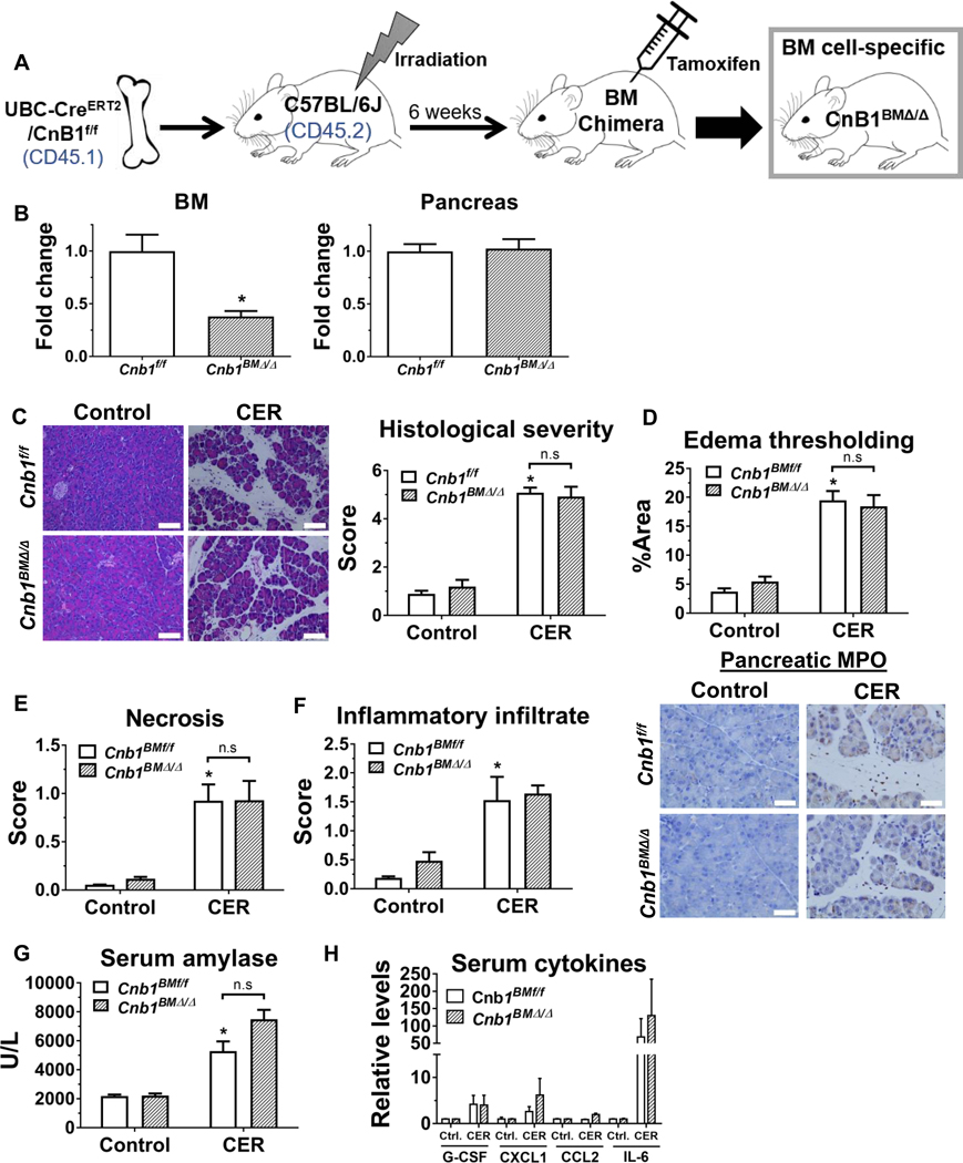
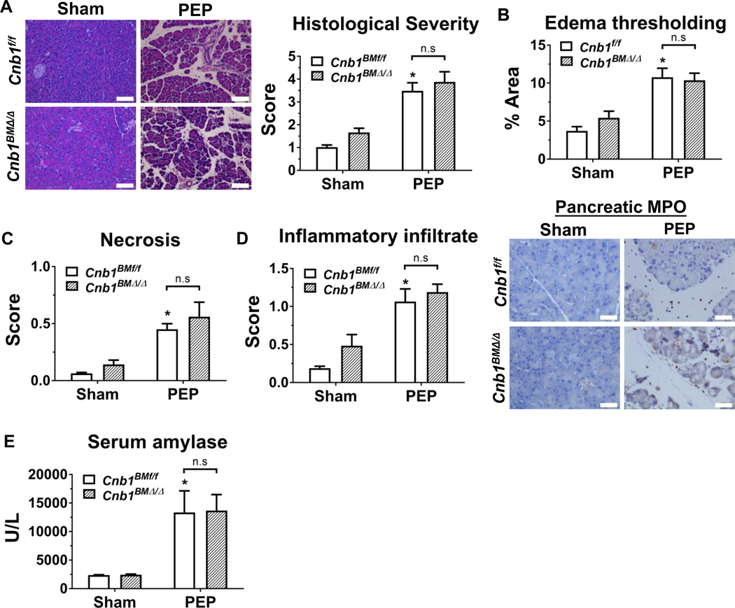
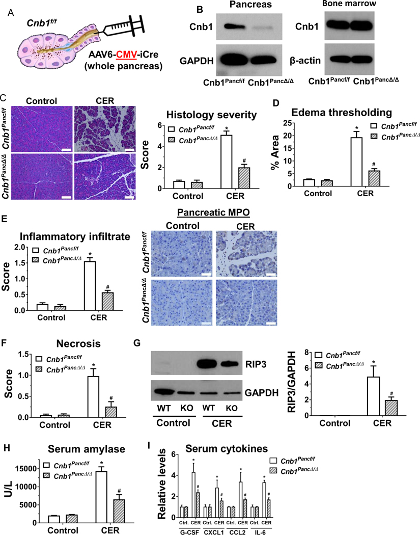
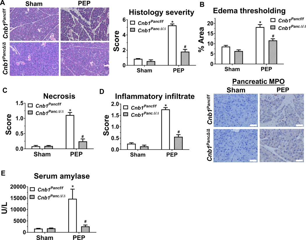
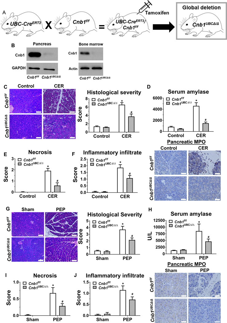
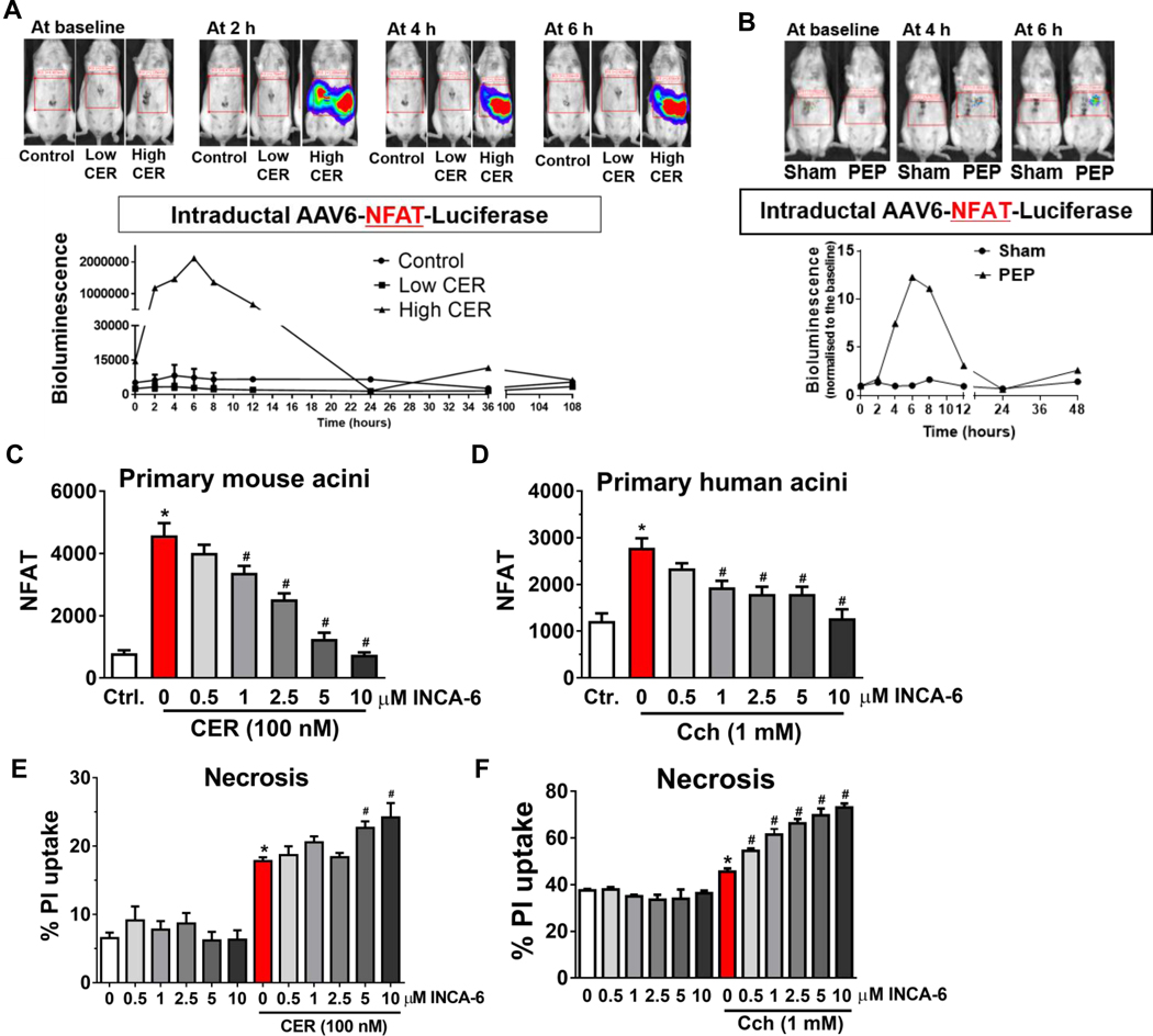
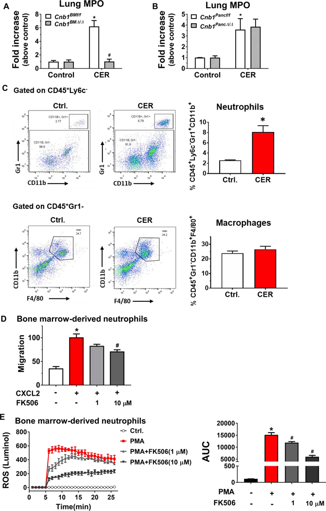
Methods: We performed studies with mice with hematopoietic-specific or pancreas-specific deletion of protein phosphatase 3, regulatory subunit B, alpha isoform (PPP3R1, also called CNB1), in mice with deletion of CNB1 (Cnb1UBC△/△) and in the corresponding controls for each deletion of CNB1. Acute pancreatitis was induced in mice by administration of caerulein or high-pressure infusion of radiocontrast into biliopancreatic ducts; some mice were also given intraductal infusions of an adeno-associated virus vector that expressed nuclear factor of activated T -cells (NFAT)-luciferase into pancreas. Pancreas, bone marrow, liver, kidney, heart, and lung were collected and analyzed by histopathology, immunohistochemistry, and immunoblots; levels of cytokines were measured in serum. Mouse and human primary pancreatic acinar cells were transfected with a vector that expressed NFAT-luciferase and incubated with an agent that blocks interaction of NFAT with calcineurin; cells were analyzed by immunofluorescence. Calcineurin-mediated neutrophil chemotaxis and reactive oxygen species production were measured in neutrophils from mice.

Results: Mice with hematopoietic-specific deletion of CNB1 developed the same level of local pancreatic inflammation as control mice after administration of caerulein or infusion of radiocontrast into biliopancreatic ducts. Cnb1UBC△/△ mice or mice with pancreas-specific deletion of CNB1 developed less severe pancreatitis and reduced pancreatic inflammation after administration of caerulein or infusion of radiocontrast into biliopancreatic ducts compared with control mice. NFAT was activated in pancreas of Swiss Webster mice given caerulein or infusions of radiocontrast into biliopancreatic ducts. Blocking the interaction between calcineurin and NFAT did not reduce pancreatic acinar cell necrosis in response to caerulein or infusions of radiocontrast. Mice with hematopoietic-specific deletion of CNB1 (but not mice with pancreas-specific deletion of CNB1) had reduced infiltration of lung tissues by neutrophils. Neutrophil chemotaxis and production of reactive oxygen species were decreased after incubation with a calcineurin inhibitor.

Conclusions: Hematopoietic and neutrophil expression of calcineurin promotes pancreatitis-associated lung inflammation, whereas pancreatic calcineurin promotes local pancreatic inflammation. The findings indicate that the protective effects of blocking or deleting calcineurin on pancreatitis are mediated by the source of its expression. This information should be used in the development of strategies to inhibit calcineurin for the prevention of pancreatitis and pancreatitis-associated lung inflammation.

Keywords: PEP; calcium signaling; drug development; targeted calcineurin inhibition.


Conflict of interest statement

Conflict of interest statement: S.Z.H has equity in Prevcon, a startup that has sublicensed a patent on the use of calcineurin inhibitors in pancreatitis. All the other authors have no conflict of interest.

Figures

Figure 1.
Figure 1.. During caerulein hyperstimulation pancreatitis (CER), hematopoietic compartment-specific deletion of CNB1 does not affect pancreatic localized injury.
(A) A hematopoietic compartment-specific calcineurin conditional knockout line (Cnb1BMΔ/Δ) was generated by adoptively transferring bone marrow (BM) cells from UBC-CreERT2/Cnb1f/f mice to the irradiated C57BL/6J mice, followed by tamoxifen administration. (B) RT-qPCR for Cnb1 expression in BM cells and the pancreas from Cnb1BMf/f and Cnb1BMΔ/Δ mice. (C) Representative H&E images of the pancreas (200X) from the control and CER conditions from Cnb1BMf/f and Cnb1BMΔ/Δ mice, along with overall histological severity. (D) Edema thresholding by ImageJ. (E) Subscoring for necrosis. (F) Subscoring for inflammatory infiltrate along with immunostaining for pancreatic myeloperoxidase (MPO) (400X). (G) Serum amylase, (H) serum cytokines assessed by Luminex multiplex assay. n=5 animals per conditions from two batches of animals; *p<0.05, compared to the controls; #p<0.05, compared to the Cnb1BMf/f group.
Figure 2.
Figure 2.. Hematopoietic-specific deletion of CNB1, protects against post-ERCP pancreatitis (PEP).
(A) Representative H&E images of the pancreas (200X) from the sham and PEP conditions from Cnb1BMf/f and Cnb1BMΔ/Δ mice along with overall histological severity scoring. (B) Edema thresholding by ImageJ. (C) Subscoring for necrosis. (D) Subscoring for inflammatory infiltrate and immunostaining for pancreatic myeloperoxidase (MPO) (400X). (E) Serum amylase. n=5 animals per groups; *p<0.05, compared to the controls; #p<0.05, compared to the Cnb1f/f groups.
Figure 3.
Figure 3.. During caerulein hyperstimulation pancreatitis (CER), pancreas-specific deletion of CNB1 largely prevents localized pancreatic injury and systemic inflammation, but it does not affect distant organ damage.
(A) Schema for intraductal infusion of AAV6-CMV-iCre into Cnb1f/f mice to specifically delete CNB1 in the whole pancreas. (B) Western blot for CNB1 from pancreas and BM lysates. AAV, adeno-associated virus. (C) Representative H&E images of the pancreas (200X) from the control and CER conditions from AAV6 control-infused (Cnb1Pancf/f) and AAV6-CMV-iCre-infused (Cnb1PancΔ/Δ) groups, along with overall histological severity scoring. (D) Edema thresholding by Image. (E) Subscoring for inflammatory infiltrate and immunostaining for pancreatic MPO (400X). (F) Subscoring for necrosis. (G) Immunoblotting and densitometry analysis for RIP3 from pancreas lysates. (I) Serum amylase. (J) Serum cytokines assessed by Luminex multiplex assay. n=5 animals per conditions from two batches of animals; *p<0.05, compared to the controls; #p<0.05, compared to the Cnb1Pancf/f group.
Figure 4.
Figure 4.. Pancreas-specific deletion of CNB1 protects against post-ERCP pancreatitis (PEP).
(A) Representative H&E images of the pancreas (200X) from the sham and PEP conditions from Cnb1pancf/f and Cnb1PancΔ/Δ mice along with overall histological severity scoring. (B) Edema thresholding by ImageJ. (C) Subscoring for necrosis. (D) Subscoring for inflammatory infiltrate and immunostaining for pancreatic MPO (400X). (E) Serum amylase. n=5 animals per groups; *p<0.05, compared to the controls; #p<0.05, compared to the Cnb1f/f groups.
Figure 5.
Figure 5.. Global deletion of CNB1 protects against two disparate models of acute pancreatitis.
(A) The global Cnb1 conditional knockout line (Cnb1UBCΔ/Δ) was induced by crossing UBC-CreERT2 mice with Cnb1f/f mice, followed by tamoxifen administration. (B) Western blotting confirms negligible Cnb1 expression in the pancreas and BM cells from the Cnb1UBCΔ/Δ mice. (C) Representative H&E images of the pancreas (200X) from the control and CER conditions from Cnb1f/f and Cnb1UBCΔ/Δ mice and overall histological severity scoring. (D) Serum amylase. (E) Subscoring for necrosis. (F) Subscoring for inflammatory infiltrate and immunostaining for pancreatic myeloperoxidase (MPO) (400X). (G) Representative H&E images of the pancreas (200X) from the sham and PEP conditions from Cnb1f/f and Cnb1UBCΔ/Δ mice and overall histological severity scoring. (H) Serum amylase. (I) Subscoring for necrosis. (J) Subscoring for inflammatory infiltrate and immunostaining for pancreatic MPO (400X). n=5 animals per conditions; *p<0.05, compared to the controls; #p<0.05, compared to the Cnb1f/f condition.
Figure 6.
Figure 6.. The calcineurin target NFAT is activated in the pancreas during acute pancreatitis, but inhibition of acinar cell NFAT does not reduce acinar necrosis.
(A) Images and quantification of the pancreatic nuclear factor of activated T cells (NFAT) signal at baseline, 2, 4, and 6 h after administration of low or high dose of caerulein. (B) Images and quantification of the pancreatic NFAT signal at baseline, 4, and 6 h after PEP induction. NFAT activity with or without INCA-6 in isolated pancreatic acinar cells from (C) mouse and (D) human. PI uptake with or without INCA-6 in isolated pancreatic acinar cells from (E) mouse and (F) human.
Figure 7.
Figure 7.. Inactivation of calcineurin modulates lung neutrophil infiltration in vivo and neutrophil migration and ROS production ex vivo.
(A) Lung MPO activity from hematopoietic compartment-specific calcineurin deletion. (B) Lung MPO activity from pancreas-specific calcineurin deletion. (C) Lung leukocytes were isolated from the control and caerulein hyperstimulation pancreatitis (CER) conditions and analyzed by flow cytometry for neutrophil (gated on CD45+Ly6c, then CD11b+Gr1+) and macrophages (gated on CD45+Gr1, then CD11b+ F4/80+) numbers. n=5 animals per condition. (D) Percentage of migrated BM derived neutrophil (BMDN) from Transwell migration assay stimulated with CXCL2 in the presence or absence of FK506 pre-treatment. (E) Neutrophil total ROS production in response to phorbol 12-myristate 13-acetate (PMA) with or without FK506 pretreatment was assayed using luminol and further quantified by an area-under-the curve. 3 independent BMDN isolations; *p<0.05, compared to the control; #p<0.05, compared to the CXCL2- or PMA-stimulated condition.

Comment in

References

    1. Lee PJ, Papachristou GI. New insights into acute pancreatitis. Nat Rev Gastroenterol Hepatol 2019. - PubMed
    1. Forsmark CE, Vege SS, Wilcox CM. Acute Pancreatitis. N Engl J Med 2016;375:1972–81. - PubMed
    1. Sah RP, Garg P, Saluja AK. Pathogenic mechanisms of acute pancreatitis. Curr Opin Gastroenterol 2012;28:507–15. - PMC - PubMed
    1. Peery AF, Crockett SD, Barritt AS, et al. Burden of Gastrointestinal, Liver, and Pancreatic Diseases in the United States. Gastroenterology 2015;149:1731–41.e3. - PMC - PubMed
    1. Lankisch PG, Apte M, Banks PA. Acute pancreatitis. Lancet 2015;386:85–96. - PubMed

Publication types

MeSH terms