. 2020 Aug 20:156:11-19.
doi: 10.1016/j.freeradbiomed.2020.05.004. Epub 2020 May 20.

Exploring the FMN binding site in the mitochondrial outer membrane protein mitoNEET

Affiliations

Exploring the FMN binding site in the mitochondrial outer membrane protein mitoNEET

Homyra Tasnim et al. Free Radic Biol Med. .

Abstract

MitoNEET is a mitochondrial outer membrane protein that hosts a redox active [2Fe-2S] cluster in the C-terminal cytosolic domain. Increasing evidence has shown that mitoNEET has an essential role in regulating energy metabolism in human cells. Previously, we reported that the [2Fe-2S] clusters in mitoNEET can be reduced by the reduced flavin mononucleotide (FMNH2) and oxidized by oxygen or ubiquinone-2, suggesting that mitoNEET may act as a novel redox enzyme catalyzing electron transfer from FMNH2 to oxygen or ubiquinone. Here, we explore the FMN binding site in mitoNEET by using FMN analogs and find that lumiflavin, like FMN, at nanomolar concentrations can mediate the redox transition of the mitoNEET [2Fe-2S] clusters in the presence of flavin reductase and NADH (100 μM) under aerobic conditions. The electron paramagnetic resonance (EPR) measurements show that both FMN and lumiflavin can dramatically change the EPR spectrum of the reduced mitoNEET [2Fe-2S] clusters and form a covalently bound complex with mitoNEET under blue light exposure, suggesting that FMN/lumiflavin has specific interactions with the [2Fe-2S] clusters in mitoNEET. In contrast, lumichrome, another FMN analog, fails to mediate the redox transition of the mitoNEET [2Fe-2S] clusters and has no effect on the EPR spectrum of the reduced mitoNEET [2Fe-2S] clusters under blue light exposure. Instead, lumichrome can effectively inhibit the FMNH2-mediated reduction of the mitoNEET [2Fe-2S] clusters, indicating that lumichrome may act as a potential inhibitor to block the electron transfer activity of mitoNEET.

Keywords: Electron transfer activity; FMN; Lumichrome; Lumiflavin; MitoNEET; Mitochondria.


Conflict of interest statement

Declaration of competing interest The authors declare no competing interests.

Figures

Figure 1.
Figure 1.. Electron transfer activity of mitoNEET at different concentrations of FMN under aerobic conditions.
A), reduction and oxidation of the mitoNEET [2Fe-2S] clusters. MitoNEET (containing 10 μM [2Fe-2S] clusters) was incubated with NADH (100 μM), and FMN (50 nM) in buffer containing NaCl (500 mM) and Tris (20 mM, pH 8.0) under aerobic conditions. The reaction was initiated by adding flavin reductase (Fre) (at a final concentration of 0.25 μM). The redox state of the mitoNEET [2Fe-2S] clusters in the reaction solutions was monitored by taking UV-Visible absorption spectra every two minutes for 20 min. B), kinetics of reduction and oxidation of the mitoNEET [2Fe-2S] clusters. MitoNEET (containing 10 μM [2Fe-2S] clusters) was incubated with NADH (100 μM) and FMN (0 to 250 nM as indicated) in buffer containing NaCl (500 mM) and Tris (20 mM, pH 8.0) under aerobic conditions. The reaction was initiated by adding flavin reductase (Fre) (at a final concentration of 0.25 μM). The relative amounts of the oxidized mitoNEET [2Fe-2S] clusters were monitored at the absorption peak of 458 nm and plotted as a function of time after addition of flavin reductase. The data were representatives from three independent experiments.
Figure 2.
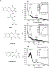
Figure 2.. Lumiflavin can substitute FMN in mediating the redox transition of the mitoNEET [2Fe-2S] clusters under aerobic conditions.
A), structure of FMN, lumiflavin, and lumichrome. B), reduction and oxidation of the mitoNEET [2Fe-2S] clusters mediated by FMN, lumiflavin, and lumichrome. MitoNEET (containing 10 μM [2Fe-2S] clusters) was incubated with NADH (100 μM), and FMN (250 nM) in buffer containing NaCl (50 mM) and Tris (20 mM, pH 8.0) under aerobic conditions. The reaction was initiated by adding flavin reductase (Fre) (at a final concentration of 0.25 μM). C), same as B), except FMN was replaced with lumiflavin (250 nM). D), same as B), except FMN was replaced with lumichrome (250 nM). The data were representatives of three independent experiments.
Figure 3.
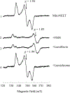
Figure 3.. Effect of FMN analogs on the EPR spectrum of the mitoNEET [2Fe-2S] clusters.
MitoNEET (containing 10 μM [2Fe-2S] clusters) (spectrum 1) was incubated with 50 μM of FMN (spectrum 2), 50 μM of lumiflavin (spectrum 3), or 50 μM of lumichrome (spectrum 4) at room temperature for 10 min under blue light exposure (50,000 Lux). The samples were then reduced with freshly prepared sodium dithionite (10 mM), transferred to EPR tubes, and frozen immediately in liquid nitrogen until EPR measurements. The reduced mitoNEET [2Fe-2S] clusters have an average g = 1.94. The reduced mitoNEET [2Fe-2S] clusters after incubation with FMN or lumiflavin under blue light exposure have a new EPR signal at g = 1.85.
Figure 4.
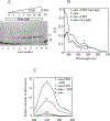
Figure 4.. MitoNEET forms a covalently bound complex with FMN under blue light exposure.
A), the SDS-PAGE analysis of mitoNEET after incubation with different concentrations of FMN under blue light exposure. MitoNEET (containing 10 μM [2Fe-2S] clusters) was incubated with FMN (0 to 40 μM) under blue light exposure (50,000 Lux) for 10 min. Aliquots were taken from the samples for the SDS-PAGE analysis. Lane 1, mitoNEET. Lanes 2–8, mitoNEET incubated with 0, 1, 2, 5, 10, 20, 40 μM of FMN under blue light exposure for 10 min, respectively. Lane 9, MitoNEET incubated with FMN (40 μM) for 10 min without blue light exposure. On right side of the gel, M, monomeric mitoNEET; D, dimeric mitoNEET.B), UV-Visible absorption spectra of mitoNEET after incubation with FMN with or without blue light exposure. MitoNEET (containing 10 μM [2Fe-2S] clusters) was incubated with or without FMN (40 μM) under blue light exposure (50,000 Lux) for 10 min. MitoNEET protein was re-purified from the incubation solutions by passing through a Desalting Column. Re-purified mitoNEET samples were subjected to the UV-Visible absorption measurements. C), quantification of FMN in the re-purified mitoNEET proteins. Re-purified mitoNEET proteins (5 μM) treated with or without 0.2% SDS were subjected to the fluorescence measurements in a spectrofluorometer (FP-6300, JASCO Co., Japan). The emission spectra of FMN were taken upon excitation at 450 nm. Spectrum 1, re-purified mitoNEET after incubation with FMN under blue light exposure was treated with 0.2% SDS. Spectrum 2, re-purified mitoNEET after incubation with FMN under blue light exposure. Spectrum 3, mitoNEET (not incubated with FMN) was treated with 0.2 % SDS. Spectrum 4, mitoNEET (control). Based on the fluorescence intensity of FMN in the mitoNEET incubated with FMN under blue light exposure and treated with 0.2% SDS, we estimated that there was at least 0.2 FMN per mitoNEET monomer (n=3).
Figure 5.
Figure 5.. Effect of FMN on other [2Fe-2S] proteins from human mitochondria.
Three human iron-sulfur proteins (Miner1, Miner2 and ferredoxin-2) were prepared and incubated with FMN under blue light exposure (50,000 Lux) for 10 min. A), purified proteins (each containing 10 μM [2Fe-2S] clusters) were reduced with freshly prepared sodium dithionite (4 mM) without incubation with FMN. Spectrum 1, mitoNEET; spectrum 2, Miner1; spectrum 3, Miner2; Spectrum 4, ferredoxin-2. B), purified protein (containing 10 μM [2Fe-2S] clusters) was incubated with FMN (50 μM) under blue light exposure for 10 min, followed by addition of sodium dithionite (4 mM). Spectrum 1, mitoNEET; spectrum 2, Miner1; spectrum 3, Miner2; Spectrum 4, ferredoxin-2. The data are representatives of three independent experiments.
Figure 6.
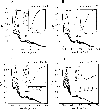
Figure 6.. Inhibition of the electron transfer activity of mitoNEET by lumichrome.
A), reduction and oxidation of the mitoNEET [2Fe-2S] clusters without lumichrome. Purified mitoNEET (containing 10 μM [2Fe-2S] clusters) was incubated with NADH (100 μM), and FMN (50 nM) in buffer containing NaCl (500 mM) and Tris (20 mM, pH 8.0) under aerobic conditions. The reaction was initiated by adding flavin reductase (Fre) (at a final concentration of 0.25 μM). The redox state of the mitoNEET [2Fe-2S] clusters was monitored by taking UV-Visible absorption spectra every 2 minutes after addition of flavin reductase. B), same as in A), except 50 nM of lumichrome was included in the incubation solution before the reaction was initiated. C), same as in A), except 100 nM of lumichrome was included in the incubation solution before the reaction was initiated. D), same as in A, except 250 nM of lumichrome was included in the incubation solution before the reaction was initiated. The data are representatives of three independent experiments.
Figure 7.
Figure 7.. A proposed model for the electron transfer activity of mitoNEET and inhibition by lumichrome.
In cytosol, NADH generated by glycolysis is oxidized by flavin reductase and FMN is reduced to FMNH2. FMNH2 interacts with mitoNEET via specific binding site and transfers its electrons to the [2Fe-2S] clusters of mitoNEET dimer which localizes on the mitochondrial outer membrane (MOM). MIM, mitochondrial inner membrane. The reduced [2Fe-2S] clusters in mitoNEET dimer transfer the electrons to oxygen or ubiquinone. Lumichrome, which has an alloxazine group as FMN, may compete with FMN for the FMN binding site in mitoNEET and inhibit the electron transfer activity of mitoNEET.

References

    1. Colca JR; McDonald WG; Waldon DJ; Leone JW; Lull JM; Bannow CA; Lund ET; Mathews WR Identification of a novel mitochondrial protein (“mitoNEET”) cross-linked specifically by a thiazolidinedione photoprobe. Am. J. Physiol. Endocrinol. Metab 286:E252–E260; 2004. - PubMed
    1. Tamir S; Paddock ML; Darash-Yahana-Baram M; Holt SH; Sohn YS; Agranat L; Michaeli D; Stofleth JT; Lipper CH; Morcos F; Cabantchik IZ; Onuchic JN; Jennings PA; Mittler R; Nechushtai R Structure-function analysis of NEET proteins uncovers their role as key regulators of iron and ROS homeostasis in health and disease. Biochim. Biophys. Acta 1853:1294–1315; 2015. - PubMed
    1. Mittler R; Darash-Yahana M; Sohn YS; Bai F; Song L; Cabantchik IZ; Jennings PA; Onuchic JN; Nechushtai R NEET Proteins: A New Link Between Iron Metabolism, Reactive Oxygen Species, and Cancer. Antioxidants & redox signaling 30:1083–1095; 2019. - PMC - PubMed
    1. Sohn YS; Tamir S; Song L; Michaeli D; Matouk I; Conlan AR; Harir Y; Holt SH; Shulaev V; Paddock ML; Hochberg A; Cabanchick IZ; Onuchic JN; Jennings PA; Nechushtai R; Mittler R NAF-1 and mitoNEET are central to human breast cancer proliferation by maintaining mitochondrial homeostasis and promoting tumor growth. Proc. Natl. Acad. Sci. U. S. A 110:14676–14681; 2013. - PMC - PubMed
    1. He QQ; Xiong LL; Liu F; He X; Feng GY; Shang FF; Xia QJ; Wang YC; Qiu DL; Luo CZ; Liu J; Wang TH MicroRNA-127 targeting of mitoNEET inhibits neurite outgrowth, induces cell apoptosis and contributes to physiological dysfunction after spinal cord transection. Sci. Rep 6:35205; 2016. - PMC - PubMed

Publication types

MeSH terms