

Structural profile of dome-shaped macula in degenerative myopia and its association with macular disorders
- PMID: 32448138
- PMCID: PMC7247247
- DOI: 10.1186/s12886-020-01473-2
Structural profile of dome-shaped macula in degenerative myopia and its association with macular disorders
Abstract
Background: To evaluate the detailed structural profile of dome-shaped macula and its association with myopic macular complications.
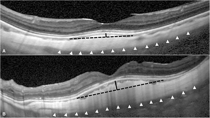
Methods: This retrospective study included 147 eyes of 93 patients who were diagnosed with degenerative myopia. The height of the scleral dome and diameter of the dome base were measured via enhanced depth imaging optical coherence tomography images with 1:1 μm setting. Spherical equivalent and best-corrected visual acuity were compared in eyes with and without dome-shaped macula. In eyes with dome-shaped macula, the height and diameter of the dome were compared in eyes with and without myopic macular complications including choroidal neovascularization, myopic foveoschisis, and macular hole.
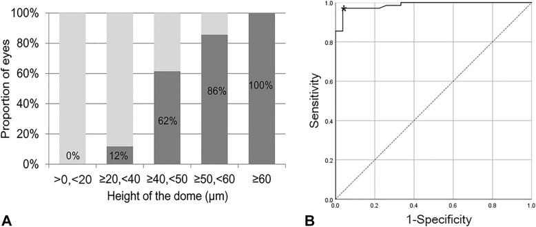
Results: Dome-shaped macula was noted in 60 eyes (40.8%) of 42 patients. The mean height of the dome in the eyes with dome-shaped macula was 126.5 ± 69.4 μm (53 to 345 μm) and the mean diameter of the dome base was 2862.1 ± 794.9 μm (1567 μm to 4886 μm). In comparing eyes with and without dome-shaped macula, eyes with dome-shaped macula had higher myopia (- 13.7 diopters vs - 12.1 diopters, P = 0.022). There was no difference in visual acuity in eyes with or without dome-shaped macula (P = 0.132). The height and diameter of the dome in eyes with and without myopic foveoschisis were 78.6 ± 20.6 μm and 134.9 ± 71.6 μm, 2499.2 ± 303.1 μm and 2969.3 ± 645.7 μm, respectively (P = 0.009 and P = 0.017). However, the height and diameter of the dome were not related to the incidence of a macular hole (P = 0.324 and P = 0.605) and choroidal neovascularization (P = 0.835 and P = 0.905).
Conclusions: The prevalence of dome-shaped macula was about 40% in the eyes with degenerative myopia. Although dome-shaped macula was associated with higher degrees of myopia, a prominent dome seemed to be protective against myopic foveoschisis.
Keywords: Degenerative myopia; Dome-shaped macula; Myopic foveoschisis.
Conflict of interest statement
The authors declare that they have no competing interests.
Figures
Similar articles
-
Two- and three-dimensional topographic analysis of pathologically myopic eyes with dome-shaped macula and inferior staphyloma by spectral domain optical coherence tomography.Graefes Arch Clin Exp Ophthalmol. 2017 May;255(5):903-912. doi: 10.1007/s00417-017-3587-z. Epub 2017 Jan 17. Graefes Arch Clin Exp Ophthalmol. 2017. PMID: 28097437
-
Factors associated with macular complications in highly myopic eyes with dome-shaped macular configuration.Graefes Arch Clin Exp Ophthalmol. 2019 Nov;257(11):2357-2365. doi: 10.1007/s00417-019-04449-1. Epub 2019 Sep 4. Graefes Arch Clin Exp Ophthalmol. 2019. PMID: 31485730
-
MORPHOLOGICAL CHARACTERISTICS AND VISUAL ACUITY OF HIGHLY MYOPIC EYES WITH DIFFERENT SEVERITIES OF MYOPIC MACULOPATHY.Retina. 2020 Mar;40(3):461-467. doi: 10.1097/IAE.0000000000002418. Retina. 2020. PMID: 30576301
-
Myopic macular diseases: A review.Clin Exp Ophthalmol. 2023 Apr;51(3):229-242. doi: 10.1111/ceo.14200. Epub 2023 Jan 11. Clin Exp Ophthalmol. 2023. PMID: 36594934 Review.
-
Insight into high myopia and the macula.Indian J Ophthalmol. 2017 Feb;65(2):85-91. doi: 10.4103/ijo.IJO_863_16. Indian J Ophthalmol. 2017. PMID: 28345561 Free PMC article. Review.
Cited by
-
Semi-Automated Analysis of Dome-Shaped Macula in Preterm and Full-Term Infants Using Handheld Swept-Source Optical Coherence Tomography.Invest Ophthalmol Vis Sci. 2024 Oct 1;65(12):35. doi: 10.1167/iovs.65.12.35. Invest Ophthalmol Vis Sci. 2024. PMID: 39441581 Free PMC article.
-
Resolution of Myopic Macular Retinoschisis and Macular Hole With Topical Medical Therapy.J Vitreoretin Dis. 2025 May 15:24741264251340107. doi: 10.1177/24741264251340107. Online ahead of print. J Vitreoretin Dis. 2025. PMID: 40384924 Free PMC article.
-
Development of a nomogram for predicting myopia risk among school-age children: a case-control study.Ann Med. 2024 Dec;56(1):2331056. doi: 10.1080/07853890.2024.2331056. Epub 2024 Mar 20. Ann Med. 2024. PMID: 38507901 Free PMC article.
-
Clinical characteristics of dome-shaped macula in mild myopic or non-myopic eyes.Eye (Lond). 2025 Jun;39(9):1787-1792. doi: 10.1038/s41433-025-03756-8. Epub 2025 Mar 17. Eye (Lond). 2025. PMID: 40097745
References
Publication types
MeSH terms
LinkOut - more resources
Full Text Sources
Medical
