

Human bone marrow mesenchymal stem cell-derived exosomes stimulate cutaneous wound healing mediates through TGF-β/Smad signaling pathway
- PMID: 32448395
- PMCID: PMC7245763
- DOI: 10.1186/s13287-020-01723-6
Human bone marrow mesenchymal stem cell-derived exosomes stimulate cutaneous wound healing mediates through TGF-β/Smad signaling pathway
Abstract
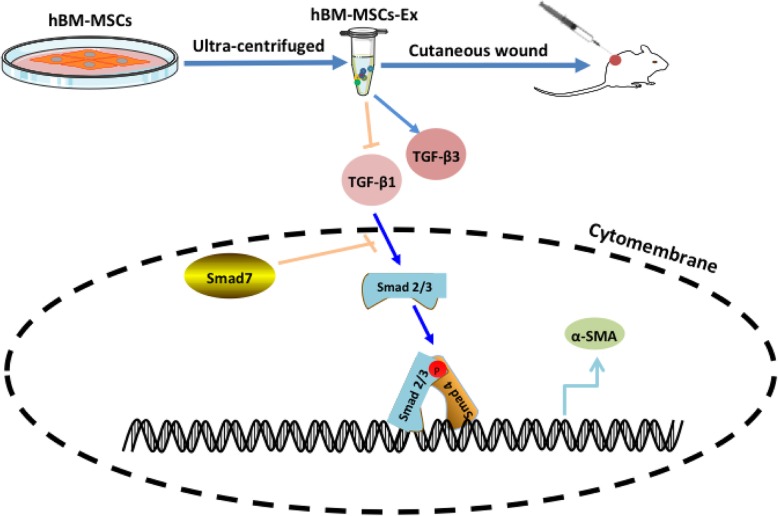
Background: Cutaneous wound healing represents a morphogenetic response to injury and is designed to restore anatomic and physiological function. Human bone marrow mesenchymal stem cell-derived exosomes (hBM-MSC-Ex) are a promising source for cell-free therapy and skin regeneration.
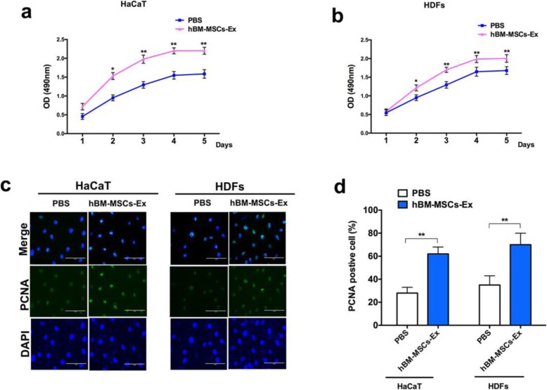
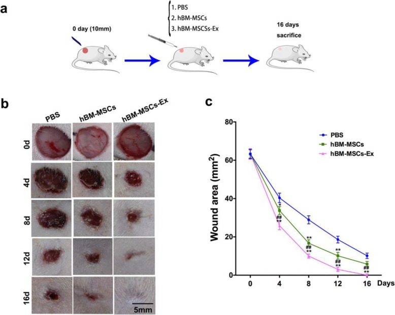
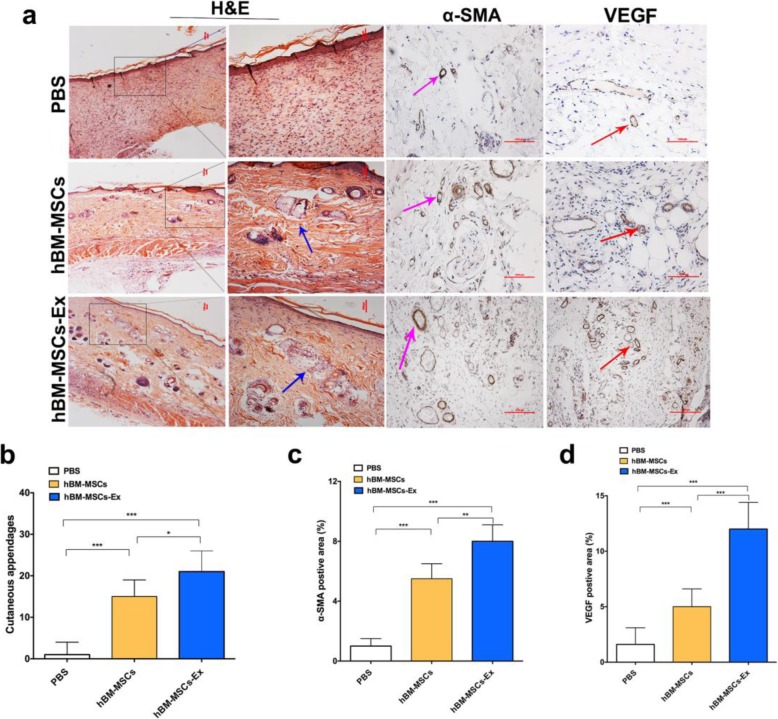
Methods: In this study, we investigated the cell regeneration effects and its underlying mechanism of hBM-MSC-Ex on cutaneous wound healing in rats. In vitro studies, we evaluated the role of hBM-MSC-Ex in the two types of skin cells: human keratinocytes (HaCaT) and human dermal fibroblasts (HDFs) for the proliferation. For in vivo studies, we used a full-thickness skin wound model to evaluate the effects of hBM-MSC-Ex on cutaneous wound healing in vivo.
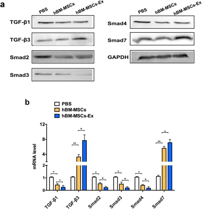
Results: The results demonstrated that hBM-MSC-Ex promote both two types of skin cells' growth effectively and accelerate the cutaneous wound healing. Interestingly, we found that hBM-MSC-Ex significantly downregulated TGF-β1, Smad2, Smad3, and Smad4 expression, while upregulated TGF-β3 and Smad7 expression in the TGF-β/Smad signaling pathway.
Conclusions: Our findings indicated that hBM-MSC-Ex effectively promote the cutaneous wound healing through inhibiting the TGF-β/Smad signal pathway. The current results provided a new sight for the therapeutic strategy for the treatment of cutaneous wounds.
Keywords: Exosomes; Human bone marrow mesenchymal stem cells; TGF-β/Smad signaling; Wound healing.
Conflict of interest statement
The authors declared no potential conflicts of interest with respect to the research, authorship, and/or publication of this article.
Figures
Similar articles
-
Exosomes derived from human umbilical cord blood mesenchymal stem cells stimulate regenerative wound healing via transforming growth factor-β receptor inhibition.Stem Cell Res Ther. 2021 Aug 3;12(1):434. doi: 10.1186/s13287-021-02517-0. Stem Cell Res Ther. 2021. PMID: 34344478 Free PMC article.
-
Hypoxic conditioned medium from human amniotic fluid-derived mesenchymal stem cells accelerates skin wound healing through TGF-β/SMAD2 and PI3K/Akt pathways.Int J Mol Sci. 2014 Jan 6;15(1):605-28. doi: 10.3390/ijms15010605. Int J Mol Sci. 2014. PMID: 24398984 Free PMC article.
-
Bone marrow mesenchymal stem cells facilitate diabetic wound healing through the restoration of epidermal cell autophagy via the HIF-1α/TGF-β1/SMAD pathway.Stem Cell Res Ther. 2022 Jul 15;13(1):314. doi: 10.1186/s13287-022-02996-9. Stem Cell Res Ther. 2022. PMID: 35841007 Free PMC article.
-
Mesenchymal stem cell-derived exosomes: A novel and potential remedy for cutaneous wound healing and regeneration.World J Stem Cells. 2022 May 26;14(5):318-329. doi: 10.4252/wjsc.v14.i5.318. World J Stem Cells. 2022. PMID: 35722196 Free PMC article. Review.
-
Stem cell-derived exosomes: emerging therapeutic opportunities for wound healing.Stem Cell Res Ther. 2023 Apr 26;14(1):107. doi: 10.1186/s13287-023-03345-0. Stem Cell Res Ther. 2023. PMID: 37101197 Free PMC article. Review.
Cited by
-
MSCs and their exosomes: a rapidly evolving approach in the context of cutaneous wounds therapy.Stem Cell Res Ther. 2021 Dec 4;12(1):597. doi: 10.1186/s13287-021-02662-6. Stem Cell Res Ther. 2021. PMID: 34863308 Free PMC article. Review.
-
Small extracellular vesicles: the origins, current status, future prospects, and applications.Stem Cell Res Ther. 2025 Apr 17;16(1):184. doi: 10.1186/s13287-025-04330-5. Stem Cell Res Ther. 2025. PMID: 40247402 Free PMC article. Review.
-
The Role of Exosomes Derived From Mesenchymal Stromal Cells in Dermatology.Front Cell Dev Biol. 2021 Apr 7;9:647012. doi: 10.3389/fcell.2021.647012. eCollection 2021. Front Cell Dev Biol. 2021. PMID: 33898436 Free PMC article. Review.
-
Stem Cell Therapy for Burns: Story so Far.Biologics. 2021 Aug 31;15:379-397. doi: 10.2147/BTT.S259124. eCollection 2021. Biologics. 2021. PMID: 34511880 Free PMC article. Review.
-
CRISPR-dCas9 Activation of TSG-6 in MSCs Modulates the Cargo of MSC-Derived Extracellular Vesicles and Attenuates Inflammatory Responses in Human Intervertebral Disc Cells In Vitro.Cell Mol Bioeng. 2025 Feb 5;18(1):83-98. doi: 10.1007/s12195-025-00843-4. eCollection 2025 Feb. Cell Mol Bioeng. 2025. PMID: 39949490 Free PMC article.
References
-
- Singer AJ, Clark RA. Cutaneous wound healing. N Engl J Med. 1999;341(10):738–746. - PubMed
-
- McFarlin K, Gao X, Liu YB, Dulchavsky DS, Kwon D, Arbab AS, Bansal M, Li Y, Chopp M, Dulchavsky SA, et al. Bone marrow-derived mesenchymal stromal cells accelerate wound healing in the rat. Wound Repair Regen. 2006;14(4):471–478. - PubMed
-
- Kim WS, Park BS, Sung JH, Yang JM, Park SB, Kwak SJ, Park JS. Wound healing effect of adipose-derived stem cells: a critical role of secretory factors on human dermal fibroblasts. J Dermatol Sci. 2007;48(1):15–24. - PubMed
-
- Francois S, Mouiseddine M, Mathieu N, Semont A, Monti P, Dudoignon N, Sache A, Boutarfa A, Thierry D, Gourmelon P, et al. Human mesenchymal stem cells favour healing of the cutaneous radiation syndrome in a xenogenic transplant model. Ann Hematol. 2007;86(1):1–8. - PubMed
Publication types
MeSH terms
Substances
LinkOut - more resources
Full Text Sources
Miscellaneous
