

A Resected Case of Follicular Cholangitis That Was Positive on 18F-fluorodeoxyglucose-positron Emission Tomography
- PMID: 32448841
- PMCID: PMC7516323
- DOI: 10.2169/internalmedicine.4611-20
A Resected Case of Follicular Cholangitis That Was Positive on 18F-fluorodeoxyglucose-positron Emission Tomography
Abstract
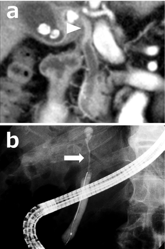
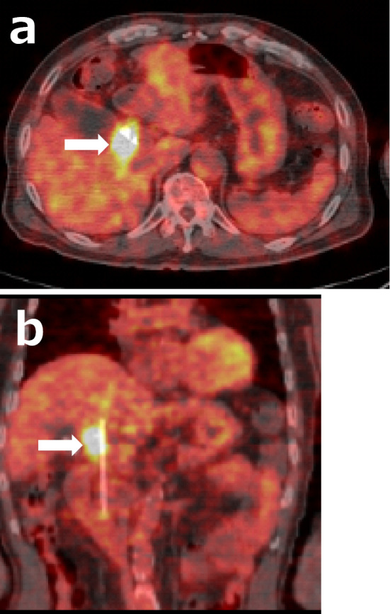
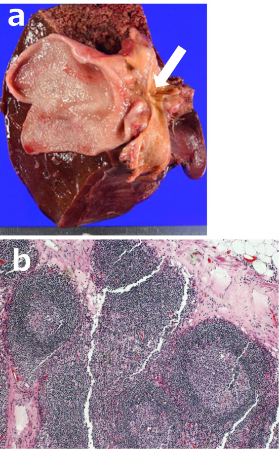
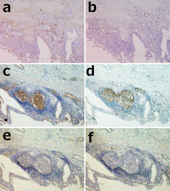
We experienced a case of follicular cholangitis that was positive on fluorodeoxyglucose-positron emission tomography (18F-FDG-PET). A 70-year-old man was admitted for jaundice. Endoscopic retrograde cholangiography showed stenosis of the middle to upper choledocus. 18F-FDG-PET depicted a localized hot spot at the stenotic lesion (maximum standardized uptake value = 8.2). Although no malignant findings were found in the cytology or on a bile duct biopsy, malignancy could not be excluded, so surgical treatment was performed. Follicular cholangitis is a new, rare disease that causes severe biliary stricture. Only 11 cases of follicular cholangitis have been reported, including the present case.
Keywords: 18F-FDG-PET; biliary stricture; cholangiocarcinoma; follicular cholangitis.
Conflict of interest statement
Figures
References
-
- Aoki T, Kubota K, Oka T, Hasegawa K, Hirai I, Makuuchi M. Follicular cholangitis: another cause of benign biliary stricture. Hepatogastroenterology 50: 639-642, 2003. - PubMed
-
- Lee JY, Lim JH, Lim HK. Follicular cholangitis mimicking hilar cholangiocarcinoma. Abdom Imaging 30: 744-747, 2005. - PubMed
-
- Fujita T, Kojima M, Kato Y, et al. Clinical and histopathological study of “follicular cholangitis”: sclerosing cholangitis with prominent lymphocytic infiltration masquerading as hilar cholangiocarcinoma. Hepatol Res 40: 1239-1247, 2010. - PubMed
-
- Zen Y, Ishikawa A, Ogiso S, Heaton N, Portmann B. Follicular cholangitis and pancreatitis - clinicopathological features and differential diagnosis of an under-recognized entity. Histopathology 60: 261-269, 2012. - PubMed
-
- Fujii M, Shiode J, Niguma T, et al. A case of follicular cholangitis mimicking hilar cholangiocarcinoma. Clin J Gastroenterol 7: 62-67, 2014. - PubMed