. 2020 May 25;68(3):17.
doi: 10.1007/s00005-020-00581-7.

Exopolysaccharide from Lactobacillus rhamnosus KL37 Inhibits T Cell-dependent Immune Response in Mice

Affiliations

Exopolysaccharide from Lactobacillus rhamnosus KL37 Inhibits T Cell-dependent Immune Response in Mice

Bernadeta Nowak et al. Arch Immunol Ther Exp (Warsz). .

Abstract

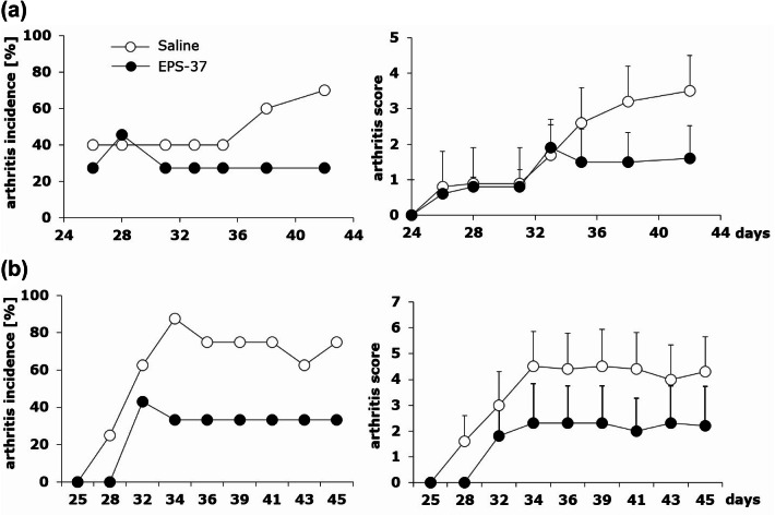
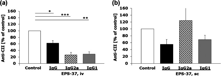
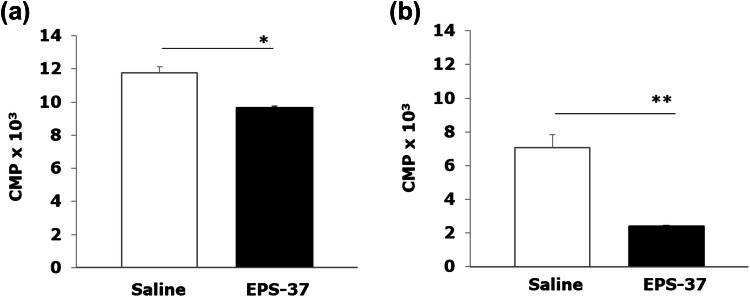
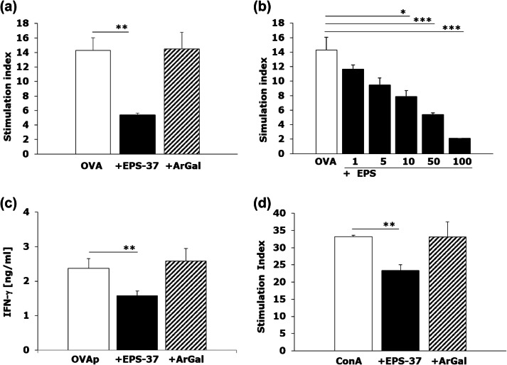
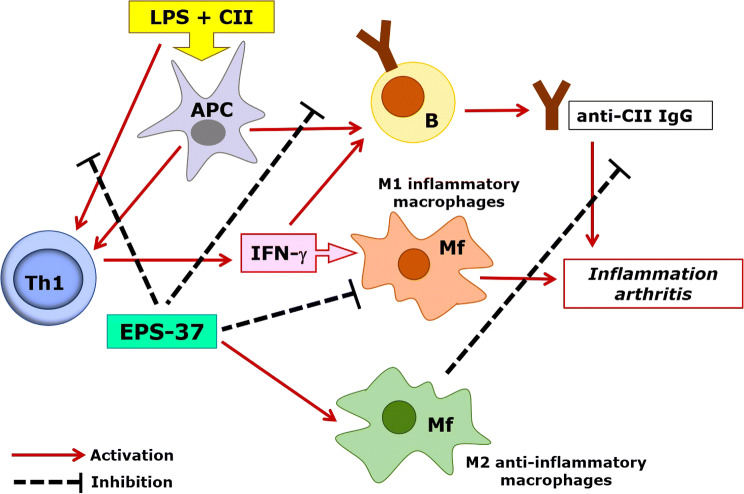
Exopolysaccharides (EPSs), major components of the bacterial biofilm, display strong strain-specific immunomodulatory properties. Previously, we have shown that crude EPS derived from Lactobacillus rhamnosus KL37 depresses the production of arthritogenic anti-collagen IgG and ameliorates collagen-induced arthritis (CIA) in DBA/1 mice, when lipopolysaccharide (LPS) was used as adjuvant. In this study, we used highly purified EPS from L. rhamnosus KL37 (EPS-37) to verify its anti-inflammatory properties and the ability to suppress T cell-dependent humoral response. We have employed the model of active CIA, in which mice immunized with type II collagen (CII) along with LPS were treated with pure EPS-37. Intravenous administration of purified EPS-37 markedly ameliorated arthritis and reduced CII-specific antibody production. EPS-37 injected subcutaneously reduced the clinical symptoms of CIA but without the reduction of arthritogenic antibodies. In addition, the effect of EPS-37 on T-cell functions was tested ex vivo and in vitro. EPS-37 inhibited the in vitro proliferation of T cells activated both in vivo (CII immunization) and in vitro (antigen/mitogen), and markedly reduced the production of interferon (IFN)-γ. These results together with other reports suggest that anti-inflammatory potential of EPS-37 depends on its ability to inhibit either one or the other or both possible inflammatory signaling pathways. Namely, Th1 → IFN-γ → M1 inflammatory macrophages → arthritis and/or Th1 → IFN-γ → B cells → arthritogenic antibodies → arthritis. We suggest that L. rhamnosus KL37 EPS might be utilized to control T cell-dependent immune responses in various inflammatory diseases. However, the most effective route of EPS-37 administration needs to be tailored for a given disorder.

Keywords: Collagen-induced arthritis; Exopolysaccharide; Immunomodulation; Inflammation; Lactobacillus rhamnosus; T cells.


Figures

Fig. 1
Fig. 1
CIA induction and examination protocol: mice were immunized with CII in the presence of CFA (day 0, first immunization) and with CII in the presence of LPS (day 21, second immunization, ip: intraperitoneal). EPS-37 (or LTA-37, or ArGal) were given to mice systemically—intravenously (iv) or subcutaneously (sc) three times a week starting on the day of second immunization (day 21) till the end of the experiment. Development of arthritis was examined by visual observation, paw thickness measurement and, finally, the level of anti-CII antibodies was measured in the mouse serum
Fig. 2
Fig. 2
Differential effects of intravenous and subcutaneous administration of pure EPS-37 on the development of CIA. Mice immunized with CII in the presence of CFA (day 0, first immunization, sc) and with CII in the presence of LPS (day 21, second immunization, ip) were given saline (open circles) or EPS-37 (black circles) systemically, intravenously (a) or subcutaneously (b) three times a week starting on the day of second immunization (day 21) till the end of the experiment. Results are expressed as arthritis incidence—the percentage of mice with signs of arthritis and arthritis score expressed in points ± SEM. Data represent one out of three similar experiments
Fig. 3
Fig. 3
Comparison of LTA-37 and ArGal capacity to affect the development of CIA. Mice immunized with CII in the presence of CFA (day 0, first immunization, sc) and with CII in the presence of LPS (day 21, second immunization, ip) were given saline (open circles) or LTA-37 (a black triangles) or ArGal (b black squares) three times a week starting on the day of second immunization (day 21) till the end of the experiment. Results are expressed as arthritis incidence—the percentage of mice with signs of arthritis and arthritis score expressed in points ± SEM. Data represent one out of three similar experiments
Fig. 4
Fig. 4
Differential effect of intravenous and subcutaneous administration of EPS-37 on the production of CII-specific IgG in the course of CIA. Mice immunized with CII in the presence of CFA (day 0, first immunization, sc) and with CII in the presence of LPS (day 21, second immunization, ip) were given EPS-37 systemically, intravenously (a) or subcutaneously (b) three times a week starting on the day of second immunization (day 21) till the end of the experiment. The level of anti-CII antibodies: IgG (black bars), IgG2a (hashed bars), IgG1 (gray bars) in serum is shown as a percentage of positive control (saline injected mice; white bars). Data represent one out of three similar experiments. Results are expressed as a mean of the measurements of each individual mouse serum ± SEM. *P < 0.05, **P < 0.01, ***P < 0.001
Fig. 5
Fig. 5
Ex vivo proliferation assay: DBA/1 mice were immunized with CII in the presence of CFA (day 0). EPS-37 (50 μg/mouse) (black bars) or saline (white bars) was injected systematically (iv) every other day (day 0, 3, 5, 7). Spleen (a) and lymph nodes (b) cells were harvested on day 8 and stimulated in vitro (5 × 105/well, in 0.2 ml) with 100-μg/ml CII for 72 h. The proliferation of cells was measured by 3H-tymidine incorporation and results, counts per minute (CPM) × 103, are expressed as a mean of triplicates of the culture wells ± SEM. Data represent one out of three similar experiments. *P < 0.05, **P < 0.01
Fig. 6
Fig. 6
EPS-37 inhibits mitogen induced and antigen specific T-cell proliferation in vitro. Spleen cells from OT II transgenic (ac) or C57BL6 mice (d) were stimulated (white bars) in vitro (5 × 105/well, in 0.2 ml) with 100 μg/ml OVA (a, b) or 1 μg/ml OVA 323–339 peptide (OVAp, c), or 1 μg/ml concanavalin A (ConA, d). EPS-37 (black bars) (30 μg/ml; except b 1–100 μg/ml) or ArGal (hashed bars) (30 μg/ml) were added to the culture. 3H-thymidine was introduced into the culture for the last 18 h. The proliferation of cells is shown as stimulation index (CPM of stimulated cell/CPM of unstimulated cells) (a, b, d). IFN-γ (c) content measured in cell culture supernatant is shown in ng/ml. Results of selected experiments, expressed as a mean of triplicates of the culture wells ± SEM are shown. *P < 0.05, **P < 0.01, ***P < 0.001
Fig. 7
Fig. 7
Hypothetical model of EPS-37 modulation of immune response in the course of CIA. Purified EPS-37 inhibits T-cell proliferation and a production of IFN-γ. Such action favors M2 macrophage polarization (an anti-inflammatory effect) and facilitates suppression of arthritogenic CII-specific IgG (T cell-dependent humoral response). Suppression = hash mark, Activation = solid line

Similar articles

Cited by

References

    1. Bermudez-Brito M, Borghuis T, Daniel C, et al. L. plantarum WCFS1 enhances Treg frequencies by activating DCs even in absence of sampling of bacteria in the Peyer patches. Sci Rep. 2018;8:1785. - PMC - PubMed
    1. Biedroń R, Konopiński MK, Marcinkiewicz J, et al. Oxidation by neutrophils-derived HOCl increases immunogenicity of proteins by converting them into ligands of several endocytic receptors involved in antigen uptake by dendritic cells and macrophages. PLoS ONE. 2015;10:e0123293. - PMC - PubMed
    1. Brand DD, Kang AH, Rosloniec EF. Immunopathogenesis of collagen arthritis. Springer Semin Immunopathol. 2003;25:3–18. - PubMed
    1. Castillo NA, Perdigon G, de Moreno de Leblanc A. Oral administration of a probiotic Lactobacillus modulates cytokine production and TLR expression improving the immune response against Salmonella enterica serovar Typhimurium infection in mice. BMC Microbiol. 2011;11:177. - PMC - PubMed
    1. Castro-Bravo N, Wells JM, Margolles A, et al. Interactions of surface exopolysaccharides from Bifidobacterium and Lactobacillus within the intestinal environment. Front Microbiol. 2018;9:2426–2440. - PMC - PubMed

MeSH terms