Case Reports
. 2020 Jul;140(1):1-6.
doi: 10.1007/s00401-020-02166-2. Epub 2020 May 24.

Neuropathology of COVID-19: a spectrum of vascular and acute disseminated encephalomyelitis (ADEM)-like pathology

Affiliations
Case Reports

Neuropathology of COVID-19: a spectrum of vascular and acute disseminated encephalomyelitis (ADEM)-like pathology

R Ross Reichard et al. Acta Neuropathol. 2020 Jul.

Abstract

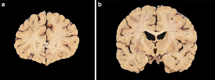
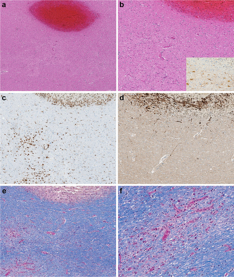
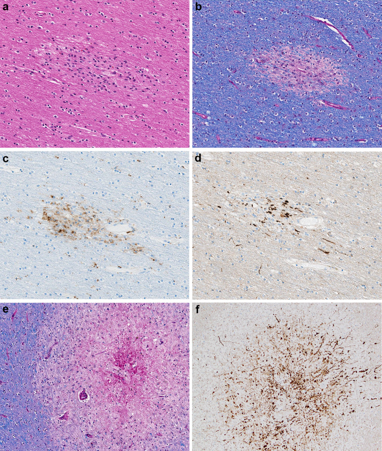
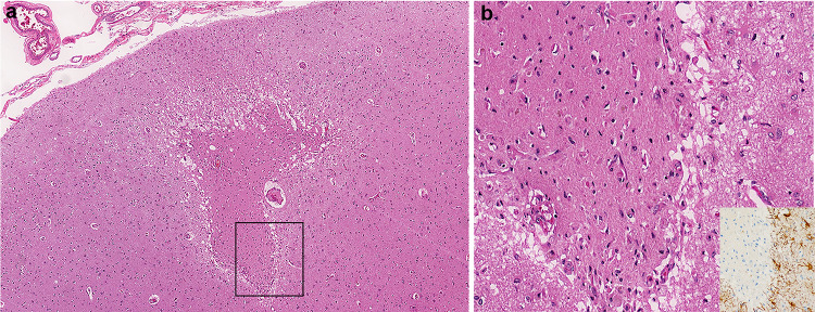
We report the neuropathological findings of a patient who died from complications of COVID-19. The decedent was initially hospitalized for surgical management of underlying coronary artery disease. He developed post-operative complications and was evaluated with chest imaging studies. The chest computed tomography (CT) imaging results were indicative of COVID-19 and he was subsequently tested for SARS-CoV-2, which was positive. His condition worsened and he died after more than 2 weeks of hospitalization and aggressive treatment. The autopsy revealed a range of neuropathological lesions, with features resembling both vascular and demyelinating etiologies. Hemorrhagic white matter lesions were present throughout the cerebral hemispheres with surrounding axonal injury and macrophages. The subcortical white matter had scattered clusters of macrophages, a range of associated axonal injury, and a perivascular acute disseminated encephalomyelitis (ADEM)-like appearance. Additional white matter lesions included focal microscopic areas of necrosis with central loss of white matter and marked axonal injury. Rare neocortical organizing microscopic infarcts were also identified. Imaging and clinical reports have demonstrated central nervous system complications in patients' with COVID-19, but there is a gap in our understanding of the neuropathology. The lesions described in this case provide insight into the potential parainfectious processes affecting COVID-19 patients, which may direct clinical management and ongoing research into the disease. The clinical course of the patient also illustrates that during prolonged hospitalizations neurological complications of COVID may develop, which are particularly difficult to evaluate and appreciate in the critically ill.

Keywords: COVID-19; Demyelinating; Infarct; Neuropathology; SARS-CoV-2; White matter.


Figures

Fig. 1
Fig. 1
a Coronal section of frontal lobes with widespread white matter hemorrhages. b Coronal section of cerebral hemispheres with widespread white matter hemorrhages
Fig. 2
Fig. 2
ad Microscopic sections of the corpus callosum genu. a H&E section of destructive hemorrhagic white matter lesion. b H&E section with white matter pallor adjacent to hemorrhagic lesion with GFAP immunoreactive reactive astrocytes evenly distributed in the white matter (inset) c CD68 immunostaining highlights a collection of macrophages at the periphery of the hemorrhagic lesion and a macrophages within an area of white matter pallor. d APP immunostain identifies axonal swellings within the hemorrhagic lesion and an absence of damaged axons within adjacent region of demyelination. e, f LFB/PAS stain distinguishes the focal area of myelin loss within the hemorrhagic lesion and adjacent PAS-positive foamy macrophages tracking along blood vessels and (f) higher magnification of the perivascular macrophages
Fig. 3
Fig. 3
a H&E section of subcortical white matter with perivascular pallor. b LFB/PAS stained section demonstrates the perivascular myelin loss within the subcortical white matter lesion. c CD68 immunostain confirms the perivascular distribution of the macrophages. d APP immunostain highlighting some damaged axons present in the region of myelin loss. e LFB/PAS-stained section of middle cerebellar peduncle with a central destructive lesion and radiating graduated myelin loss. f APP immunostained section of middle cerebellar peduncle lesion highlighting the marked axonal injury
Fig. 4
Fig. 4
a Microscopic H&E section of frontal lobe cortex with organizing infarct. b Higher magnification of the infarct with reactive blood vessels and adjacent GFAP immunoreactive reactive astrocytes (inset)

References

    1. Barton LM, Duval EJ, Stroberg E, Ghosh S, Mukhopadhyay S. COVID-19 autopsies, Oklahoma, USA. Am J Clin Pathol. 2020 doi: 10.1093/ajcp/aqaa062. - DOI - PMC - PubMed
    1. Bender SJ, Weiss SR. Pathogenesis of murine coronavirus in the central nervous system. J Neuroimmune Pharmacol. 2010;5:336–354. doi: 10.1007/s11481-010-9202-2. - DOI - PMC - PubMed
    1. Dong E, Du H, Gardner L. An interactive web-based dashboard to track COVID-19 in real time. Lancet Infect Dis. 2020 doi: 10.1016/s1473-3099(20)30120-1. - DOI - PMC - PubMed
    1. Fazakerley JK, Walker R. Virus demyelination. J Neurovirol. 2003;9:148–164. doi: 10.1080/13550280390194046. - DOI - PMC - PubMed
    1. Gu J, Korteweg C. Pathology and pathogenesis of severe acute respiratory syndrome. Am J Pathol. 2007;170:1136–1147. doi: 10.2353/ajpath.2007.061088. - DOI - PMC - PubMed

Publication types