. 2020 Dec;27(6):579-588.
doi: 10.1007/s10140-020-01787-0. Epub 2020 May 25.

Imaging evaluation of COVID-19 in the emergency department

Affiliations

Imaging evaluation of COVID-19 in the emergency department

Amir Pakray et al. Emerg Radiol. 2020 Dec.

Abstract

Purpose: The purpose of this study is to elucidate the chest imaging findings of suspected COVID-19 patients presenting to the emergency department and the relationship with their demographics and RT-PCR testing results.

Methods: Patients presenting to the ED between March 12 and March 28, 2020, with symptoms suspicious for COVID-19 and subsequent CXR and/or CT exam were selected. Patients imaged for other reasons with findings suspicious for COVID-19 were also included. Demographics, laboratory test results, and history were extracted from the medical record. Descriptive statistics were used to explore the relationship between imaging and these factors.

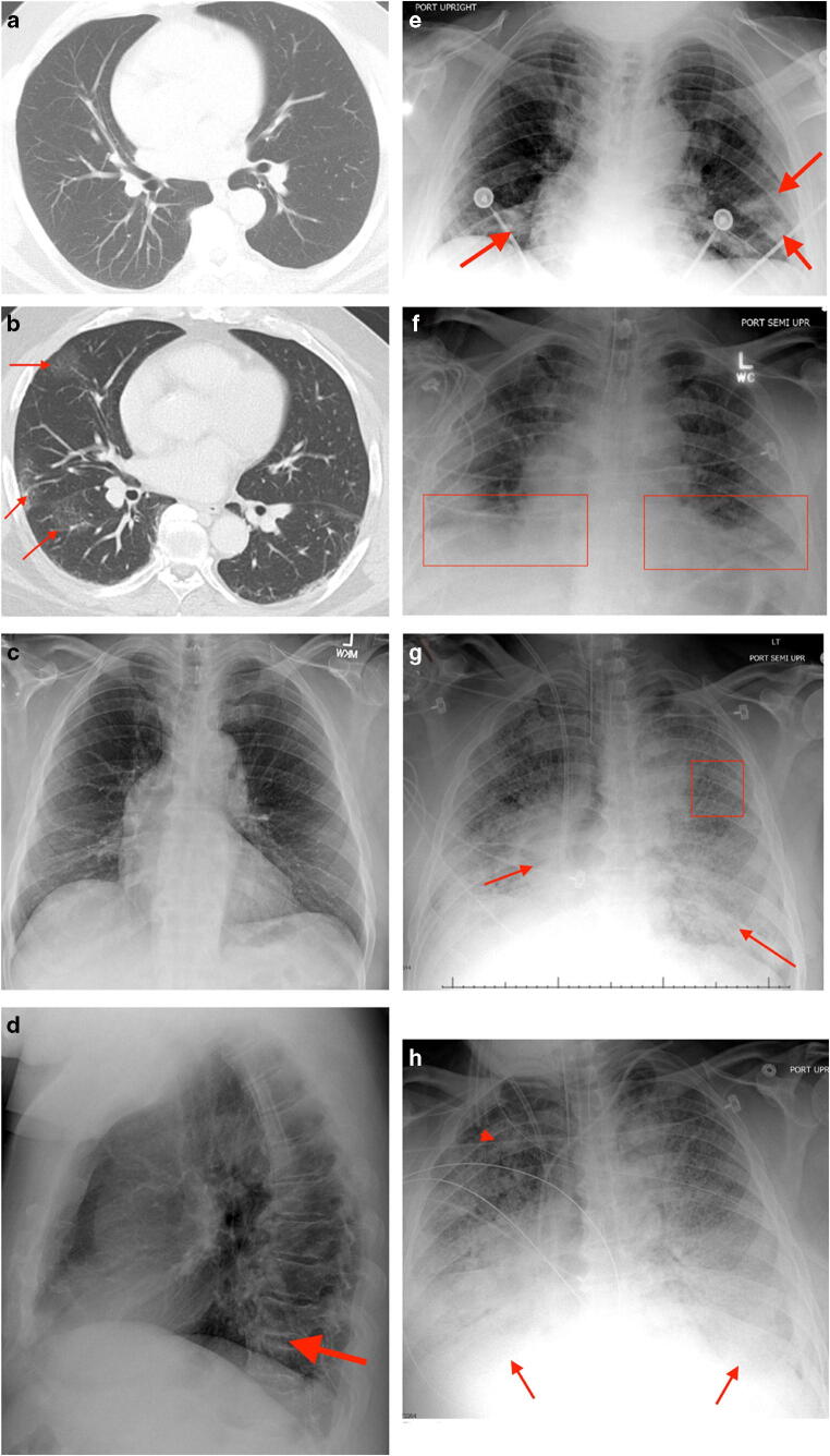
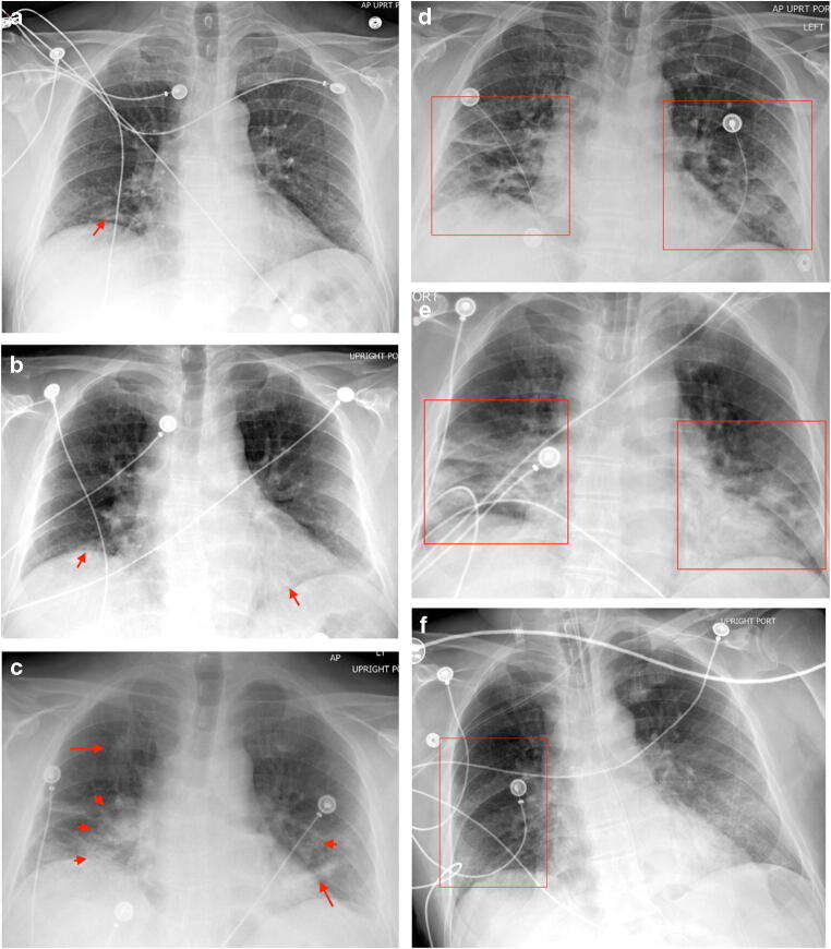
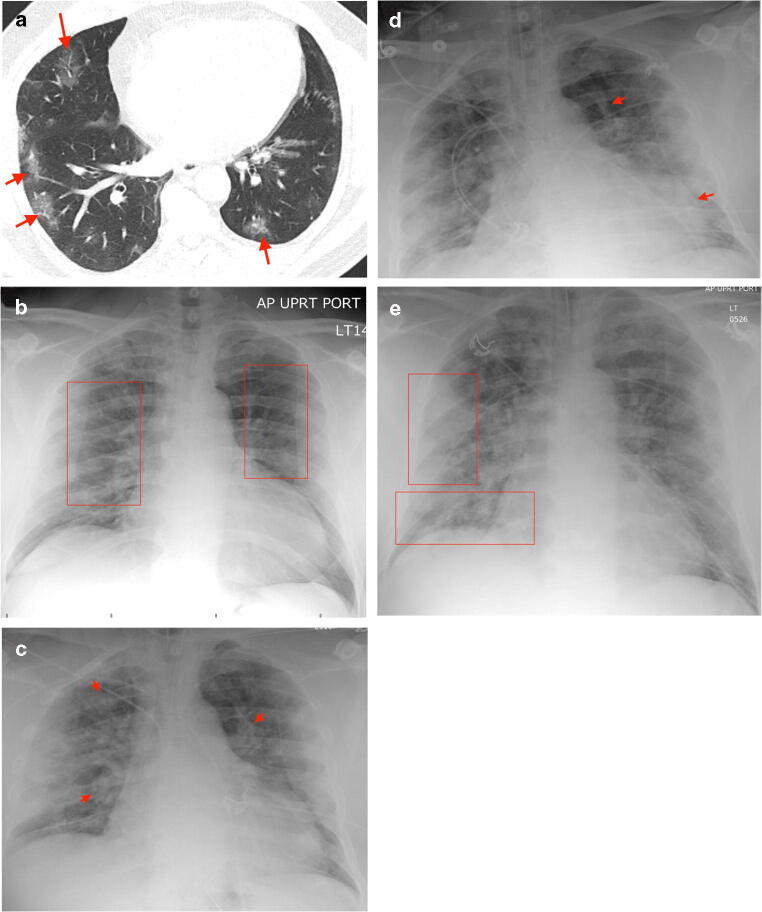
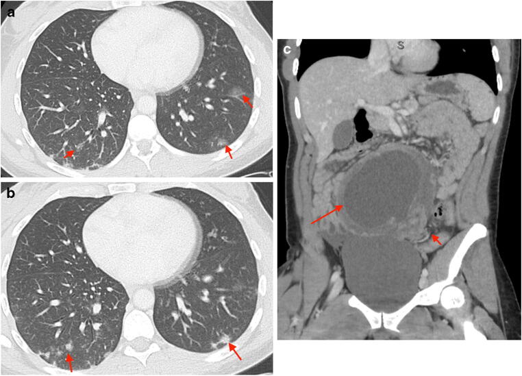
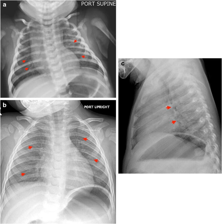
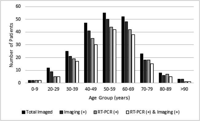
Results: A total of 227 patients from the emergency department were analyzed (224 CXRs and 25 CTs). Of the 192 patients with COVID-19 results, 173 (90.1%) had COVID-19 RT-PCR (+). Abnormal imaging (CXR, 85.7% and/or CT, 100%) was noted in 155 (89.6%) of COVID-19 RT-PCR (+) cases. The most common imaging findings were mixed airspace/interstitial opacities (39.8%) on CXR and peripheral GGOs on CT (92%). The most common demographic were African Americans (76.8%). Furthermore, 97.1% of African Americans were RT-PCR (+) compared to 65.8% of Caucasians.

Conclusion: We found a similar spectrum of thoracic imaging findings in COVID-19 patients as previous studies. The most common demographic were African Americans (76.8%). Furthermore, 97.1% of African Americans were RT-PCR (+) compared to 65.8% of Caucasians. Both CT and CXR can accurately identify COVID-19 pneumonitis in 89.6% of RT-PCR (+) cases, 89.5% of false negatives, and 72.7% of cases with no RT-PCR result.

Keywords: COVID-19; Coronavirus; Detroit; Emergency radiology; Ground glass opacities.


Conflict of interest statement

The authors declare that they have no conflict of interest.

Figures

Fig. 1
Fig. 1
Progression of disease on imaging in a 61-year-old male presenting with abdominal pain, fever, headache, and myalgias. CT chest w/o IV contrast 7 years prior to the current presentation demonstrates normal lung parenchyma (a). CT abdomen/pelvis w/ IV contrast on day 1 displays peripheral GGOs at the right lung base (arrows) (b). Frontal PA CXR on day 1 demonstrates interstitial opacities at the lung bases (c) and a subtle retrocardiac airspace opacity on the lateral CXR (arrow) (d). Portable AP frontal radiograph on day 3 demonstrates basal and peripheral airspace opacities (arrows) (e), becoming more confluent on day 6 (boxes) (f), with continued progression to diffuse interstitial opacities (box) and increasing airspace consolidation (arrows) on day 9, concerning for ARDS (g). Portable AP frontal radiograph on day 11 demonstrates interval increase in consolidation at the lung bases (arrows) and right upper lobe (arrowhead) (h). The patient expired on day 12. GGO: ground glass opacity, ARDS: acute respiratory distress syndrome
Fig. 2
Fig. 2
CXRs of a 57-year-old male with FN COVID-19 RT-PCR results. On day 1, the patient presented with cough and dyspnea for two days with a frontal AP radiograph demonstrating bilateral basal interstitial and airspace opacities (arrow) (a). The patient tested RT-PCR (−) on day 1 and was subsequently sent home on azithromycin. Frontal AP radiograph on day 2 after readmission for continued symptoms demonstrates increasing bibasilar airspace opacities (arrows), with the report suggesting a viral pneumonia pattern (b). Repeat RT-PCR on day 2 was again negative for COVID-19. Frontal AP radiograph on day 4 displays progression of right cardiophrenic airspace opacity (right-sided arrowheads), interval right upper lobe airspace opacity (right-sided arrow), and increasing left basal and mid lung airspace opacities (left-sided arrow and arrowhead) (c). The patient was empirically treated for viral pneumonitis and sent home on medications for a presumed asthma exacerbation. Frontal AP radiograph on day 6 after readmission with a fever displays increasing peripheral and basal airspace opacities (boxes) (d). The patient’s third repeat RT-PCR was negative for COVID-19. Frontal AP radiograph on day 8 after intubation displays increasing perihilar, basal, and peripheral airspace consolidation (boxes) with the report suggesting COVID-19 associated pneumonitis (e). On day 9, the fourth and final RT-PCR came back positive for COVID-19. Frontal AP radiograph on day 11 demonstrates improved aeration of the right hemithorax (box) (f). FN: false negative, RT-PCR: reverse transcriptase polymerase chain reaction, ICU: intensive care unit
Fig. 3
Fig. 3
Imaging of a 54-year-old male with a FN COVID-19 RT-PCR. On day 1, the patient presented with abdominal pain, subsequent negative RT-PCR, and an axial CT abdomen/pelvis w/ IV contrast image that displays bilateral peripheral GGOs (arrows) (a). Inflammatory changes around an appendiceal mucocele were also noted (not displayed). Frontal AP radiograph on day 1 demonstrates bilateral perihilar interstitial prominence (boxes) (b), with interval worsening by day 2 of the diffuse interstitial and peripheral airspace opacities (arrows) and a radiology report expressing high concern for COVID-19 (c). Repeat RT-PCR later that day was positive for COVID-19. Frontal AP radiograph on day 4 demonstrates mild interval progression of peripheral and basilar airspace consolidation bilaterally (arrows) (d), with continued progression of the peripheral (superior box) and basilar (inferior box) airspace opacities on day 6 (e). FN: false negative, RT-PCR: reverse transcriptase polymerase chain reaction
Fig 4
Fig 4
Incidental COVID-19 findings in a 33-year-old female with abdominal pain, emesis, and fever. Axial CT abdomen/pelvis w/ IV contrast displays bilateral peripheral GGOs (arrows) (a) and more consolidated peripheral opacities (arrows) (b) as a peripheral finding. A diagnosis of COVID-19 was suggested on imaging with subsequent positive testing. The patient was taken to the operating room for abdominal findings under appropriate COVID-19 precautions. Coronal CT abdomen/pelvis w/ IV contrast displays ovarian torsion, with an enlarged ovary (long arrow) and twisting of adnexal vasculature (short arrow) (c). GGO: ground glass opacity
Fig 5
Fig 5
Pediatric COVID-19 CXR findings. Frontal AP radiograph of a 6-week-old male with congenital adrenal insufficiency and RT-PCR COVID-19 (+) demonstrates streaky bilateral perihilar opacities (arrowheads) (a). In another pediatric patient, frontal AP radiograph of a 16-month-old male with congenitally absent kidney and RT-PCR COVID-19 (+) demonstrates prominent bilateral interstitial markings (arrows) in a pattern compatible with small-airway disease or viral pneumonitis (b) with peribronchial cuffing on the lateral CXR (arrows) (c). RT-PCR: reverse transcriptase polymerase chain reaction
Fig 6
Fig 6
Age distribution of total imaged patients compared with RT-PCR (+) (no patients presented from age range 10–19 years)

References

    1. Hui DS, Azhar EI, Madani TA, Ntoumi F, Kock R, Dar O, Ippolito G et al (2020) The Continuing 2019-NCoV Epidemic Threat of Novel Coronaviruses to Global Health — The Latest 2019 Novel Coronavirus Outbreak in Wuhan, China. Int J Infect Dis 91:264–266. 10.1016/j.ijid.2020.01.009 - PMC - PubMed
    1. Zhou F, Yu T, Ronghui D, Fan G, Liu Y, Liu Z, Xiang J et al (2020) Clinical Course and Risk Factors for Mortality of Adult Inpatients with COVID-19 in Wuhan, China: A Retrospective Cohort Study. Lancet 395(10229):1054–1062. 10.1016/S0140-6736(20)30566-3 - PMC - PubMed
    1. Huang C, Wang Y, Li X, Ren L, Zhao J, Hu Y, Zhang L et al (2020) Clinical Features of Patients Infected with 2019 Novel Coronavirus in Wuhan, China. Lancet 395(10223):497–506. 10.1016/S0140-6736(20)30183-5 - PMC - PubMed
    1. Lin C, Ding Y, Xie B, Sun Z, Li X, Chen Z, Niu M (2020) Asymptomatic Novel Coronavirus Pneumonia Patient Outside Wuhan: The Value of CT Images in the Course of the Disease. Clin Imaging 63:7–9. 10.1016/j.clinimag.2020.02.008 - PMC - PubMed
    1. Fang Y, Zhang H, Xie J, Lin M, Ying L, Pang P, Ji W (2020) Sensitivity of Chest CT for COVID-19: Comparison to RT-PCR. Radiology 200432. 10.1148/radiol.2020200432 - PMC - PubMed

MeSH terms