. 2020 Dec;108(6):1841-1850.
doi: 10.1002/JLB.3AB0320-156R. Epub 2020 May 25.

Effect of extracellular vesicles from S. aureus-challenged human neutrophils on macrophages

Affiliations

Effect of extracellular vesicles from S. aureus-challenged human neutrophils on macrophages

Edwina R Allen et al. J Leukoc Biol. 2020 Dec.

Abstract

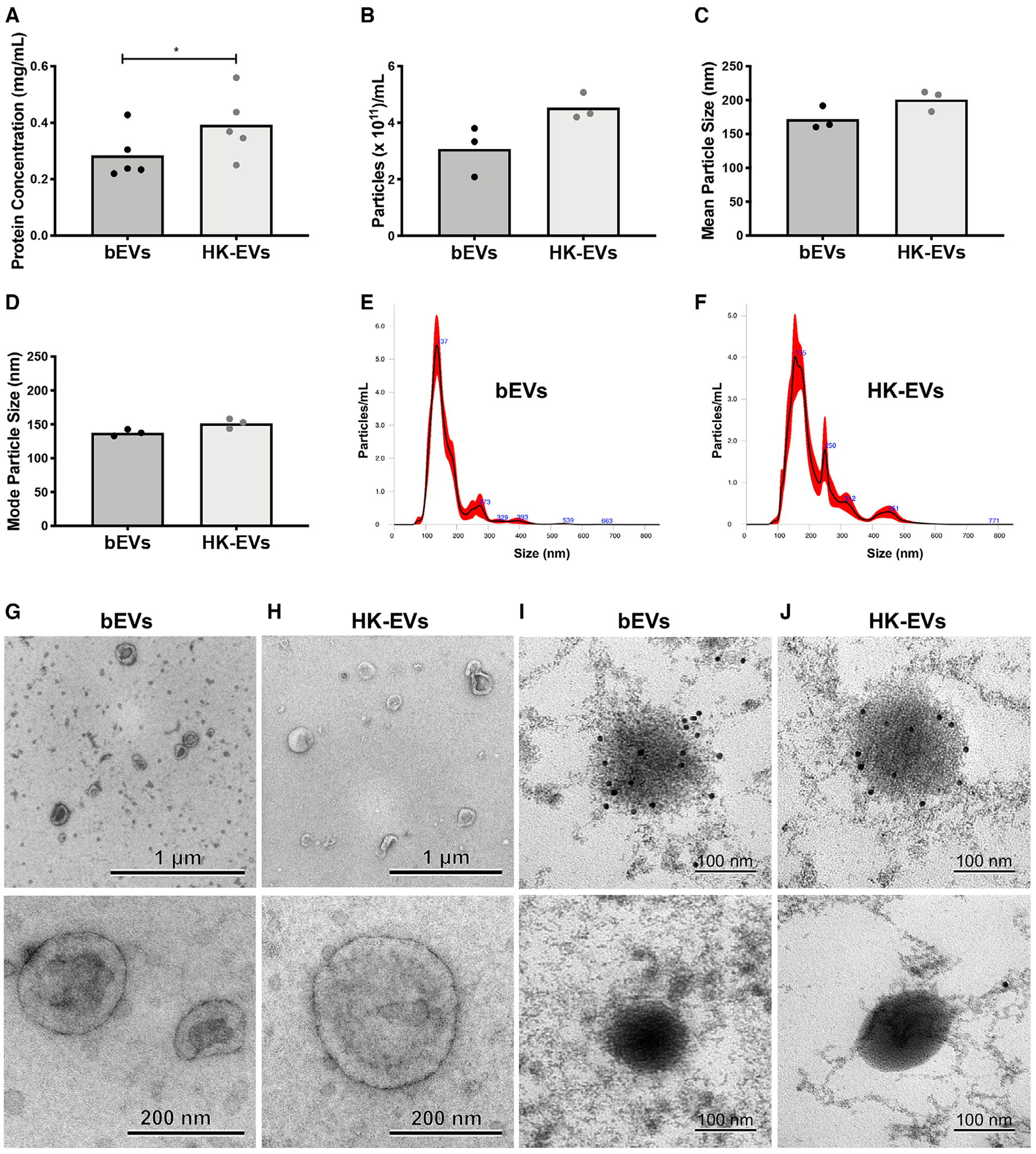
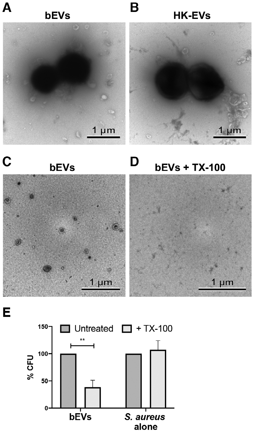
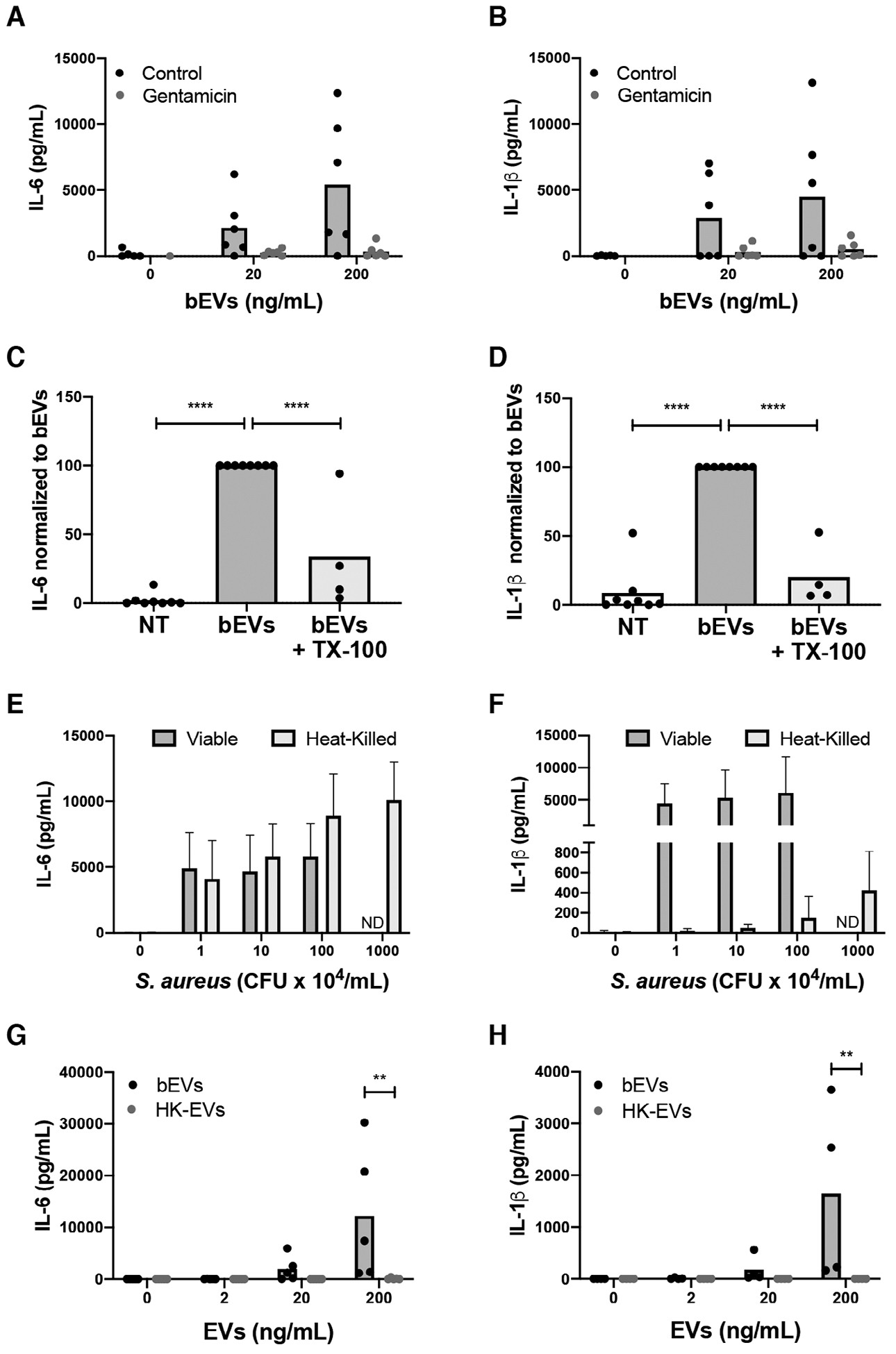
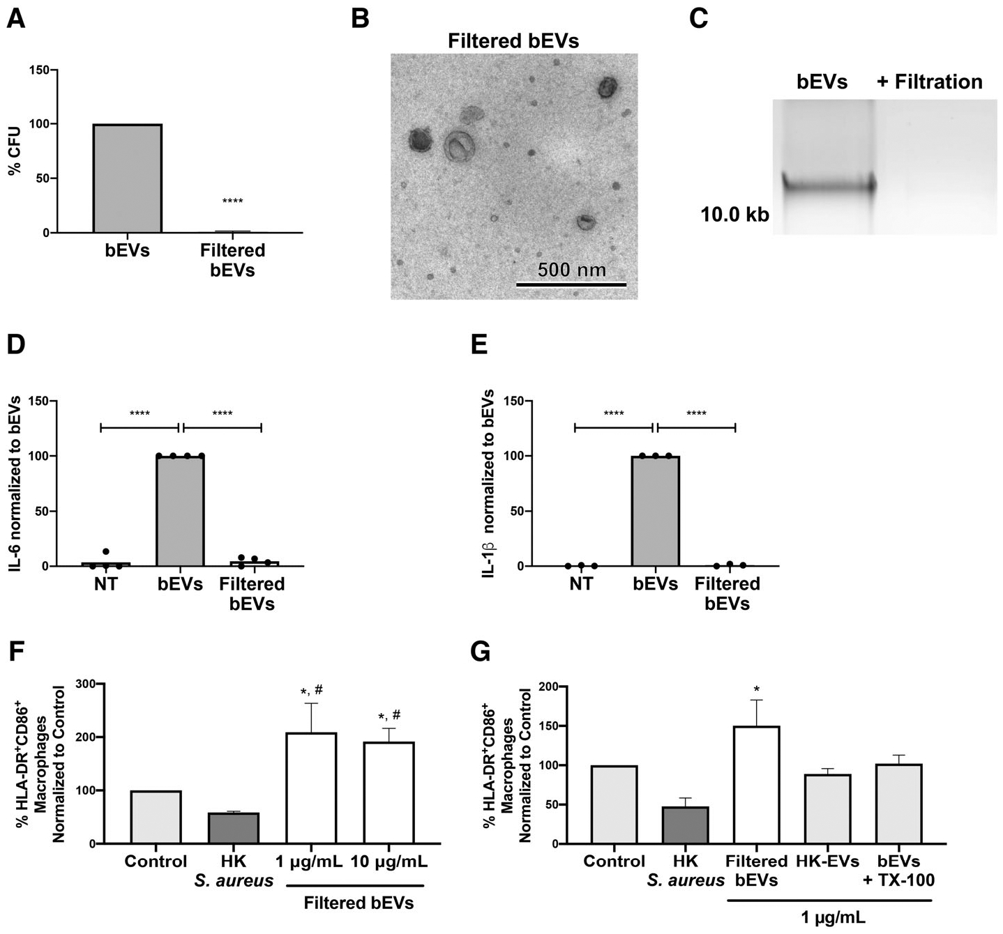
Staphylococcus aureus enhances neutrophil extracellular vesicle (EV) production. To investigate whether S. aureus viability influences EV biogenesis, EVs were isolated from human neutrophils incubated with viable bacteria (bEVs) or heat-killed bacteria (heat-killed EVs). Protein analysis, nanoparticle tracking and transmission electron microscopy showed comparable EV production between subsets, and both viable and nonviable bacteria were also detected in respective EV subsets. As anticipated, S. aureus, as well as bEVs with viable bacteria, were proinflammatory, and killing bacteria with gentamicin reduced cytokine production to baseline levels. Although heat-killed bacteria induced macrophage IL-6 production, heat-killed EVs did not. Additionally, we found that human and bacterial DNA associated with bEVs, but not heat-killed EVs, and that the DNA association could be partially decreased by disrupting electrostatic interactions. We investigated the potential for DNA isolated from EVs (EV-DNA) or EVs to cause inflammation. Although liposomal encapsulation of EV-DNA increased IL-6 production from baseline by 7.5-fold, treatment of bEVs with DNase I had no effect on IL-6 and IL-1β production, suggesting that the DNA did not contribute to the inflammatory response. Filtered EVs, which lacked DNA and associated bacteria, exhibited less proinflammatory activity relative to bEVs, and enhanced macrophage expression of CD86 and HLA-DR. Ultimately, we show that bEVs isolated by differential centrifugation co-purify with bacteria and DNA, and studying their concerted activity and relative contribution to immune response is important to the study of host-pathogen interactions.

Keywords: CD86 and HLA-DR; MRSA; ectosomes; exosomes; extracellular DNA; microparticles.


Conflict of interest statement

DISCLOSURES

The authors declare no conflicts of interest.

Figures

FIGURE 1
FIGURE 1. Viable bacteria are not required for extracellular vesicle (EV) biogenesis.
Protein concentration for the two EV subsets was determined by bicinchoninic acid (BCA) (A, n = 5). Nanoparticle tracking analysis (NTA) was used to determine particle concentration (B), mean particle size (C), and mode particle size (D) of EV samples (n = 3). Symbols represent individual data points and bars represent the mean. P-values were determined using a paired Student’s t-test (*P < 0.05). Representative histograms collected from NTA are shown in (E) and (F). Representative transmission electron microscopy micrographs are shown at 10,000× (top panels, scale bar = 1 μm) and 60,000× magnification (bottom panels, scale bar = 200 nm) for positive-negative stained bEV (G) and heat-killed EV (H) subsets (n = 3). For immunogold labeling, gp91 bound to primary antibody was labeled with secondary antibody-conjugated gold particles, then stained and visualized. Shown are representative images from two experiments (scale bar = 100 nm, I and J, top panels). In a single experiment, the primary antibody to gp91 was omitted to confirm specificity of immunogold staining (scale bar = 100 nm, I and J, bottom panels)
FIGURE 2
FIGURE 2. Differential centrifugation enriches for EVs and bacteria.
Shown are representative transmission electron microscopy (TEM) images from 3 experiments of bacteria in bEV (A) and heat-killed EV (B) preparations, as well as TEM images of paired preparations of bEVs (C) and Triton X-100 (TX-100) lysed bEVs (D) (10,000× magnification, scale bar = 1 μm). Colony forming unit were determined from both untreated and TX-100-treated bEVs, as well as S. aureus, and normalized to the paired untreated sample. Bars represent an average of at least three experiments ± SEM. P-values were determined for each group using a 2-way repeated measures ANOVA and Sidak’s multiple comparisons posttest (**P < 0.01; E)
FIGURE 3
FIGURE 3. Viable bacteria in bEV preparations cause proinflammatory cytokine production.
Macrophages were treated with vehicle, 20, or 200 ng/mL of bEVs with and without 10 μg/mL gentamicin. Supernatants were collected after 18 h at 37°C and analyzed by ELISA for IL-6 (A) and IL-1β (B). Symbols represent individual data points, and bars represent the mean (n = 6). Macrophages were treated with vehicle, bEVs (200 ng/mL), or lysed bEVs (200 ng/mL), and IL-6 (C) and IL-1β (D) in supernatants were analyzed by ELISA. Cytokine production was normalized to bEVs. Symbols represent individual data points, and bars represent the mean (n ≥ 4). P-values were determined using a 1-way ANOVA of values normalized to bEVs and Dunnett’s posttest (*P < 0.05 and **P < 0.01 vs. bEVs). Macrophages were cultured with buffer alone, 1–100 × 104 CFU/mL (colony forming unit) of viable S. aureus (n = 6), or 1–1000 × 104 CFU/mL heat-killed S. aureus (n = 5). After 18 h at 37°C, and IL-6 (E) and IL-1β (F) in supernatants were assayed by ELISA. Bars represent the average cytokine production ± SEM. After macrophages were treated with 0, 2, 20, and 200 ng/mL of bEVs and heat-killed extracellular vesicles (EVs) for 18 h at 37°C, supernatants were assayed by ELISA for IL-6 (G) and IL-1β (H). Symbols represent individual data points, and bars represent the mean (n ≥ 4). P-values were determined for each group using a 2-way repeated measures ANOVA and Sidak’s multiple comparisons posttest (**P < 0.01)
FIGURE 4
FIGURE 4. DNA is present and associated electrostatically with bEVs, but does not contribute significantly to IL-6 and IL-1β production.
DNA extracted from 50μL of bEVs and heat-killed extracellular vesicles (EVs) was separated by electrophoresis on a 0.7% agarose gel stained with Gel Red (n = 3, A). E. coli DNA (EC-DNA, 1 μg), bEVs, or EC-DNA encapsulated in DOTAP were treated with PBS or DNase I (5 units), then DNA was resolved by gel electrophoresis (n = 3, B). EVs were conjugated to beads, stained with CellVue, and then treated with 0 M, 0.1 M, or 1 M NaCl. After washing, EVs were then stained with either propidium iodide or CD63 and analyzed by flow cytometry (n ≥ 3, C). Bars represent average percentage of positive events ± SEM. P-values were determined for each group using a 1-way ANOVA and Dunnett’s posttest (*P < 0.05 vs. 0 M NaCl). Extracted DNA from untreated or lysed bEVs was assessed as described in A (n = 6, D). Macrophages were treated with either free or DOTAP-encapsulated purified EV-DNA or EC-DNA (1 μg/mL) for 18 h at 37°C before supernatants were analyzed by ELISA for IL-6 (E). Bars represent average values ± SEM (n = 3). P-values were determined for each group using a 2-way ANOVA and multiple comparisons posttest (**P < 0.01). bEVs (200 ng/mL) were incubated with either vehicle or DNase I (5 units) for 1 h at 37°C followed by DNase inactivation with 5 mM EDTA. Macrophages were treated with vehicle or manipulated bEVs for 18–20 h, then supernatants were collected and analyzed by ELISA for IL-6 (F) and IL-1β (G). Symbols represent individual data points, and bars represent the average of at least 24 experiments. P-values were determined using a 1-way ANOVA and Dunnett’s posttest (*P < 0.05 and **P < 0.01 vs. bEVs + vehicle)
FIGURE 5
FIGURE 5. Filtered extracellular vesicles (EVs) are not proinflammatory in macrophages, and enhance macrophage expression of CD86 and HLA-DR.
bEVs and filtered bEVs were plated on tryptic soy agar (TSA) overnight at 37°C and colony forming units were enumerated. For each preparation, bacterial quantity was normalized to bEVs and bars represent an average of four experiments. P-values were determined using an unpaired t-test (****P < 0.0001; A). A representative transmission electron microscopy micrograph of filtered bEVs is shown at 10,000× magnification (scale bar = 500 nm; n = 3; B). DNA from bEVs or filtered bEVs was separated on a 0.7% agarose gel stained with Gel Red (n = 3; C). Macrophages were treated with either PBS, bEVs, or filtered bEVs at 200 ng/mL, then IL-6 (D) and IL-1β (E) in supernatants were analyzed by ELISA. Cytokine production was normalized to bEVs. Symbols represent individual data points, and bars represent the mean (n ≥ 4). P-values were determined using a 1-way ANOVA of values normalized to bEVs and Dunnett’s posttest (*P < 0.05 and **P < 0.01 vs. bEVs). Macrophages were pretreated with gentamicin (10 μg/mL), and left in buffer alone (control) or treated with heat-killed S. aureus (75 μg/mL), filtered bEVs, EVs isolated from neutrophils stimulated with heat-killed S. aureus, or Triton X-100 (TX-100) lysed bEVs at indicated concentrations. CD14+macrophages were analyzed by flow cytometry for CD86 and HLA-DR (n ≥ 3, F, G). Bars represent average percentage of HLA-DR+CD86+ events of CD14+ ± SEM. P-values were determined using a 1-way ANOVA and Tukey’s posttest (*P < 0.05 vs. control and #P < 0.05 vs. heat-killed S. aureus)

References

    1. Hassoun A, Linden PK, Friedman B. Incidence, prevalence, and management of MRSA bacteremia across patient populations-a review of recent developments in MRSA management and treatment. Crit Care. 2017;21(1):211. - PMC - PubMed
    1. DeLeo FR, Diep BA, Otto M. Host defense and pathogenesis in Staphylococcus aureus infections. Infect Dis Clin North Am. 2009;23:17–34. - PMC - PubMed
    1. Cheng AG, DeDent AC, Schneewind O, Missiakas D. A play in four acts: Staphylococcus aureus abscess formation. Trends Microbiol. 2011;19:225–232. - PMC - PubMed
    1. Monaco M, Pimentel de Araujo F, Cruciani M, Coccia EM, Pantosti A. Worldwide epidemiology and antibiotic resistance of Staphylococcus aureus. Curr Top Microbiol Immunol. 2017;409:21–56. - PubMed
    1. Mölne L, Verdrengh M, Tarkowski A. Role of neutrophil leukocytes in cutaneous infection caused by Staphylococcus aureus. Infect Immun. 2000;68:6162–6167. - PMC - PubMed

Publication types

MeSH terms