. 2020 Jul;24(14):7850-7861.
doi: 10.1111/jcmm.15418. Epub 2020 May 25.

FOXO1 contributes to diabetic cardiomyopathy via inducing imbalanced oxidative metabolism in type 1 diabetes

Affiliations

FOXO1 contributes to diabetic cardiomyopathy via inducing imbalanced oxidative metabolism in type 1 diabetes

Dan Yan et al. J Cell Mol Med. 2020 Jul.

Abstract

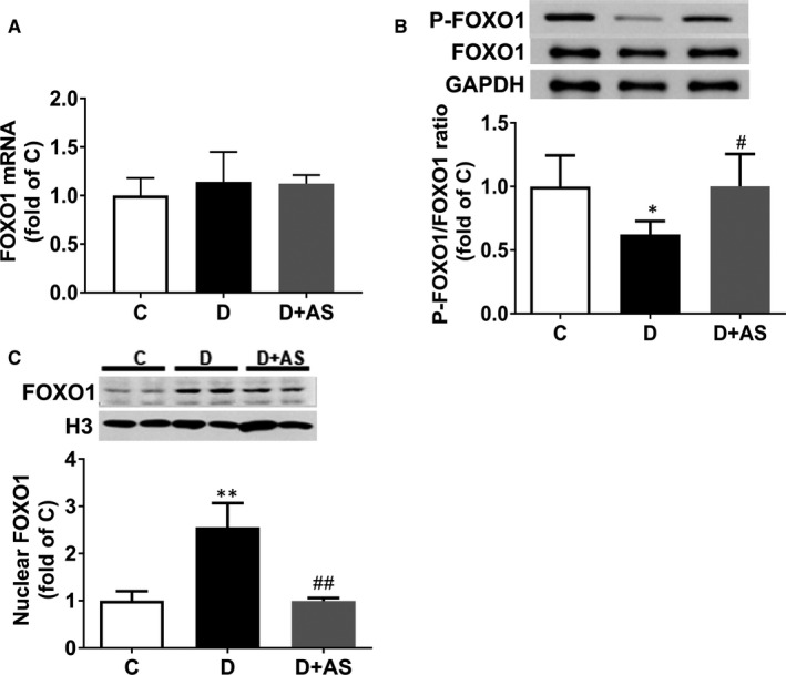
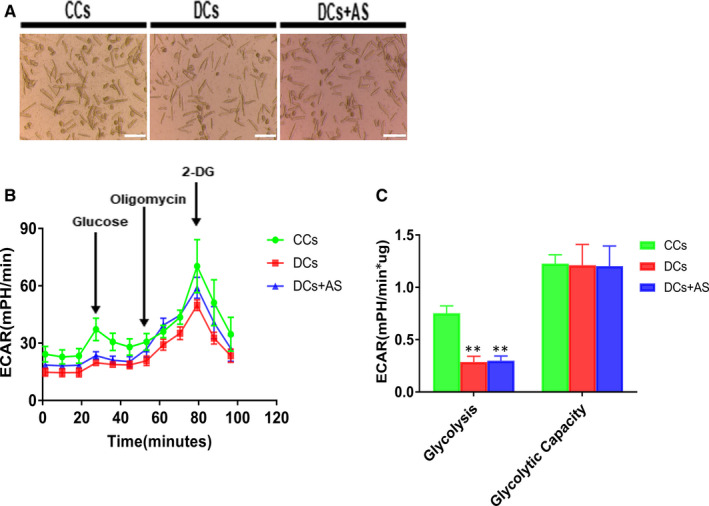
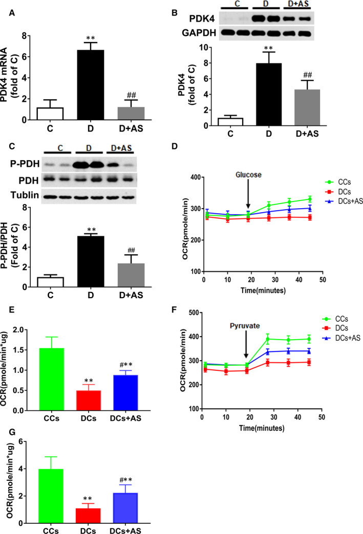
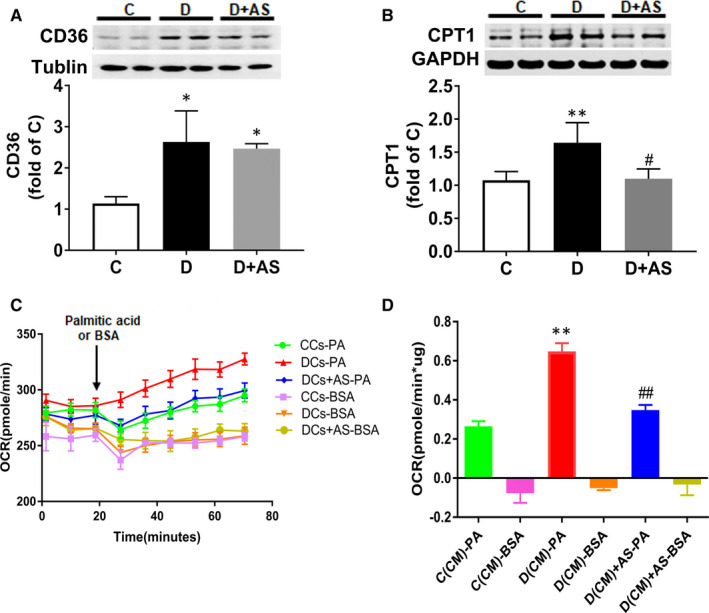
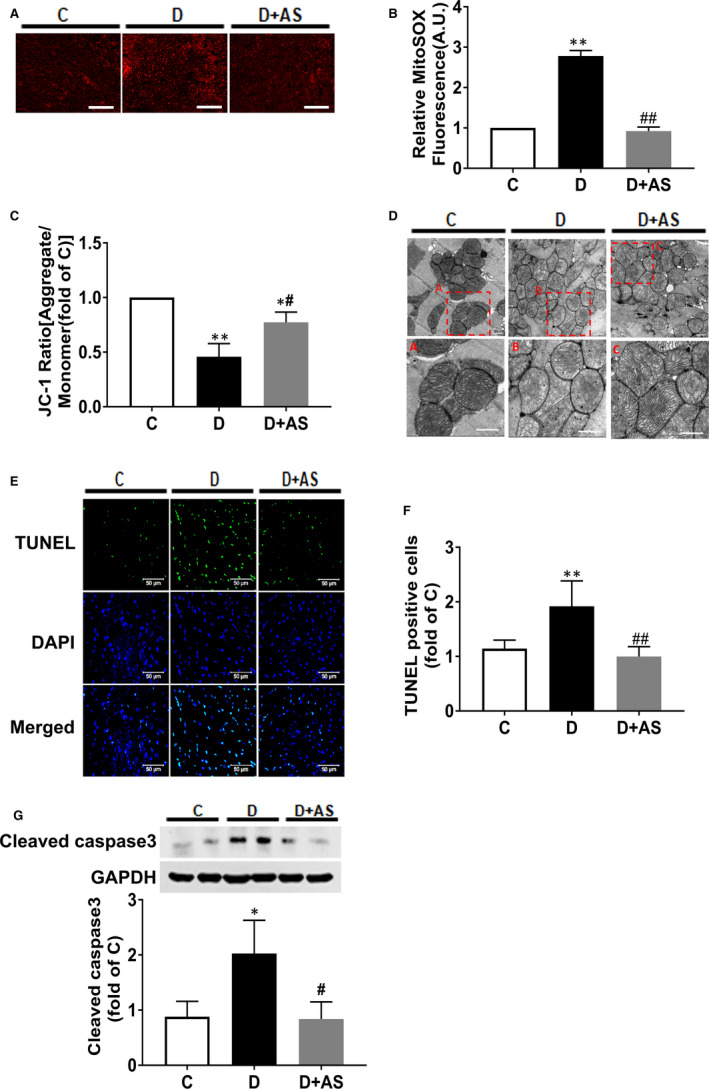
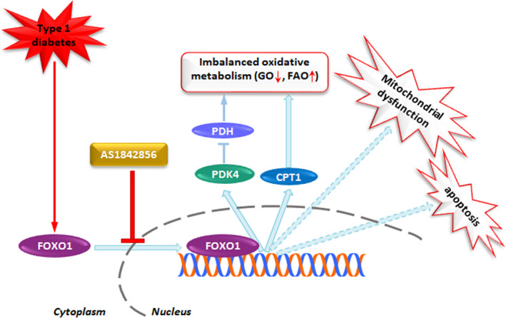
Forkhead box protein O1 (FOXO1), a nuclear transcription factor, is preferably activated in the myocardium of diabetic mice. However, its role and mechanism in the development of diabetic cardiomyopathy in non-obese insulin-deficient diabetes are unclear. We hypothesized that cardiac FOXO1 over-activation was attributable to the imbalanced myocardial oxidative metabolism and mitochondrial and cardiac dysfunction in type 1 diabetes. FOXO1-selective inhibitor AS1842856 was administered to streptozotocin-induced diabetic (D) rats, and cardiac functions, mitochondrial enzymes PDK4 and CPT1 and mitochondrial function were assessed. Primary cardiomyocytes isolated from non-diabetic control (C) and D rats were treated with or without 1 µM AS1842856 and underwent Seahorse experiment to determine the effects of glucose, palmitate and pyruvate on cardiomyocyte bioenergetics. The results showed diabetic hearts displayed elevated FOXO1 nuclear translocation, concomitant with cardiac and mitochondrial dysfunction (manifested as elevated mtROS level and reduced mitochondrial membrane potential) and increased cell apoptosis (all P < .05, D vs C). Diabetic myocardium showed impaired glycolysis, glucose oxidation and elevated fatty acid oxidation and enhanced PDK4 and CPT1 expression. AS1842856 attenuated or prevented all these changes except for glycolysis. We concluded that FOXO1 activation, through stimulating PDK4 and CPT1, shifts substrate selection from glucose to fatty acid and causes mitochondrial and cardiac dysfunction.

Keywords: FOXO1; diabetic cardiomyopathy; oxidative metabolism.


Conflict of interest statement

The authors confirm that there are no conflicts of interest regarding the publication of this article.

Figures

Figure 1
Figure 1
AS supplementation reduced myocardial P‐FOXO1/FOXO1 and nuclear presence of FOXO1 in 5‐wk diabetic rats. A, Cardiac mRNA level of FOXO1, B, P‐FOXO1/FOXO1 ratio and C, nuclear FOXO1 protein level were detected in non‐diabetes control (C), diabetic (D) and diabetic rats treated with AS (D + AS) groups. AS, AS1842856. Data are expressed as means ± SEM (n = 6) *P < .05 vs C; **P < .01 vs C; # P < .05 vs D.## P < .05 vs D
Figure 2
Figure 2
AS treatment did not affect glycolytic flux and glycolytic capacity in isolated diabetic cardiomyocytes. A, Isolated cardiomyocytes were seeded onto matrix gel–coated Seahorse XF24 Cell Culture Plates (8000 cells/well) treated with or without 1 μM AS for 24 h. Scale bar, 200 μm. B, Kinetic extracellular acidification rate (ECAR) responses of isolated cardiomyocytes to glucose (10 mM), oligomycin (1 μM) and 2‐DG (100 mM). C, Calculated glycolytic flux and glycolytic capacity. The former one is calculated by the ECAR increase (ECAR value of measurement 3 subtracted by measurement 4) normalized with cell protein content. The latter one is calculated by the ECAR increase (ECAR value of measurement 3 subtracted by measurement 9) normalized with cell protein content. Control cardiomyocytes (CCs); diabetic cardiomyocytes (DCs); AS‐treated diabetic cardiomyocytes (DCs + AS); AS, AS1842856; 2‐DG, 2‐deoxy‐glucose. Data are expressed as means ± SEM (n = 6). **P < .01 vs CCs
Figure 3
Figure 3
AS treatment reduced PDK4 and P‐PDH expression and restored glucose oxidation in diabetic myocardium. A‐C, Cardiac PDK4 mRNA level, PDK4 protein level and p‐PDH protein level. D, Kinetic oxygen consumption rate (OCR) responses of isolated cardiomyocytes to 10 mM glucose. E, Calculated glucose oxidation rate. F, Kinetic OCR responses of isolated cardiomyocytes to 1 mM pyruvate. G, Calculated pyruvate oxidation rate. The glucose or pyruvate oxidation rate was calculated by the OCR increase (OCR value of measurement 3 subtracted by measurement 6) normalized with cell protein content. AS, AS1842856. Data are expressed as means ± SEM (n = 6). *P < .05 vs C or CCs; **P < .01 vs C or CCs; #P < .05 vs D or DCs; ##P < .01 vs D or DCs
Figure 4
Figure 4
AS treatment significantly reduced diabetic myocardial CPT1 expression and concomitantly reduced increased palmitate oxidation in diabetic cardiomyocytes. A and B, Cardiac CD36 protein level and CPT1 protein level. C, Kinetic OCR responses of isolated cardiomyocytes to glucose 1 mM palmitate acid. D, Calculated palmitate acid oxidation. Palmitate acid oxidation is calculated by the OCR increase (OCR value of measurement 3 subtracted by measurement 9) normalized with cell protein content. OCR, oxygen consumption rate; AS, AS1842856. Data are expressed as means ± SEM (n = 6). *P < .05 vs C or CCs; **P < .01 vs C or CCs; #P < .05 vs D or DCs; ##P < .01 vs D or DCs
Figure 5
Figure 5
AS supplementation restored mitochondrial dysfunction and attenuated myocyte apoptosis in diabetic heart. A and B, mtROS was assessed by MitoSOX Red staining in frozen sections of heart tissues. Scale bar, 200 μM. C, Mitochondrial membrane potential by JC‐1 staining in mitochondria isolated from heart tissues. D, Representative electron photomicrographs of mitochondria (5200 × magnification) in heart tissue, and the given area (red rectangle) in original image (upper layer) was cropped and magnified (lower layer). Scale bar, 500 nM. E, F and G, Myocardial cell apoptosis assessed by cleaved caspase 3 protein expression and terminal deoxynucleotidyl transferase dUTP nick‐end labelling (TUNEL) assay. mtROS, mitochondrial reactive oxygen species; AS, AS1842856. Data are shown as means ± SEM, with n = 6 animals per group. *P < .05 vs C, **P < .01 vs C; # P < .05 vs D, ## P < .01 vs D
Figure 6
Figure 6
Schematic illustration of proposed signalling mechanism involved in diabetes‐induced FOXO1 activation promotes imbalanced oxidative metabolism, mitochondrial dysfunction and cardiac dysfunction in heart. Under the condition of diabetes, FOXO1, by enhancing PDK4 and CPT1 expression, induces imbalanced oxidative metabolism manifested as decreased glucose oxidation (GO) and elevated fatty acid oxidation (FAO), which was concomitant with mitochondrial dysfunction and cardiac dysfunction. However, AS1842856 treatment attenuated or prevented all the changes via inhibition of diabetes‐induced FOXO1 nuclear translocation

References

    1. Jia G, Hill MA, Sowers JR. Diabetic cardiomyopathy: an update of mechanisms contributing to this clinical entity. Circ Res. 2018;122(4):624‐638. - PMC - PubMed
    1. Varma U, Koutsifeli P, Benson VL, et al. Molecular mechanisms of cardiac pathology in diabetes ‐ Experimental insights. Biochim Biophys Acta‐Mol Basis Dis. 2018;1864(5):1949‐1959. - PubMed
    1. Holscher ME, Bode C, Bugger H. Diabetic cardiomyopathy: does the type of diabetes matter? Int J Mol Sci. 2016;17(12):2136. - PMC - PubMed
    1. De Blasio MJ, Huynh N, Deo M, et al. Defining the progression of diabetic cardiomyopathy in a mouse model of type 1 diabetes. Front Physiol. 2020;11:124. - PMC - PubMed
    1. Herrero P, Peterson LR, McGill JB, et al. Increased myocardial fatty acid metabolism in patients with type 1 diabetes mellitus. J Am Coll Cardiol. 2006;47(3):598‐604. - PubMed

Publication types

MeSH terms

Substances