. 2020 Aug;93(1112):20200243.
doi: 10.1259/bjr.20200243. Epub 2020 Jun 2.

Chest CT-based differential diagnosis of 28 patients with suspected corona virus disease 2019 (COVID-19)

Affiliations

Chest CT-based differential diagnosis of 28 patients with suspected corona virus disease 2019 (COVID-19)

Sidong Xie et al. Br J Radiol. 2020 Aug.

Abstract

Objectives: The chest CT findings that can distinguish patients with corona virus disease 2019 (COVID-19) from those with clinically suspected COVID-19 but subsequently found to be COVID-19 negative have not previously been described in detail. The purpose of this study was to determine the distinctions among patients with COVID-19 by comparing the imaging findings of patients with suspected confirmed COVID-19 and those of patients initially suspected to have COVID-19 who were ultimately negative for the disease.

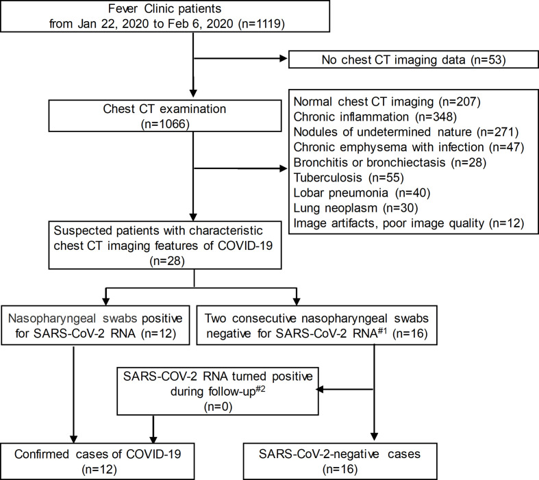
Methods: 28 isolated suspected in-patients with COVID-19 were enrolled in this retrospective study from January 22, 2020 to February 6, 2020. 12 patients were confirmed to have positive severe acute respiratory syndrome corona virus 2 (SARS-CoV-2) RNA results, and 16 patients had negative results. The thin-section CT imaging findings and clinical and laboratory data of all the patients were evaluated.

Results: There were no significant differences between the 12 confirmed COVID-19 (SARS-Cov-2-positive) patients and 16 SARS-CoV-2-negative patients in epidemiology and most of the clinical features or laboratory data. The CT images showed that the incidence of pure/mixed ground-glass opacities (GGOs) was not different between COVID-19 and SARS-CoV-2-negative patients [9/12 (75.0%) vs 10/16 (62.5%), p = 0.687], but pure/mixed GGOs in the peripheral were more common in patients with COVID-19 [11/12 (91.7%) vs 6/16 (37.5%), p = 0.006]. There were no significant differences in the number of lesions, bilateral lung involvement, large irregular/patchy opacities, rounded opacities, linear opacities, crazy-paving patterns, halo signs, interlobular septal thickening or air bronchograms.

Conclusions: Although peripheral pure/mixed GGOs on CT may help distinguish patients with COVID-19 from clinically suspected but negative patients, CT cannot replace RT-PCR testing.

Advances in knowledge: Peripheral pure/mixed GGOs on-chest CT findings can be helpful in distinguishing patients with COVID-19 from those with clinically suspected COVID-19 but subsequently found to be COVID-19 negative.


Conflict of interest statement

Conflict of Interest: The authors of this manuscript declare no relationships with any companies, whose products or services may be related to the subject matter of the article.

Figures

Figure 1.
Figure 1.
Flowchart of the study population. Note: #1 The interval between two times was more than 24 h; #2 The last follow-up was on February 20, 2020; CT = computed tomography; COVID-19 = corona virus disease 2019; SARS-CoV-2=severe acute respiratory syndrome corona virus 2.
Figure 2.
Figure 2.
(a-b) An axial CT image obtained without intravenous contrast in a 43-year-old male showed peripheral/subpleural patchy consolidation in the anterior of the left upper lobe, with interlobular septal thickening, air bronchogram and linear opacities. He had a history of living in Xiaogan, Hubei Province and presented with fever for 1 week. Nasopharyngeal swabs for SARS-CoV-2 RNA were positive.
Figure 3.
Figure 3.
(a-b) A31-year-old male with a history of travelling on a cruise presented with fever, diarrhoea, sore throat and fatigue. (a) Axial thin-section non-contrast CT image on February 1, 2020, showed peripheral/subpleural, patchy, rounded, mixed GGOs in the right lower lobe, with interlobular septal thickening and crazy-paving patterns. (b) Follow-up CT image on February 9, 2020, showed that the lesions were obviously resolved after anti-infection therapy. Nasopharyngeal swabs for SARS-CoV-2 RNA were negative.
Figure 4.
Figure 4.
(a-b) A 56-year-old female with a history of travelling to Guangzhou from Wuhan presented with fever, cough and diarrhoea for 2 days. (a) (b) Axial thin-section non-contrast CT image showed bilateral peripheral/subpleural, patchy, pure/mixed GGOs with interlobular septal thickening and crazy-paving patterns. Two consecutive nasopharyngeal swabs for SARS-CoV-2 RNA(the interval between the two swabs more than 24 h) were negative.

References

    1. Xu X, Chen P, Wang J, Feng J, Zhou H, Li X, et al. . Evolution of the novel coronavirus from the ongoing Wuhan outbreak and modeling of its spike protein for risk of human transmission. Sci China Life Sci 2020; 63: 457–60. doi: 10.1007/s11427-020-1637-5 - DOI - PMC - PubMed
    1. Huang C, Wang Y, Li X, Ren L, Zhao J, Hu Y, et al. . Clinical features of patients infected with 2019 novel coronavirus in Wuhan, China. Lancet 2020; 395: 497–506. doi: 10.1016/S0140-6736(20)30183-5 - DOI - PMC - PubMed
    1. Song F, Shi N, Shan F, Zhang Z, Shen J, Lu H, et al. . Novel coronavirus (2019-nCoV) pneumonia. RADIOLOGY 2019; 295: 210–72020. - PMC - PubMed
    1. Xie X, Zhong Z, Zhao W, Zheng C, Wang F, Liu J. Chest CT for typical 2019-nCoV pneumonia: relationship to negative RT-PCR testing. Radiology 2020; 200343: 200343. doi: 10.1148/radiol.2020200343 - DOI - PMC - PubMed
    1. Kanne JP. Chest CT findings in 2019 novel coronavirus (2019-nCoV) infections from Wuhan, China: key points for the radiologist. Radiology 2020; 295: 16–17. doi: 10.1148/radiol.2020200241 - DOI - PMC - PubMed