. 2020 May 25;20(1):132.
doi: 10.1186/s12866-020-01802-x.

LipF increases rifampicin and streptomycin sensitivity in a Mycobacterium tuberculosis surrogate

Affiliations

LipF increases rifampicin and streptomycin sensitivity in a Mycobacterium tuberculosis surrogate

Ana Leticia Arriaga-Guerrero et al. BMC Microbiol. .

Abstract

Background: Mortality due to tuberculosis (TB) has increased due to the development of drug resistance, the mechanisms of which have not been fully elucidated. Our research group identified a low expression of lipF gene in Mycobacterium tuberculosis clinical isolates with drug resistance. The aim of this work was to evaluate the effect of lipase F (LipF) expression on mycobacterial drug resistance.

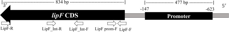
Results: The effects of expressing lipF from Mycobacterium tuberculosis in Mycobacterium smegmatis on resistance to antituberculosis drugs were determined with resazurin microtiter assay plate and growth kinetics. Functionality of ectopic LipF was confirmed. LipF expression reduced the rifampicin (RIF) and streptomycin (STR) minimum inhibitory concentration (MIC) from 3.12 μg/mL to 1.6 μg/mL and 0.25 μg/mL to 0.06 μg/mL respectively, moreover a reduced M. smegmatis growth in presence of RIF and STR compared with that of a control strain without LipF expression (p < 0.05 and p < 0.01) was shown.

Conclusions: LipF expression was associated with increased RIF and STR sensitivity in mycobacteria. Reduced LipF expression may contribute to the development of RIF and STR resistance in Mycobacterium species. Our findings provide information pertinent to understanding mycobacterial drug resistance mechanisms.

Keywords: LipF, lipases; Mycobacterium; Rifampicin-resistance; Streptomycin-resistance; Tuberculosis.


Conflict of interest statement

The authors declare that they have no competing interests.

Figures

Fig. 1
Fig. 1
Genomic organization of lipF in M. tuberculosis. Promoter and coding sequence (CDS) are indicated. Coordinates are relative to the translation start site. Small arrows represent the primers used in cloning and in sequencing assays
Fig. 2
Fig. 2
Ectopic expression of LipF in M. smegmatis. a Total RNA was extracted from pMV261-lipF transformants and RT-PCR assays were performed to confirm lipF expression in M. smegmatis. Lane 1 MW: 1 Kb plus DNA ladder. Lane 2–5: treatment with or without DNase and RT. Lane 6: negative control nuclease free water. Lane 7: positive control (pMV261-lipF DNA). b LipF protein (arrow) in M. smegmatis transformed with pMV261-lipF
Fig. 3
Fig. 3
Enzymatic activity of LipF in M. smegmatis. Lipolytic activity assessed with cleavage of polyoxyethylene sorbitan monolaurate (Tween 20) and polyoxyethylene sorbitan monooleate (Tween 80) at pH 7.5. Assays were performed in triplicate. Error bars show standard errors, * p ≤ 0.05
Fig. 4
Fig. 4
Growth kinetics of M. smegmatis expressing LipF in the presence of RIF and STR. Squares represent LipF-expressing M. smegmatis, circles represent M. smegmatis-pMV261 control, and triangles represent wild-type M. smegmatis mc2155 strain. a Light gray lines correspond to bacterial growth in medium with 3.12 μg/mL RIF; black lines correspond to growth in medium with 1.6 μg/mL RIF; and dark gray lines correspond to growth in medium with 0.8 μg/mL RIF. b Light gray lines correspond to bacterial growth in medium with 0.25 μg/mL; STR black lines correspond to growth in medium with 0.12 μg/mL STR; and dark gray lines correspond to growth in medium with 0.06 μg/mL STR. Growth kinetics were determined in 7H9 medium supplemented with 10% ADC, 20 μg/mL kanamycin and RIF or STR at the indicated concentrations. Growth kinetics of M. smegmatis expressing LipF and its control (M. smegmatis-pMV261) are shown in the upper left of each drug. Solid lines represent 7H9 medium supplemented with 10% ADC and 20 μg/mL kanamycin (selection antibiotic) and discontinuous lines represent medium without kanamycin. Assays were performed in duplicate. Errors bars show standard erros, *, p ≤ 0.05 and ** p ≤ 0.01

Similar articles

Cited by

References

    1. WHO . Global Tuberculosis Report 2018. 2018.
    1. Bloom BR, Murray CJ. Tuberculosis: commentary on a reemergent killer. Science. 1992;257(5073):1055–1064. doi: 10.1126/science.257.5073.1055. - DOI - PubMed
    1. Janin YL. Antituberculosis drugs: ten years of research. Bioorg Med Chem. 2007;15(7):2479–2513. doi: 10.1016/j.bmc.2007.01.030. - DOI - PubMed
    1. Honore N, Cole ST. Streptomycin resistance in mycobacteria. Antimicrob Agents Chemother. 1994;38(2):238–242. doi: 10.1128/AAC.38.2.238. - DOI - PMC - PubMed
    1. WHO consolidated guidelines on drug-resistant tuberculosis treatment. WHO consolidated guidelines on drug-resistant tuberculosis treatment. Geneva; 2019. - PMC - PubMed

Publication types

MeSH terms

LinkOut - more resources