

Epigenetic deregulation of lamina-associated domains in Hutchinson-Gilford progeria syndrome
- PMID: 32450911
- PMCID: PMC7249329
- DOI: 10.1186/s13073-020-00749-y
Epigenetic deregulation of lamina-associated domains in Hutchinson-Gilford progeria syndrome
Abstract
Background: Hutchinson-Gilford progeria syndrome (HGPS) is a progeroid disease characterized by the early onset of age-related phenotypes including arthritis, loss of body fat and hair, and atherosclerosis. Cells from affected individuals express a mutant version of the nuclear envelope protein lamin A (termed progerin) and have previously been shown to exhibit prominent histone modification changes.
Methods: Here, we analyze the possibility that epigenetic deregulation of lamina-associated domains (LADs) is involved in the molecular pathology of HGPS. To do so, we studied chromatin accessibility (Assay for Transposase-accessible Chromatin (ATAC)-see/-seq), DNA methylation profiles (Infinium MethylationEPIC BeadChips), and transcriptomes (RNA-seq) of nine primary HGPS fibroblast cell lines and six additional controls, two parental and four age-matched healthy fibroblast cell lines.
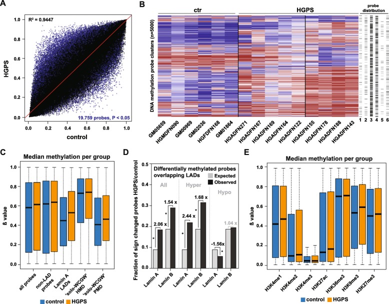
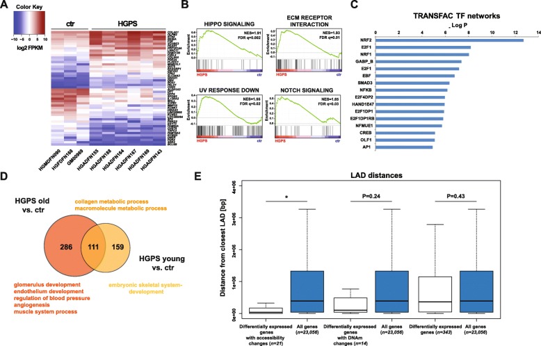
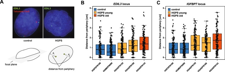
Results: Our ATAC-see/-seq data demonstrate that primary dermal fibroblasts from HGPS patients exhibit chromatin accessibility changes that are enriched in LADs. Infinium MethylationEPIC BeadChip profiling further reveals that DNA methylation alterations observed in HGPS fibroblasts are similarly enriched in LADs and different from those occurring during healthy aging and Werner syndrome (WS), another premature aging disease. Moreover, HGPS patients can be stratified into two different subgroups according to their DNA methylation profiles. Finally, we show that the epigenetic deregulation of LADs is associated with HGPS-specific gene expression changes.
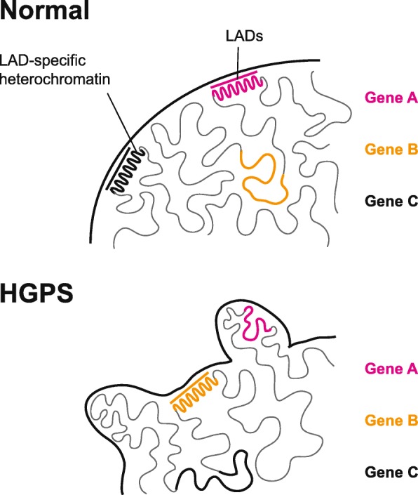
Conclusions: Taken together, our results strongly implicate epigenetic deregulation of LADs as an important and previously unrecognized feature of HGPS, which contributes to disease-specific gene expression. Therefore, they not only add a new layer to the study of epigenetic changes in the progeroid syndrome, but also advance our understanding of the disease's pathology at the cellular level.
Keywords: Aging; Chromatin accessibility; DNA methylation; Epigenetics; Hutchinson-Gilford progeria syndrome; Lamina-associated domains (LADs).
Conflict of interest statement
The authors declare that they have no competing interests.
Figures


Similar articles
-
Identification of mitochondrial dysfunction in Hutchinson-Gilford progeria syndrome through use of stable isotope labeling with amino acids in cell culture.J Proteomics. 2013 Oct 8;91:466-77. doi: 10.1016/j.jprot.2013.08.008. Epub 2013 Aug 20. J Proteomics. 2013. PMID: 23969228
-
Epigenetic involvement in Hutchinson-Gilford progeria syndrome: a mini-review.Gerontology. 2014;60(3):197-203. doi: 10.1159/000357206. Epub 2014 Feb 28. Gerontology. 2014. PMID: 24603298 Review.
-
Reprogramming progeria fibroblasts re-establishes a normal epigenetic landscape.Aging Cell. 2017 Aug;16(4):870-887. doi: 10.1111/acel.12621. Epub 2017 Jun 8. Aging Cell. 2017. PMID: 28597562 Free PMC article.
-
DNA methylation signatures in Blood DNA of Hutchinson-Gilford Progeria syndrome.Aging Cell. 2022 Feb;21(2):e13555. doi: 10.1111/acel.13555. Epub 2022 Jan 19. Aging Cell. 2022. PMID: 35045206 Free PMC article.
-
Hutchinson-Gilford progeria syndrome through the lens of transcription.Aging Cell. 2013 Aug;12(4):533-43. doi: 10.1111/acel.12070. Epub 2013 Apr 19. Aging Cell. 2013. PMID: 23496208 Review.
Cited by
-
The Potentials of Methylene Blue as an Anti-Aging Drug.Cells. 2021 Dec 1;10(12):3379. doi: 10.3390/cells10123379. Cells. 2021. PMID: 34943887 Free PMC article. Review.
-
Disorganized chromatin hierarchy and stem cell aging in a male patient of atypical laminopathy-based progeria mandibuloacral dysplasia type A.Nat Commun. 2024 Nov 20;15(1):10046. doi: 10.1038/s41467-024-54338-3. Nat Commun. 2024. PMID: 39567511 Free PMC article.
-
Potential Impact of Parental Origin of Inheritance on the Clinical Presentation of Familial Partial Lipodystrophy Type 2 Syndrome.Clin Endocrinol (Oxf). 2025 Oct;103(4):504-512. doi: 10.1111/cen.15303. Epub 2025 Jul 16. Clin Endocrinol (Oxf). 2025. PMID: 40671313 Free PMC article.
-
Probe-free optical chromatin deformation and measurement of differential mechanical properties in the nucleus.Elife. 2024 Jan 12;13:e76421. doi: 10.7554/eLife.76421. Elife. 2024. PMID: 38214505 Free PMC article.
-
Alterations to Genome Organisation in Stem Cells, Their Differentiation and Associated Diseases.Results Probl Cell Differ. 2022;70:71-102. doi: 10.1007/978-3-031-06573-6_3. Results Probl Cell Differ. 2022. PMID: 36348105
References
-
- Gerace L, Blobel G. The nuclear envelope lamina is reversibly depolymerized during mitosis. Cell. 1980;19:277–287. - PubMed
-
- Höger TH, Zatloukal K, Waizenegger I, Krohne G. Characterization of a second highly conserved B-type lamin present in cells previously thought to contain only a single B-type lamin. Chromosoma. 1990;99:379–390. - PubMed
-
- Capell BC, Collins FS. Human laminopathies: nuclei gone genetically awry. Nat Rev Genet. 2006;7:940–952. - PubMed
-
- Hennekam RC. Hutchinson-Gilford progeria syndrome: review of the phenotype. Am J Med Genet. 2006;140:2603–2624. - PubMed
Publication types
MeSH terms
Substances
LinkOut - more resources
Full Text Sources
Molecular Biology Databases
