Review
. 2020 Aug;28(8):682-695.
doi: 10.1016/j.tim.2020.02.004. Epub 2020 Mar 26.

The Helicobacter pylori Cag Type IV Secretion System

Affiliations
Review

The Helicobacter pylori Cag Type IV Secretion System

Timothy L Cover et al. Trends Microbiol. 2020 Aug.

Abstract

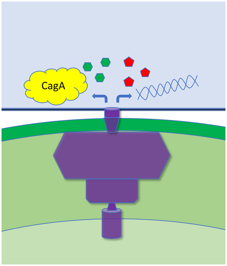
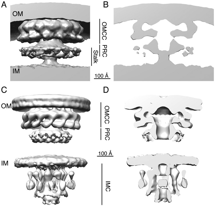
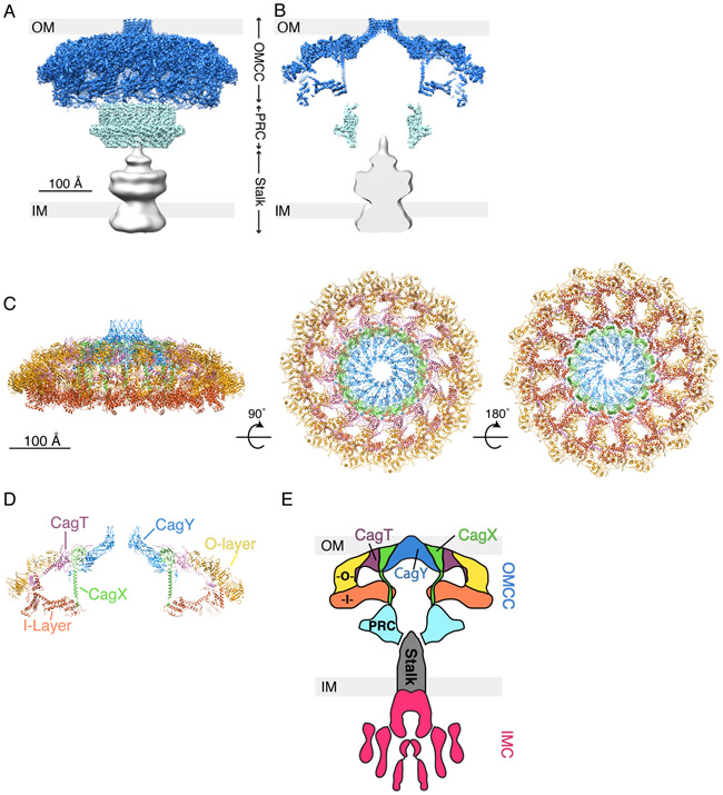
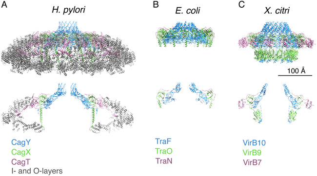
Colonization of the human stomach with Helicobacter pylori strains containing the cag pathogenicity island is a risk factor for development of gastric cancer. The cag pathogenicity island contains genes encoding a secreted effector protein (CagA) and components of a type IV secretion system (Cag T4SS). The molecular architecture of the H. pylori Cag T4SS is substantially more complex than that of prototype T4SSs in other bacterial species. In this review, we discuss recent discoveries pertaining to the structure and function of the Cag T4SS and its role in gastric cancer pathogenesis.

Keywords: bacterial nanomachines; bacterial protein secretion; cryo-electron microscopy; cryo-electron tomography; gastric adenocarcinoma; gastric cancer; macromolecular structures; peptic ulcer disease; type IV secretion system.


Figures

Fig. 1.
Fig. 1.
Schematic depicting an H. pylori bacterium adhering to a host cell. The Cag T4SS (purple) facilities delivery of the CagA effector protein into host cells, as well as intracellular entry of H. pylori LPS metabolites, peptidoglycan and DNA.
Fig. 2.
Fig. 2.
Cryo-ET analyses of the H. pylori Cag T4SS. Spatial relationships between the T4SS, the bacterial outer membrane (OM) and inner membrane (IM) are shown. (A,B) Cryo-ET analysis of H. pylori attached to AGS gastric cells, showing the 3D structure and a central axial slice view of the 3D structure (EMD-7474)(13). Scale bar, 100 Å. (C,D). Cryo-ET analysis of H. pylori grown in pure culture (without AGS gastric cells) showing the 3D structure and a central axial slice view of the 3D structure (EMD- 0634 and 0635)(14). Scale bar, 100 Å. The outer membrane core complex (OMCC), periplasmic ring complex (PRC), stalk, and inner membrane complex (IMC) are labeled.
Fig. 3.
Fig. 3.
3D structure of Cag T4SS particles extracted from H. pylori and determined by single particle cryo-EM. (A) 3D reconstruction of the Cag T4SS with the predicted position of the bacterial outer membrane (OM) and inner membrane (IM) shown (EMD-20023, 20020, and 20021) (16). Outer membrane core complex (OMCC), blue; periplasmic ring complex (PRC), cyan; and stalk, grey. (B) Central axial slice view of the 3D structure in panel A. (C) Secondary structure model of the Cag T4SS OMCC showing the positions of CagT (purple, PDB-6OEE), the C-terminal portion of CagX (green, PDB-6OEG), and the C-terminal portion of CagY (blue, PDB-6ODI), as well as unassigned outer-layer (O-layer, yellow, PDB-6OEF) and inner-layer (I-layer, orange, PDB-6OEH) densities (16). The structure is rotated 90° and then 180° around the X-axis. (D) Central slice view of the structure shown in panel C (left image). (E) Schematic summarizing our current understanding of Cag T4SS architecture, based on cryo-ET and single particle EM studies (13, 14, 16). The Cag T4SS is composed of an OMCC, periplasmic ring complex (PRC, cyan), stalk (grey), and inner membrane complex (IMC, magenta). The positions of CagT (purple) and portions of CagX (green) and CagY (blue) have been mapped into the OMCC. There are outer and inner layer densities (O, yellow and I, orange) that have yet to be assigned a specific T4SS component. The identities and position of T4SS components in the PRC and stalk also have not yet been structurally determined.
Fig. 4.
Fig. 4.
Structural comparison of the H. pylori Cag T4SS OMCC with minimized T4SSs from other bacterial species. (A-C) Upper panel, secondary structural models of the complexes. Lower panel, Central axial slice views of the secondary structural models. Scale bar, 100 Å. (A) OMCC from the H. pylori Cag T4SS (16). CagY (PDB-6ODI, blue), CagX (PDB-6OEG, green), CagT (PDB-6OEE, purple), and outer- and inner-layer proteins (PDB-6OEF and -6OEH, grey). (B) Conjugation system (encoded by pKM101) (66). TraF (PDB-3JQO, blue), TraO (PDB-3JQO, green), and TraN (PDB-3JQO, purple). (C) Xanthomonas citri T4SS (67). VirB10 (PDB-6GYB, blue), VirB9 (PDB-6GYB, green), and VirB7 (PDB-6GYB, purple).

References

    1. Hooi JKY, Lai WY, Ng WK, Suen MMY, Underwood FE, Tanyingoh D, Malfertheiner P, Graham DY, Wong VWS, Wu JCY, Chan FKL, Sung JJY, Kaplan GG, Ng SC. 2017. Global Prevalence of Helicobacter pylori Infection: Systematic Review and Meta-analysis. Gastroenterology doi:10.1053/j.gastro.2017.04.022. - DOI - PubMed
    1. Cover TL, Blaser MJ. 2009. Helicobacter pylori in health and disease. Gastroenterology 136:1863–73. - PMC - PubMed
    1. Bray F, Ferlay J, Soerjomataram I, Siegel RL, Torre LA, Jemal A. 2018. Global cancer statistics 2018: GLOBOCAN estimates of incidence and mortality worldwide for 36 cancers in 185 countries. CA Cancer J Clin 68:394–424. - PubMed
    1. Censini S, Lange C, Xiang Z, Crabtree JE, Ghiara P, Borodovsky M, Rappuoli R, Covacci A. 1996. cag, a pathogenicity island of Helicobacter pylori, encodes type I-specific and disease-associated virulence factors. Proc Natl Acad Sci 93:14648–14653. - PMC - PubMed
    1. Akopyants NS, Clifton SW, Kersulyte D, Crabtree JE, Youree BE, Reece CA, Bukanov NO, Drazek ES, Roe BA, Berg DE. 1998. Analyses of the cag pathogenicity island of Helicobacter pylori. Molecular Microbiology 28:37–53. - PubMed

Publication types

MeSH terms

Substances