Review
. 2020 Jul;41(7):881-894.
doi: 10.1038/s41401-020-0425-3. Epub 2020 May 25.

Nanomedicine-mediated alteration of the pharmacokinetic profile of small molecule cancer immunotherapeutics

Affiliations
Review

Nanomedicine-mediated alteration of the pharmacokinetic profile of small molecule cancer immunotherapeutics

Simon Van Herck et al. Acta Pharmacol Sin. 2020 Jul.

Abstract

The advent of immunotherapy is a game changer in cancer therapy with monoclonal antibody- and T cell-based therapeutics being the current flagships. Small molecule immunotherapeutics might offer advantages over the biological drugs in terms of complexity, tissue penetration, manufacturing cost, stability, and shelf life. However, small molecule drugs are prone to rapid systemic distribution, which might induce severe off-target side effects. Nanotechnology could aid in the formulation of the drug molecules to improve their delivery to specific immune cell subsets. In this review we summarize the current efforts in changing the pharmacokinetic profile of small molecule immunotherapeutics with a strong focus on Toll-like receptor agonists. In addition, we give our vision on limitations and future pathways in the route of nanomedicine to the clinical practice.

Keywords: cancer immunotherapy; nanomedicine; pharmacokinetic profile; small molecule drugs; stimulator of interferon genes (STING); toll-like receptors.


Conflict of interest statement

The authors declare no competing interests.

Figures

Fig. 1
Fig. 1
Chemical structure of TLR2 agonists Pam3CSK4 and diprovocim-1
Fig. 2
Fig. 2
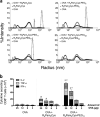
Protein antigen complex of OVA with branched TLR2 agonist R4Pam2Cys-PEG11. a Size distribution profiles of electrostatic complexation of OVA with different pegylated agonists. Increasing PEG length helped in solubilization and decreased particle size. b CD8+ T cell responses were measured by inoculating C57BL/6 mice (n = 3 per group) with OVA alone or OVA formulated with lipopeptide at 1:3 molar ratio. Spleens were obtained 10 days later and OVA257-264-specific IFN-γ, TNF-α, and IL-2 secreting CD8+ T cells enumerated by ICS assay. Each bar graph depicts the total number (±SD) of CD8+ T cells secreting each cytokine. Reproduced from reference [43]. Reprinted with permission from Elsevier
Fig. 3
Fig. 3
TLR4 ligands MPLA, Ugi compound AZ617, pyrimido [5,4,b]indole 26 and amphotericin B
Fig. 4
Fig. 4
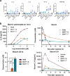
Influence of LeX modified liposomes on cellular uptake and MPLA delivery. a Preferential uptake of LeX-liposomes by DCs in comparison to other cell lines. b Intracellular fate of liposomes. Colocalisation with lyposomal stain LAMP1-DiD indicated endocytosis. c Targeting DC with LewisX- and MPLA-modified liposomes enhances antigen presentation to CD8+ T cells. HLA-A2+ DC were exposed to various concentrations of non-modified or LeX- and/or MPLA-modified liposomes, all loaded with gp100280–288 peptide, for 1 h, in the presence or absence of soluble MPLA. Data reproduced from reference [74]. Reprinted with permission from Elsevier
Fig. 5
Fig. 5
a Intracellular destination of hydrophobic payload encapsulated in responsive NPs imaged by confocal microscopy. b Cytotoxicity analysis of free and encapsulated ampB incubated with RAW264.7 macrophages. Cell viability was analysed via MTT assay after 24 h incubation. c End-point titers after prime and boost immunization against SHe peptide. Reprinted with permission from ACS [89]
Fig. 6
Fig. 6
TLR7/8 agonists resiquimod, loxoribine, UC-1V150, IMDQ
Fig. 7
Fig. 7
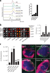
a Bioluminescence imaging of inguinal lymph nodes (LN), femur, and tibia in BALB/c mice (n = 3) after i.v. injection of Luc-LPX or naked Luc-RNA. b Splenic localization of CD11c and Cy3 double-positive cells in BALB/c mice (n = 2) 1 h after i.v. injection of Cy3-labeled RNA-LPX. Scale bar, 100 μm. MZ marginal zone; RP red pulp; WP white pulp. c Bioluminescence imaging of BALB/c mice (n = 3) after i.v. injection of Luc-LPX at various charge ratios. Pie charts show relative contribution of each organ to total signal. Data reproduced from reference. Reprinted with permission from Springer Nature [99]
Fig. 8
Fig. 8
a Systemic cytokines after intratumoral injection of 1V270 [109]. b Assessment of pharmacokinetic properties of SMIPs after intramuscular injection. Reprinted with permission from The American Society for Clinical Investigation
Fig. 9
Fig. 9
Biodistribution visualized by luminescence images of luciferase reporter mice (IFNβ + /Δβ-luc). a images taken 4 and 24 h after footpad injection of soluble IMDQ, amphiphilic polymer conjugated IMDQ (amphIMDQ), or hydrophilic polymer conjugated IMDQ (hydroIMDQ) [114]. Reprinted with permission from ACS. b Images taken before and 4, 24, and 48 h after peritumoral injection of soluble IMDQ (IMDQsoluble) or nanogel conjugated IMDQ (IMDQnano) [117]. Reprinted with permission from Wiley
Fig. 10
Fig. 10
a Size exclusion elugram of CpG-lipids alone or after incubation with serum proteins. b, c IVIS fluorescence imaging of excised draining LNs from C57BL/6 mice (n = 4 LNs per group) injected with Lipid-CpGs (3.3 nmol) in soluble form, emulsified in IFA, entrapped in liposomes, or as amphiphile conjugates. b IVIS images and quantification from inguinal and axillary nodes at 24 h. c left: IVIS quantification of CpG in LNs 24 h after injection of G-quadruplex-forming Lipo-Gn-CpGs. Right: Immunohistochemistry of inguinal LNs 24 h after injection (CD3, blue; B220, pink; CpG, green). Data reproduced from reference [127]. Reprinted with permission from Springer Nature
Fig. 11
Fig. 11
STING agonists cGAMP, DMXAA, amidobenzimidazole (ABZI) compound 3 and BNBC
Fig. 12
Fig. 12
NP-cdGMP enhances LN uptake of CDNs. a Distribution of cdGMP after injected in soluble form or liposomal NP measured by fluorescence spectroscopy. b Percentage of cdGMP positive APCs in the inguinal and axillary lymph node [140]. Reprinted with permission from The American Society for Clinical Investigation

References

    1. Xin Yu J, Hubbard-Lucey VM, Tang J. Immuno-oncology drug development goes global. Nat Rev Drug Discov. 2019;18:899–900. - PubMed
    1. Tang J, Hubbard-Lucey VM, Pearce L, O’Donnell-Tormey J, Shalabi A. The global landscape of cancer cell therapy. Nat Rev Drug Discov. 2018;17:465–6. - PubMed
    1. Nam J, Son S, Park KS, Zou W, Shea LD, Moon JJ. Cancer nanomedicine for combination cancer immunotherapy. Nat Rev Mater. 2019;4:398–414.
    1. Adams JL, Smothers J, Srinivasan R, Hoos A. Big opportunities for small molecules in immuno-oncology. Nat Rev Drug Discov. 2015;14:603–22. - PubMed
    1. Weinmann H. Cancer immunotherapy: selected targets and small-molecule modulators. ChemMedChem. 2016;11:450–66. - PubMed

MeSH terms