. 2020 Jul;23(7):892-902.
doi: 10.1038/s41593-020-0647-1. Epub 2020 May 25.

Cross-hemispheric gamma synchrony between prefrontal parvalbumin interneurons supports behavioral adaptation during rule shift learning

Affiliations

Cross-hemispheric gamma synchrony between prefrontal parvalbumin interneurons supports behavioral adaptation during rule shift learning

Kathleen K A Cho et al. Nat Neurosci. 2020 Jul.

Abstract

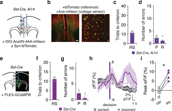
Organisms must learn new strategies to adapt to changing environments. Activity in different neurons often exhibits synchronization that can dynamically enhance their communication and might create flexible brain states that facilitate changes in behavior. We studied the role of gamma-frequency (~40 Hz) synchrony between prefrontal parvalbumin (PV) interneurons in mice learning multiple new cue-reward associations. Voltage indicators revealed cell-type-specific increases of cross-hemispheric gamma synchrony between PV interneurons when mice received feedback that previously learned associations were no longer valid. Disrupting this synchronization by delivering out-of-phase optogenetic stimulation caused mice to perseverate on outdated associations, an effect not reproduced by in-phase stimulation or out-of-phase stimulation at other frequencies. Gamma synchrony was specifically required when new associations used familiar cues that were previously irrelevant to behavioral outcomes, not when associations involved new cues or for reversing previously learned associations. Thus, gamma synchrony is indispensable for reappraising the behavioral salience of external cues.


Figures

Extended Data Fig. 1:
Photometry signals from PV interneurons and behavior during rule shifts in mice used for photometry experiments. a, PV-Cre mice had a unilateral FLEX-GCaMP6f injection and fiber-optic implant in mPFC for photometry (scale bar, 100 μm). b, Rule-shift (RS) performance of PV-Cre mice (n = 8) used for photometry experiments. c, Numbers of perseverative (P) or random (R) errors during the rule shift of PV-Cre mice (n = 8). d, Averaged PV interneuron photometry signal (dF/F), aligned to the start of each trial, for correct (white line) vs. incorrect trials (black line; n = 8). e, Averaged PV interneuron photometry signal (dF/F), aligned to the start of the intertrial interval (ITI), for correct (white line) vs. incorrect trials (black line; n = 8) for the first ten seconds of ITIs. f, Peak dF/F during the entire intertrial interval (ITI), usually lasting around two minutes. Signals are significantly higher on incorrect rule shift trials than correct rule shift trials (n = 8 mice; two-way ANOVA; main effect of outcome: F1,14 = 26.53, ***P = 0.0001; task X outcome interaction: F1,14 = 7.47, *P = 0.016; RS correct versus incorrect, post hoc t(14) = 5.58, ***P = 0.0001; IA correct versus incorrect, post hoc t(14) = 1.71, P = 0.22). g, Mouse behavior scored at the time of the peak dF/F signal during rule shift error trials (n = 31 error trials of 8 mice). h, Mouse behavior scored at the time of the peak dF/F signal during initial association error ITI (n = 21 error ITIs of 8 mice). i, Mouse behavior scored at the time of the peak dF/F signal during rule shift error ITI (n = 31 error ITIs of 8 mice). Data are shown as means (b, c); error bars (b, c) and shading (d, e) denote s.e.m. Two-way ANOVA followed by Bonferroni post hoc comparisons were used. Comparisons were not significant unless otherwise noted.
Extended Data Fig. 2:
Cross-hemispheric synchrony of PV interneurons at non-zero phase lag. a, Schematic: analysis to measure out-of-phase synchrony. In this case, one Ace-mNeon is signal is shifted 90 degrees out-of-phase relative to the other signals, before following the procedure outlined in Figure 2e. bd, Out-of-phase 30–50 Hz synchrony (n = 12 mice) did not differ between the baseline period and RS correct trials (two-way ANOVA; frequency X condition interaction: F2,33 = 0.16, P = 0.86; post hoc t(33) = 1.35, P = 0.56), baseline period and RS incorrect trials (two-way ANOVA; frequency X condition interaction: F2,33 = 0.06, P = 0.94; post hoc t(33) = 0.05, P > 0.99), or correct and incorrect trials (two-way ANOVA; frequency X condition interaction: F2,33 = 0.05, P = 0.95; post hoc t(33) = 1.53, P = 0.41). Two-way ANOVA followed by Bonferroni post hoc comparisons were used. Comparisons were not significant.
Extended Data Fig. 3:
Learning during rule shifts in mice used for TEMPO measurements from Sst interneurons, and photometry signals from Sst interneurons during rule shifts. a, Sst-Cre, Ai14 mice had bilateral AAV-DIO-Ace2N-4AA-mNeon ± AAV-Syn-tdTomato injections and fiber-optic implants in mPFC. b, Representative images of tdTomato (red) and Ace-mNeon (green) fluorescence in a coronal section of mPFC (left), alongside a high power image (right). Scale bars: 100 μm and 25 μm, respectively. c, Rule-shift (RS) performance of Sst-Cre, Ai14 mice (n = 5) used for dual-site TEMPO imaging. d, Number of perseverative (P) and random (R) errors during the rule shift of Sst-Cre, Ai14 mice (n = 5). e, Sst-Cre mice had a unilateral FLEX-GCaMP6f injection and fiber-optic implant in mPFC for photometry (scale bar, 100 μm). f, Rule-shift (RS) performance of Sst-Cre mice (n = 4) used for photometry experiments. g, Numbers of perseverative (P) or random (R) errors during the rule shift Sst-Cre mice (n = 4). h, Averaged Sst interneuron photometry signal (dF/F), aligned to the time of dig, which indicates a decision, for correct (white line) vs. incorrect trials (purple line; n = 4). i, Peak dF/F during the 4–8 sec following the decision (this was the time at which peak Sst activity occurred). Sst interneuron photometry signals are significantly higher on incorrect than correct trials (n = 4 mice; two-tailed, paired t-test; t(3) = 4.65, *P = 0.02). Data are shown as means (c, d, f, g, h); error bars (c, d, f, g) and shading (h) denote s.e.m.
Extended Data Fig. 4:
Extended Data Fig. 4:. Cross-hemispheric synchrony between prefrontal PV interneurons for various frequency bands and types of trials.
a, PV-Cre, Ai14 mice had bilateral AAV-DIO-Ace2N-4AA-mNeon ± AAV-Syn-tdTomato injections and fiber-optic implants in mPFC. Experimental design: Day 1: Initial association (IA) followed by rule shift (RS) or rule reversal (RR); Day 2: IA followed by the rule change (RS or RR) that was not performed on Day 1. b, During learning of the IA that preceded the RS, synchrony was not different after correct decisions vs. during the baseline period (n = 7 mice; two-way ANOVA; main effect of condition: F1,18 = 0.51, P = 0.48; frequency X condition interaction: F2,18 = 0.20, P = 0.82). c, During learning of this IA, synchrony was not different after incorrect decisions vs. during the baseline period (n = 7 mice; two-way ANOVA; main effect of condition: F1,18 = 0.39, P = 0.54; frequency X condition interaction: F2,18 = 0.07, P = 0.94). d, IA performance was not different across days (n = 7 mice; two-tailed, paired t-test; t(6) = 1.29, P = 0.25). e, There was no difference in synchrony after correct trials during learning of the IA on Day 1 vs. 2 (n = 7 mice; two-way ANOVA; main effect of day: F1,18 = 0.02, P = 0.89; frequency X condition interaction: F2,18 = 1.48, P = 0.26). f, There was no difference in synchrony after incorrect trials during learning of the IA on Day 1 vs. 2 (n = 7 mice; two-way ANOVA; main effect of day: F1,18 = 3.05, P = 0.10; frequency X condition interaction: F2,18 = 0.03, P = 0.97). g, During the RR, synchrony was not different after correct decisions vs. during the baseline period (n = 7 mice; two-way ANOVA; main effect of condition: F1,18 = 0.28, P = 0.60; frequency X condition interaction: F2,18 = 1.24, P = 0.31). h, During the RR, synchrony was not different after incorrect decisions vs. the baseline period (n = 7 mice; two-way ANOVA; main effect of condition: F1,18 = 0.09, P = 0.77; frequency X condition interaction: F2,18 = 0.30, P = 0.74). i, Synchrony after correct decisions did not differ between the IA vs. RS (n = 7 mice; two-way ANOVA; main effect of condition: F1,18 = 0.13, P = 0.73; frequency X condition interaction: F2,18 = 1, P = 0.39). j, Synchrony after correct decisions did not differ between the RR vs. RS (n = 7 mice; two-way ANOVA; main effect of condition: F1,18 = 0.16, P = 0.70; frequency X condition interaction: F2,18 = 2.55, P = 0.11). k, The plot shows the average gamma synchrony on correct vs. incorrect trials, during the first 2 (‘early’) or next 3 (‘late’) trials of a rule shift (RS) or rule reversal (RR). In order to average together values from different mice (n = 7), each synchrony value was computed relative to the average gamma synchrony measured during the first 5 RS and RR trials from the same mouse. We performed ANOVA on the gamma synchrony from each of the first 5 trials during a rule shift (RS) or rule reversal (RR), including the following factors and interaction terms: mouse (F6,59 = 1.72, P = 0.13), type of rule change (RS vs. RR) (F1,59 = 3.74, P = 0.06), correct vs. incorrect trial outcome (F1,59 = 2.64, P = 0.11), an interaction of correct-incorrect X RS-RR (F1,59 = 11.12, **P = 0.0015), and an interaction of correct-incorrect X RS-RR X early vs. late trials (i.e. first 2 vs. next 3 trials) (F1,59 = 4.28, *P = 0.043). l, Following errors, synchrony during the intertrial interval (ITI) was specifically higher in the 30–50 Hz band during the RS than during the IA (n = 7 mice; two-way ANOVA; condition X frequency interaction: F4,36 = 3.217, *P = 0.023; 30–50 Hz post hoc t(6) = 3.55, *P = 0.036) or RR (two-way ANOVA; 30–50 Hz post hoc t(6) = 3.97, *P = 0.022). m, Mouse behavior scored during the initial association error ITI for the first 5 IA dual-site TEMPO trials (n = 10 error ITIs of 8 mice). n, Mouse behavior scored during the rule shift error ITI for the first 5 RS dual-site TEMPO trials (n = 17 error ITIs of 8 mice). There is no difference in seconds of movement between IA and RS (two-tailed, paired t-test; t(6) = 0.52, P = 0.62) nor in seconds of not moving between IA and RS (two-tailed, paired t-test; t(6) = 0.56, P = 0.59). Two-way ANOVA followed by Bonferroni post hoc comparisons were used in panels bc and ej, l. Comparisons were not significant unless otherwise noted.
Extended Data Fig. 5:
Extended Data Fig. 5:. Types of errors during rule shifts (RS) in the presence of various forms of optogenetic stimulation in PV-Cre mice.
a, d, PV-Cre, Ai14 mice had bilateral AAV-DIO-eYFP or AAV-DIO-ChR2 injections and fiber-optic implants in mPFC. Experimental design: Day 1: out-of-phase 40 Hz stimulation during the rule shift (RS); Day 2: no stimulation. b, e, Representative images showing mPFC expression of eYFP (b) or ChR2-eYFP (e) (scale bar, 100 μm). c, f, Optogenetic stimulation increases perseverative errors in ChR2-expressing mice (n = 5) compared to eYFP-expressing (n = 5 mice) controls (two-way ANOVA; main effect of day: F1,8 = 20.8, **P = 0.0018; main effect of virus: F1,8 = 8.96, *P = 0.017; day X virus interaction: F1,8 = 14.89, **P = 0.0048). There is no change in random errors (two-way ANOVA; main effect of day: F1,8 = 0, P > 0.99; main effect of virus: F1,8 = 0, P > 0.99; day X virus interaction: F1,8 = 0.89, P = 0.37). c, Light delivery does not affect the number of perseverative or random errors in eYFP-expressing controls (perseverative: post hoc t(8) = 0.50, P > 0.99; random: post hoc t(8) = 0.67, P > 0.99). f, Optogenetic stimulation of PV interneurons on Day 1 increased the number of perseverative errors compared to no stimulation on Day 2 (post hoc t(8) = 5.95, ***P = 0.0007), but does not affect random errors (post hoc t(8) = 0.67, P > 0.99). g, PV-Cre mice had bilateral AAV-DIO-ChR2-eYFP injections and fiber-optic implants in mPFC. Experimental design: Day 1: out-of-phase 20 Hz stimulation; Day 2: out-of-phase 40 Hz stimulation; Day 3: in-phase 40 Hz stimulation. h, Representative image showing mPFC ChR2 expression (scale bar, 100 μm). i, Out-of-phase 40 Hz stimulation (Day 2) increases perseverative errors but does not affect random errors, relative to out-of-phase 20 Hz stimulation (Day 1) or in-phase 40 Hz stimulation (Day 3) (n = 5 mice; two-way ANOVA; main effect of day: F2,16 = 13.5, ***P = 0.0004; Day 1 vs. Day 2 perseverative: ***P = 0.0002, Day 2 vs. Day 3 perseverative: ****P = 0.00003, Day 1 vs. Day 3 perseverative: P > 0.99; Day 1 vs. Day 2 random: P > 0.99, Day 2 vs. Day 3 random: P > 0.99, Day 1 vs. Day 3 random: P > 0.99). Two-way ANOVA followed by Bonferroni post hoc comparisons were used.
Extended Data Fig. 6:
Extended Data Fig. 6:. Behavior and types of errors during rule shifts (RS) in the presence of various forms of optogenetic stimulation in Dlx5/6+/−, Sst-Cre and Sst-Cre mice.
a, e, Sst-Cre mice had bilateral injections of AAV-DIO-eYFP (a, ‘Sst-eYFP’) or AAV-DIO-ChR2-eYFP (e, ‘Sst-ChR2’) along with fiber-optic implants in mPFC. Experimental design: Day 1: no light stimulation; Day 2: out-of-phase 20 Hz stimulation; Day 3: out-of-phase 40 Hz stimulation. b, Representative image showing mPFC ChR2 expression (scale bar, 100 μm). c, g, Light delivery did not affect performance in either Sst-eYFP (c; n = 4 mice) or Sst-ChR2 (g; n = 5) mice (two-way ANOVA; main effect of day: F2,14 = 2.53, P = 0.12; main effect of virus: F1,7 = 0.01, P = 0.92; day X virus interaction: F2,14 = 0.59, P = 0.57). d, Sst-eYFP mice showed no change in perseverative or random errors from Day 1 to Day 2 to Day 3 (n = 4 mice; two-way ANOVA; day X type of error interaction: F2,9 = 1.18, P = 0.35). h, Sst-ChR2 mice showed no change in perseverative or random errors from Day 1 to Day 2 to Day 3 (n = 5 mice; two-way ANOVA; day X type of error interaction: F2,16 = 0.81, P = 0.46). i,m, Dlx5/6+/−, Sst-Cre mice had bilateral control virus (AAV-DIO-eYFP or AAV-i12b-DIO-eYFP) or AAV-DIO-ChR2-eYFP (m) injections and fiber-optic implants in mPFC. Experimental design: Day 1: no stimulation; Day 2: 40 Hz stimulation. j,n, Representative eYFP (j) and ChR2-eYFP (n) expression in the mPFC of Dlx5/6+/−, Sst-Cre mice (scale bar, 100 μm). k,o, Light delivery did not affect performance in Sst-eYFP-expressing (k; n = 4 mice) or Sst-ChR2-expressing (o; n = 8) mutant mice (two-way ANOVA; main effect of day: F1,10 = 0.08, P = 0.79; main effect of virus: F1,10 = 0.002, P = 0.96; day X virus interaction: F1,10 = 2.69, P = 0.13). l, Sst-eYFP expressing mutants showed no change in perseverative or random errors from Day 1 to Day 2 (n = 4 mice; two-way ANOVA; day X type of error interaction: F1,6 = 0.16, P = 0.70). p, Sst-ChR2 expressing mutants showed no change in perseverative or random errors from Day 1 to Day 2 (n = 8 mice; two-way ANOVA; day X type of error interaction: F1,14 = 0.64, P = 0.44). Two-way ANOVA followed by Bonferroni post hoc comparisons were used. Comparisons were not significant unless otherwise noted.
Extended Data Fig. 7:
Extended Data Fig. 7:. Single unit recordings from PV interneurons and regular spiking neurons in the mPFC of awake, head-fixed mice.
a, Schematic of opto-silicon probe recording PFC in awake head-fixed mice (top panel). Histology of the recording electrode (bottom panel, scale = 1mm). b, Example raster plot of a putative PV cell responding to ChR2 activation. Stimulation at 1 Hz (5 ms illumination, 0.25 mW). The blue line represents the duration of ChR2 stimulation (5 ms). c, Average peristimulus time histogram (PSTH) of PV cell responses during 40 Hz ChR2 stimulation (n = 8 PV cells, time bin = 1 ms). The blue line indicates the period of ChR2 stimulation (5 ms flash) at 40 Hz. PV cells fired 0.49 ± 10 spikes per 40 Hz cycle at a latency of 1.82 ± 0.34 ms following the onset of each light flash. d, Baseline subtracted and peak normalized activity of all putative PV cells (green line; n = 8 cells, 3 mice) and all the regular-spiking (RS) cells (black line; n = 237 cells, 4 mice). Mean firing rate of RS cells was 4.9 vs. 2.1 spikes/sec at the peak vs. trough of each cycle, respectively. The blue line indicates the period of ChR2 stimulation (5 ms flash) at 40 Hz. Data are shown as means (c, d) and shading (c, d) denote s.e.m.
Extended Data Fig. 8:
Extended Data Fig. 8:. Changes in the power spectra of prefrontal LFPs elicited by in- vs. out-of-phase stimulation of PV interneurons.
ad, Difference between the power spectra for LFPs recorded during light stimulation vs. at baseline for control mice (PV-Cre mice injected with AAV-DIO-eYFP, n = 6 recordings from 3 mice) receiving (a) in- or (c) out-of-phase stimulation, or for PV-Cre mice (n = 12 recordings from 6 mice) injected with AAV-DIO-ChR2-eYFP receiving (b) in- or (d) out-of-phase stimulation. Positive (negative) values correspond to higher power during periods of stimulation (at baseline). e, Quantification of average change in 40 Hz power in various conditions (relative to baseline). The change was significantly different from 0 for PV-ChR2 in-phase condition (n = 12 recordings from 6 mice; two-tailed, paired t-test; t(11) = 2.24, *P = 0.047) and for PV-ChR2 out-of-phase condition (n = 12 recordings from 6 mice; two-tailed, paired t-test; t(11) = 2.34, *P = 0.039). f, Quantification of average change in 150–200 Hz power in various conditions (relative to baseline). The change was significantly different from 0 for PV-ChR2 in-phase condition (n = 12 recordings from 6 mice; two-tailed, paired t-test; t(11) = 2.40, *P = 0.036) and for PV-ChR2 out-of-phase condition (n = 12 recordings from 6 mice; two-tailed, paired t-test; t(11) = 2.65, *P = 0.023). Two-tailed, paired t-tests were used. Data are shown as means; shading (ad) and error bars (e,f) denote s.e.m.
Extended Data Fig. 9:
Extended Data Fig. 9:. Out-of-phase 40 Hz stimulation does not disrupt the ability of PV-Cre mice to revert to an initial association.
a, Schematic for task: The mouse learns an initial association (IA), then a rule shift (RS). After the mouse learns the RS, the task reverts to the original rule, i.e., the rule learned during the IA with out-of-phase 40 Hz stimulation. b, PV-Cre mice had bilateral AAV-DIO-eYFP (‘PV-eYFP’) or AAV-DIO-ChR2-eYFP (‘PV-ChR2’) injections and fiber-optic implants in mPFC. Experimental design: no stimulation during learning of the IA or RS. Then, when the rule reverts to the IA, out-of-phase 40 Hz stimulation is delivered. c, In PV-eYFP mice (n = 5), there was no difference in the number of trials needed to reach the criterion during initial learning of the IA (when no light was delivered) vs. when reverting to the IA after learning the RS (when light stimulation was delivered; two-tailed, paired t-test; t(4) = 1.31, P = 0.261). d, In PV-ChR2 mice (n = 5), there was no difference in the number of trials needed to reach the criterion during initial learning of the IA (when no light was delivered) vs. when reverting to the IA after learning the RS (when light stimulation was delivered; two-tailed, paired t-test; t(4) = 0.466, P = 0.666). Two-tailed, paired t-tests were used. Comparisons were not significant unless otherwise noted.
Extended Data Fig. 10:
Extended Data Fig. 10:. Behavior and cross-hemispheric synchrony between prefrontal PV or Sst interneurons during rule shifts, baseline periods, or learning of initial associations in Dlx5/6+/− mice and wild-types.
a, Rule-shift (RS) performance for photometry experiments in Dlx5/6+/−, PV-Cre and Dlx5/6+/+, PV-Cre mice. Compared to wild-type littermates (n = 8), mutant mice (n = 7) make more perseverative errors (two-way ANOVA; main effect of genotype: F1,13 = 43.6, ****P = 0.00002; main effect of error type: F1,13 = 24.7, ***P = 0.0003; error type X genotype interaction: F1,13 = 10.5, **P = 0.006; post hoc t(26) = 6.55, ****P = 0.000001), but similar numbers of random errors (post hoc t(26) = 1.36, P = 0.37). b, Rule-shift performance for mice used in dual-site TEMPO experiments. Compared to wild-type mice (n = 12), mutant mice (n = 8) make more perseverative errors (two-way ANOVA; main effect of genotype: F1,18 = 89.4, ****P =0.00000002; main effect of error type: F1,13 = 137.3, ****P = 0.0000000007; type of error X genotype interaction: F1,18 = 46.5, ****P = 0.000002; post hoc t(36) = 11.6, ****P = 0.0000000000002), and random errors (post hoc t(36) = 2.43, *P = 0.04). c, Compared to wild-type mice, Dlx5/6+/−, Sst-Cre, Ai14 mice make more perseverative errors (n = 5 mice in each cohort; two-way ANOVA; main effect of genotype: F1,8 = 42.3, ***P = 0.0002; main effect of error type: F1,8 = 30.7, ***P = 0.0005; error type X genotype interaction: F1,8 = 17.0, **P = 0.003; post hoc t(16) = 7.12, ****P = 0.000005), but numbers of random errors are comparable (post hoc t(16) = 0.38, P > 0.99). d, Synchrony was not different between Dlx5/6++-, Sst-Cre, Ai14 and Dlx5/6+/−, Sst-Cre, Ai14 mice during the baseline period (n = 5 mice in each cohort; two-way ANOVA; main effect of genotype: F1,8 = 0.77, P = 0.41; frequency X genotype interaction: F2,16 = 0.05, P = 0.95). e, Dlx5/6+/−, PV-Cre mice had bilateral AAV-DIO-ChR2-eYFP injections and fiber-optic implants in mPFC. Experimental design: Day 1: no stimulation; Day 2: in-phase 40 Hz stimulation during the first 5 RS trials. f, In-phase 40 Hz stimulation on Day 2 reduces perseverative errors relative to no stimulation on Day 1 (n = 6 mice; two-way ANOVA; main effect of day: F1,10 = 18.32, **P = 0.0016; main effect of error type: F1,10 = 49.9, ****P =0.000034; post hoc t(10) = 3.98, **P = 0.005); there was no change in random errors (post hoc t(10) = 2.07, P = 0.13). g, Dlx5/6+/−, PV-Cre mice had bilateral AAV-DIO-ChR2-eYFP injections and fiber-optic implants in mPFC. Experimental design: Day 1: out-of-phase 20 Hz stimulation; Day 2: out-of-phase 40 Hz stimulation; Day 3: in-phase 40 Hz stimulation. h, Perseverative errors are reduced by in phase 40 Hz stimulation on Day 3, compared to either out-of-phase 20 Hz stimulation on Day 1 or out-of-phase 40 Hz stimulation on Day 2 (n = 5 mice; two-way ANOVA; main effect of day: F2,16 = 11.6, ***P = 0.0008; main effect of error type: F1,8 = 31.5, ***P = 0.0005; Day 1 vs. Day 2 perseverative: P > 0.99, Day 2 vs. Day 3 perseverative: ***P = 0.0005, Day 1 vs. Day 3 perseverative: **P = 0.0016). There are no changes in random errors across days (post hoc Day 1 vs. Day 2: P > 0.99, Day 2 vs. Day 3: P > 0.99, Day 1 vs. Day 3: P = 0.33). i, Dlx5/6+/−, PV-Cre, Ai14 mice had bilateral AAV-DIO-Ace2N-4AA-mNeon ± AAV-Syn-tdTomato injections and fiber-optic implants in mPFC. j, Representative images of tdTomato (red) and Ace-mNeon (green) fluorescence within PV interneurons in a coronal section of mPFC (left), alongside a high power image (right). Scale bars: 100 μm and 25 μm, respectively. k, In mutants, PV interneuron synchrony was not different after correct decisions vs. during the baseline period (n = 8 mice; two-way ANOVA; main effect of condition: F1,21 = 1.89, P = 0.18; frequency X condition interaction: F2,21 = 0.35, P = 0.71). l, PV interneuron synchrony was not different after incorrect decisions vs. during the baseline period (n = 8 mice; two-way ANOVA; main effect of condition: F1,21 = 3.31, P = 0.083; frequency X condition interaction: F2,21 = 0.04, P = 0.96). m, Dlx5/6+/−, Sst-Cre, Ai14 mice had bilateral AAV-DIO-Ace2N-4AA-mNeon ± AAV-Syn-tdTomato injections and fiber-optic implants in mPFC. n, Representative images of tdTomato (red) and Ace-mNeon (green) fluorescence in Sst interneurons within a coronal section of mPFC (left) from a Dlx5/6+/−, Sst-Cre, Ai14 mouse, alongside a high power image (right). Scale bars: 100 μm and 25 μm, respectively. o, In mutants, Sst interneuron synchrony was not different after correct decisions vs. during the baseline period (n = 5 mice; two-way ANOVA; main effect of frequency: F2,12 = 9.88, **P = 0.003; frequency X condition interaction: F2,12 = 0.58, P = 0.58). p, Sst interneuron synchrony was not different after incorrect decisions vs. during the baseline period (n = 5 mice; two-way ANOVA; main effect of frequency: F2,12 = 4.95, *P = 0.027; frequency X condition interaction: F2,12 = 0.44, P = 0.66). q, Learning of an initial association (IA) was similar in mutants (n = 6) and their wild-type (n = 11) littermates (two-tailed, unpaired t-test; n = 11; t(15) = 0.202, P = 0.842). r, There was no difference in cross-hemispheric PV interneuron synchronization between mutant (n = 6 mice) and wild-type (n = 11 mice) littermates at baseline (two-way ANOVA; main effect of genotype: F1,15 = 0.45, P = 0.51; genotype X frequency interaction F2,30 = 1.11, P = 0.34). s, During learning of an initial association, changes in PV interneuron synchrony following errors (relative to synchrony after correct decisions) is similar in mutants (n = 6 mice) and their wild-type (n = 11 mice) littermates (two-way ANOVA; main effect of genotype: F1,15 = 0.07, P = 0.80; genotype X frequency interaction: F2,30 = 0.16, P = 0.86). Data are shown as means (ac); error bars (ac) denote s.e.m. Two-way ANOVA followed by Bonferroni post hoc comparisons were used. Comparisons were not significant unless otherwise noted.
Fig. 1:
Fig. 1:. Prefrontal PV interneurons are recruited after errors during rule shifts.
a, Rule shift task schematic. On each trial, a mouse chooses one of two bowls, each scented with a different odor (O1 or O2) and filled with a different textured digging medium (TA or TB), to find a food reward. Mice first learn an initial association (IA) between one of these cues (e.g., odor O1) and food reward (the cue associated with reward is indicated in orange). Once mice reach the learning criterion (8/10 consecutive trials correct), this association undergoes an extra-dimensional rule shift (RS; e.g., from O1 to TA). b, Rule reversal task schematic. Mice learn an initial association (IA) between one cue (e.g., odor O1) and food reward (the rewarded cue is indicated in orange). Once mice reach the learning criterion, this association undergoes an intra-dimensional rule reversal (RR), e.g., from O1 to O2. c, Trial timeline. A mouse begins each trial by entering the home cage, then makes a decision, indicated by digging in one bowl. If the mouse is correct, food reward is consumed. The mouse is then transferred to the holding cage until the next trial. The intertrial interval is longer after incorrect choices. d, Representative image showing mPFC FLEX-GCaMP6f expression in a PV-Cre mouse (scale bar, 100 μm). e, Averaged PV interneuron photometry signal (dF/F), aligned to the time of dig, which indicates a decision, for correct (white line) vs. incorrect trials (black line; n = 8 mice). f, Peak dF/F during the 4 sec following the decision. Signals are significantly higher on incorrect than correct trials (n = 8 mice; two-tailed, paired t-test; t(7) = 3.93, **P = 0.006). Data are shown as means (e); shading (e) denotes s.e.m.
Fig. 2:
Fig. 2:. Cross-hemispheric gamma synchrony of PV interneurons increases after errors during rule shifts.
a, PV-Cre, Ai14 mice had bilateral AAV-DIO-Ace2N-4AA-mNeon ± AAV-Syn-tdTomato injections and fiber-optic implants in mPFC. b, Representative images of tdTomato (red) and Ace-mNeon (green) fluorescence in a coronal section of mPFC (left), alongside a high-power image (right). Scale bars: 100 μm and 25 μm, respectively. c, Schematic for dual-site TEMPO measurements. Each fiber-optic implant, for delivering illumination and collecting fluorescence, connects to a mini-cube coupled to two LEDs and two photoreceivers (PR) to separately excite and collect emitted fluorescence from Ace-mNeon and tdTomato. Two lock-in amplifiers modulate LED output and demodulate PR signals, which are then acquired by a multichannel real-time signal processor. d, Initial association (IA) and rule shift (RS) performance in this cohort (n = 12 mice). e, Overview of dual-site TEMPO analysis: tdTomato and Ace-mNeon fluorescence signals from each hemisphere are filtered around a frequency of interest, then both tdTomato signals and one Ace-mNeon signal are used to model the second Ace-mNeon signal. Performance is compared to models based on shuffled versions of the first Ace-mNeon signal. f, R2 values, measuring zero-phase lag ~40 Hz cross-hemispheric PV interneuron synchrony, during the last 3 IA trials and the first 5 RS trials in one mouse. g, Synchrony was not different after correct decisions vs. during the baseline period (n = 12 mice; two-way ANOVA; condition X frequency interaction: F2,33 = 1.05, P = 0.36). hi, 30–50 Hz synchronization was specifically higher after RS errors than during the baseline period (n = 12 mice; two-way ANOVA; main effect of condition: F1,33 = 10.51, **P = 0.003; frequency X condition interaction: F2,33 = 8.23, **P = 0.001; 15–25 Hz: post hoc t(33) = 0.43, P > 0.99; 30–50 Hz: post hoc t(33) = 5.18, ****P = 0.00003; 50–70 Hz: post hoc t(33) = 0.007, P > 0.99) or after RS correct decisions (n = 12 mice; two-way ANOVA; condition X frequency interaction: F2,33 = 7.32, **P = 0.002; post hoc t(33) = 4.36, ***P = 0.0004). j, R2 values, measuring zero-phase lag ~40 Hz cross-hemispheric Sst interneuron synchrony during the last 3 IA trials and the first 5 RS trials in one mouse. km, Cross-hemispheric Sst synchrony (n = 5 mice) was not different between the baseline period and RS correct trials (two-way ANOVA; main effect of condition: F1,12 = 0.07, P = 0.79; main effect of frequency: F2,12 = 6.07, *P = 0.015; condition X frequency interaction F2,12 = 0.10, P = 0.90), baseline period and RS incorrect trials (two-way ANOVA; main effect of condition: F1,12 = 0.47, P = 0.51; main effect of frequency: F2,12 = 7.34, **P = 0.008; condition X frequency interaction: F2,12 = 0.26, P = 0.78), nor correct and incorrect trials (two-way ANOVA; main effect of condition: F1,12 = 0.25, P = 0.63; main effect of frequency: F2,12 = 4.66, *P = 0.03; condition X frequency interaction: F2,12 = 0.034, P = 0.97). n, In a cohort of PV-Cre mice used for simultaneous dual-site TEMPO measurements and LFP recordings (n = 5 mice), 30–50 Hz synchronization between left and right PV interneuron TEMPO signals was specifically higher after RS errors than after RS correct decisions (two-way ANOVA; frequency X condition interaction: F2,12 = 7.13, **P = 0.009; post hoc t(12) = 2.83, *P = 0.045). o, Difference in zero-phase-lag LFP wavelet coherence following errors relative to after correct decisions (i.e., incorrect – correct; n = 5 mice). p, LFP wavelet coherence was higher at 20 and 40 Hz following RS errors than after correct decisions (n = 5 mice; two-way ANOVA; main effect of condition: F1,12 = 21.40, ***P = 0.0006; main effect of frequency: F2,12 = 1.86, P = 0.199; condition X frequency: F2,12 = 0.17, P = 0.85; 20 Hz post hoc t(12) = 2.85, *P = 0.04; 40 Hz post hoc t(12) = 2.96, *P = 0.04). Data are shown as means (d, o); error bars (d, o) denote s.e.m. Two-way ANOVA followed by Bonferroni post hoc comparisons were used, unless otherwise noted. Comparisons were not significant, unless otherwise noted.
Fig. 3:
Fig. 3:. Cross-hemispheric synchrony does not increase during initial associations and rule reversals.
a, Averaged PV interneuron photometry signal (dF/F), aligned to the time of dig, which indicates a decision, for correct (brown line) and incorrect trials (yellow line) during the initial association (IA) (n = 8 mice). b, Peak dF/F during the 4 sec following the decision. Signals are comparable during correct and incorrect IA trials (n = 8 mice; two-tailed, paired t-test; t(7) = 0.44, P = 0.67). c, Averaged PV interneuron photometry signal (dF/F), aligned to the time of dig, which indicates a decision, for incorrect trials during the IA (yellow line) or rule shift (RS) (black line; n = 8 mice). d, Peak dF/F during the 4 sec following the decision. Signals on incorrect trials are significantly lower during the IA than the RS (n = 8 mice; two-tailed, paired t-test; t(7) = 2.87, *P = 0.024). (Note: to be conservative and include all data, we did not exclude one datapoint which appeared to be an outlier; however, had this datapoint been excluded, this P value would have been 0.0096). e, PV-Cre, Ai14 mice had bilateral injections of AAV-DIO-Ace2N-4AA-mNeon ± AAV-Syn-tdTomato in mPFC and fiber-optic implants in mPFC. Experimental design: Day 1: IA followed by RS or RR; Day 2: IA followed by the task not performed on Day 1. f, PV-Cre, Ai14 mice performed rule shifts (RS) and rule reversals (RR) in a similar number of trials (n = 7 mice; two-tailed, paired t-test; t(6) = 0.92, P = 0.39). g, During the IA, synchrony did not differ following correct vs. incorrect trials (n = 7 mice; two-way ANOVA; main effect of condition: F1,18 = 0.0007, P = 0.98; condition X frequency interaction: F2,18 = 0.068, P = 0.93). h, During the RR, synchrony did not differ following correct vs. incorrect trials (n = 7 mice; two-way ANOVA; main effect of condition: F1,18 = 0.10, P = 0.76; condition X frequency interaction F2,18 = 4.55, *P = 0.025; 15–25 Hz: post hoc t(18) = 0.25, P > 0.99; 30–50 Hz: post hoc t(18) = 1.71, P = 0.32; 50–70 Hz: post hoc t(18) = 2.50, P = 0.07). i, Following errors, synchrony was specifically higher for the 30–50 Hz band during RS than IA (n = 7 mice; two-way ANOVA; condition X frequency interaction: F2,18 = 6.02, **P = 0.0099; 30–50 Hz post hoc t(18) = 3.42, **P = 0.009). j, Following errors, synchrony was specifically higher in the 30–50 Hz band RS than RR (two-way ANOVA; condition X frequency interaction F2,18 = 3.96, *P = 0.038; 30–50 Hz post hoc t(18) = 2.64, *P = 0.0499). Data are shown as means (a, c); shading (a, c) denotes s.e.m. Two-way ANOVA followed by Bonferroni post hoc comparisons, unless otherwise noted.
Fig. 4.
Fig. 4.. Out-of-phase, but not in-phase, gamma-frequency stimulation of PV interneurons disrupts learning during rule shifts but not during initial associations or rule reversals.
a, PV-Cre, Ai14 mice had injections of AAV-DIO-eYFP or AAV-DIO-ChR2-eYFP in mPFC and fiber-optic implants bilaterally in mPFC. Experimental design: Day 1: out-of-phase 40 Hz stimulation during the rule shift (RS); Day 2: no stimulation. bc, Out of phase 40 Hz stimulation impairs rule shift performance in ChR2-expressing mice compared to eYFP-expressing controls (n = 5 mice in each cohort; two-way ANOVA; main effect of day: F1,8 = 40.2, ***P = 0.0002; main effect of virus: F1,8 = 32.5, ***P = 0.0005; day X virus interaction: F1,8 = 47.3, ***P = 0.0001). b, Performance of eYFP-expressing controls did not change from Day 1 to 2 (n = 5 mice; post hoc t(8) = 0.38, P > 0.99). c, Out-of-phase 40 Hz stimulation of PV interneurons across hemispheres during the RS on Day 1 impaired rule shifts in ChR2-expressing mice, compared to no stimulation on Day 2 (n = 5 mice; post hoc t(8) = 9.34, ****P = 0.00003). d, PV-Cre mice had bilateral injections of AAV-DIO-ChR2-eYFP and fiber-optic implants in mPFC. Experimental design: Day 1: out-of-phase 20 Hz stimulation; Day 2: out-of-phase 40 Hz stimulation; Day 3: in-phase 40 Hz stimulation. e, Out-of-phase 40 Hz stimulation (Day 2) impairs rule shifts relative to out-of-phase 20 Hz stimulation (Day 1) or in-phase 40 Hz stimulation (Day 3) (n = 5 mice; one-way repeated measures ANOVA followed by Tukey’s multiple comparisons test, main effect of treatment: F1.294,5.175 = 25.3, **P = 0.003; Day 1 vs. Day 2: *P = 0.025, Day 2 vs. Day 3: **P = 0.006, Day 1 vs. Day 3: P = 0.47). f, PV-Cre, Ai14 mice had bilateral injections of AAV-DIO-eYFP or AAV-DIO-ChR2-eYFP and fiber-optic implants in mPFC. Experimental design: Day 1: out-of-phase 40 Hz stimulation during the initial association (IA); Day 2: no stimulation during the IA, followed by out-of-phase 40 Hz stimulation during the rule reversal (RR). g, Out-of-phase 40 Hz stimulation does not affect the ability of ChR2-expressing mice to learn an IA (n = 5 mice in each cohort; two-tailed, unpaired t-test; compared to control eYFP-expressing mice; t(8) = 0.69, P = 0.51). h, Out-of-phase 40 Hz stimulation does not affect the ability of ChR2-expressing mice to learn a RR (n = 5 mice in each cohort; two-tailed, unpaired t-test; compared to control eYFP-expressing mice; t(8) = 0.89, P = 0.40). Data are shown as means (g, h); error bars (g, h) denote s.e.m. Two-way ANOVA followed by Bonferroni post hoc comparisons, unless otherwise noted.
Fig. 5:
Fig. 5:. Cross-hemispheric gamma synchrony fails to increase during rule shifts in mutant mice.
a, Representative FLEX-GCaMP6f expression in a Dlx5/6+/−, PV-Cre mouse (scale bar, 100 μm). b, Rule shift (RS) performance is impaired in mutant mice (blue; n = 7 mice) compared wild-type (Dlx5/6+/+) littermates (black; n = 8 mice; two-tailed, unpaired t-test; t(13) = 7.82, ****P = 0.000003). c, Averaged dF/F from PV interneurons in mutant (blue; n = 7 mice) vs. wild-type (black; n = 8 mice) mice, aligned to the time of correct decisions. d, Peak PV interneuron dF/F values during the 4 sec following correct decisions during rule shifts were similar in Dlx5/6+/− (blue; n = 7 mice) vs. wild-types (black; n = 8 mice; two-tailed, unpaired t-test; t(13) = 0.79, P = 0.44). e, Averaged dF/F from PV interneurons in mutant (blue; n = 7 mice) vs. wild-types (black; n = 8 mice), aligned to the time of incorrect decisions. f, Peak dF/F from PV interneurons during the 4 sec following incorrect decisions is significantly decreased in Dlx5/6+/− mice (blue; n = 7 mice) compared to wild-types (black; n = 8 mice) (two-tailed, unpaired t-test; t(8.085) = 3.18, *P = 0.01). g, R2 values, measuring zero-phase lag ~40 Hz cross-hemispheric interneuron synchronization between TEMPO signals from PV interneurons in mutant mice during the last 3 IA and first 5 RS trials in one Dlx5/6+/− mouse. h, In Dlx5/6+/− mice, cross-hemispheric PV interneuron synchronization was not different following errors vs. correct decisions (n = 8 mice; two-way ANOVA; main effect of condition: F1,21 = 0.09, P = 0.77; condition X frequency interaction: F2,21 = 0.29, P = 0.75). i, Rule shift performance is impaired in mutant mice (blue; n = 8 mice) compared to wild-type (black) littermates (n = 12 mice; two-tailed, unpaired t-test; t(8.071) = 7.40, ****P = 0.00007). j, Increases in PV interneuron synchrony following errors (relative to synchrony after correct decisions) are significantly attenuated in mutants (n = 8 mice) compared to wild-type littermates (n = 12 mice), specifically in the 30–50 Hz frequency band (two-way ANOVA; genotype X frequency interaction: F2,36 = 3.98, *P = 0.028; 15–25 Hz: post hoc t(54) = 1.15, P = 0.76; 30–50 Hz: post hoc t(54) = 2.67, *P = 0.03; 50–70 Hz: t(54) = 0.63, P > 0.99). k, R2 values, measuring zero-phase lag ~40 Hz cross-hemispheric Sst interneuron synchronization during the last 3 IA and first 5 RS trials in one Dlx5/6+/− mouse. l, In mutants (n = 5 mice), cross-hemispheric Sst interneuron synchrony is similar following correct vs. incorrect decisions (two-way ANOVA; main effect of condition: F1,12 = 0.70, P = 0.42; main effect of frequency: F2,12 = 2.16, P = 0.16; condition X frequency interaction: F2,12 = 0.71, P = 0.51). m, Rule shift performance is impaired in mutants (n = 5 mice) compared to wild-type littermates (n = 5 mice; two-tailed, unpaired t-test; t(8) = 8.64, ****P = 0.00003). n, Changes in Sst interneuron synchrony following errors (relative to synchrony after correct decisions) are not different in mutants (n = 5 mice) vs. wild-types (n = 5 mice; two-way ANOVA; main effect of genotype: F1,8 = 0.06, P = 0.82; genotype X frequency interaction: F2,16 = 0.54, P = 0.59). Data are shown as means (bf, ij, mn); error bars (b, d, f, ij, mn) and shading (c, e) denote s.e.m. Two-way ANOVA followed by Bonferroni post hoc comparisons were used, unless otherwise noted.
Fig. 6:
Fig. 6:. Restoring cross-hemispheric PV interneuron gamma synchrony is required to rescue rule shift performance in Dlx5/6+/− mutant mice.
a, Dlx5/6+/−, PV-Cre mice had bilateral AAV-DIO-ChR2-eYFP injections and fiber-optic implants in mPFC. Experimental design: Day 1: no stimulation; Day 2: in-phase 40 Hz stimulation during the first 5 RS trials. b, Representative ChR2-eYFP expression in the mPFC of a Dlx5/6+/−, PV-Cre mouse (scale bar, 100 μm). c, In-phase 40 Hz stimulation on Day 2 normalizes rule shift performance in mutant mice (n = 6 mice; two-tailed, paired t-test; t(5) = 10.3, ***P = 0.0001). d, Dlx5/6+/−, PV-Cre mice had bilateral AAV-DIO-ChR2-eYFP injections and fiber-optic implants in mPFC. Experimental design: Day 1: out-of-phase 20 Hz stimulation; Day 2: out-of-phase 40 Hz stimulation; Day 3: in-phase 40 Hz stimulation. e, Representative ChR2-eYFP expression in the mPFC of a Dlx5/6+/−, PV-Cre mouse (scale bar, 100 μm). f, In mutants (n = 5 mice), in-phase 40 Hz stimulation (Day 3), but not out-of-phase 40 Hz stimulation (Day 2), rescues rule shift performance (one-way repeated measures ANOVA followed by Tukey’s multiple comparisons test, main effect of treatment: F1.451,5.806 = 12.98, **P = 0.009; Day 1 vs. Day 2: P = 0.98, Day 2 vs. 3: *P = 0.016, Day 1 vs. 3: *P = 0.01). Two-way ANOVA followed by Bonferroni post hoc comparisons were used, unless otherwise noted.
Fig. 7:
Fig. 7:. Low-dose clonazepam increases cross-hemispheric gamma synchrony during rule shifts in mutant mice.
a, Dlx5/6+/−, PV-Cre, Ai14 mice (n = 5 mice) had bilateral AAV-DIO-Ace2N-4AA-mNeon ± AAV-Syn-tdTomato injections and fiber-optic implants in mPFC. Experimental design: all mice received vehicle only (veh) on Day 1. On Day 2, some mice received clonazepam (clz; n = 3 mice); others received vehicle. On Day 3, we administered clz to those mice that received veh on Day 2 (n = 2 mice). b, Low-dose clonazepam normalizes rule shift performance in mutants (n = 5 mice; two-tailed, paired t-test; t(4) = 8.07, **P = 0.0013). c, Low-dose clonazepam decreases perseverative and random errors (n = 5 mice; two-tailed, paired t-test; t(4) = 6.15, **P = 0.0036 for perseverative, t(4) = 6.53, **P = 0.0028 for random). d, R2 values, measuring zero-phase lag ~40 Hz cross-hemispheric interneuron synchronization between TEMPO signals from PV interneurons in one Dlx5/6+/− mouse, during the last 3 IA and first 5 RS trials, in the vehicle condition. e, R2 values, measuring zero-phase lag ~40 Hz cross-hemispheric interneuron synchronization between TEMPO signals from PV interneurons in the same mutant mouse, during the last 3 IA and first 5 RS trials in the clonazepam condition. f, During the baseline period, synchrony did not differ between the vehicle and clonazepam conditions (n = 5 mice; two-way ANOVA; main effect of treatment: F1,12 = 1.37, P = 0.26; frequency X treatment interaction: F2,12 = 3.22, P = 0.08). g, Synchrony did not differ following correct trials in the vehicle and clonazepam conditions (n = 5 mice; two-way ANOVA; main effect of treatment: F1,12 = 0.08, P = 0.78; interaction F2,12 = 1.60, P = 0.24; 15–25 Hz: post hoc t(12) = 0.19, P > 0.99; 30–50 Hz: post hoc t(12) = 1.57, P = 0.43; 50–70 Hz: post hoc t(12) = 0.88, P > 0.99). h, Following RS errors, synchrony was specifically higher in the clonazepam condition for the 30–50 Hz band (n = 5 mice; two-way ANOVA; treatment X frequency interaction: F2,12 = 8.63, **P = 0.005; 30–50 Hz post hoc t(12) = 3.73, **P = 0.009). Two-way ANOVA followed by Bonferroni post hoc comparisons were used, unless otherwise noted.

References

    1. Young DA, Davila R, & Scher H. Unawareness of illness and neuropsychological performance in chronic schizophrenia. Schizophr. Res. 10, 117–124 (1993). - PubMed
    1. Shad MU, Tamminga CA, Cullum M, Haas GL, & Keshavan MS Insight and frontal cortical function in schizophrenia: a review. Schizophr. Res. 86, 54–70 (2006). - PubMed
    1. von der Malsburg C. The correlation theory of brain function. Internal Report 81–82, Dept. Neurobiology, Max-Planck-Institute for Biophysical Chemistry (1981).
    1. Abeles M, Prut Y, Bergman H, & Vaadia E. Synchronization in neuronal transmission and its importance for information processing. Prog. Brain Res. 102, 395–404 (1994). - PubMed
    1. Singer W. Neuronal synchrony: a versatile code for the definition of relations? Neuron 49, 111–125 (1999). - PubMed

Publication types