Review
. 2020 Jul;37(3):89-94.
doi: 10.1007/s10014-020-00365-z. Epub 2020 May 25.

Diffuse midline glioma of the cervical spinal cord with H3 K27M genotype phenotypically mimicking anaplastic ganglioglioma: a case report and review of the literature

Affiliations
Review

Diffuse midline glioma of the cervical spinal cord with H3 K27M genotype phenotypically mimicking anaplastic ganglioglioma: a case report and review of the literature

Theo F J Kraus et al. Brain Tumor Pathol. 2020 Jul.

Abstract

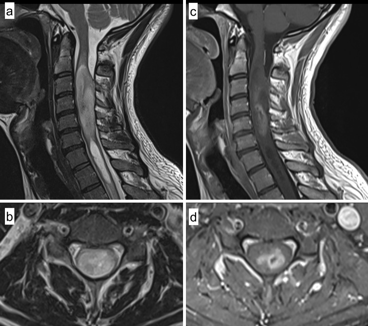
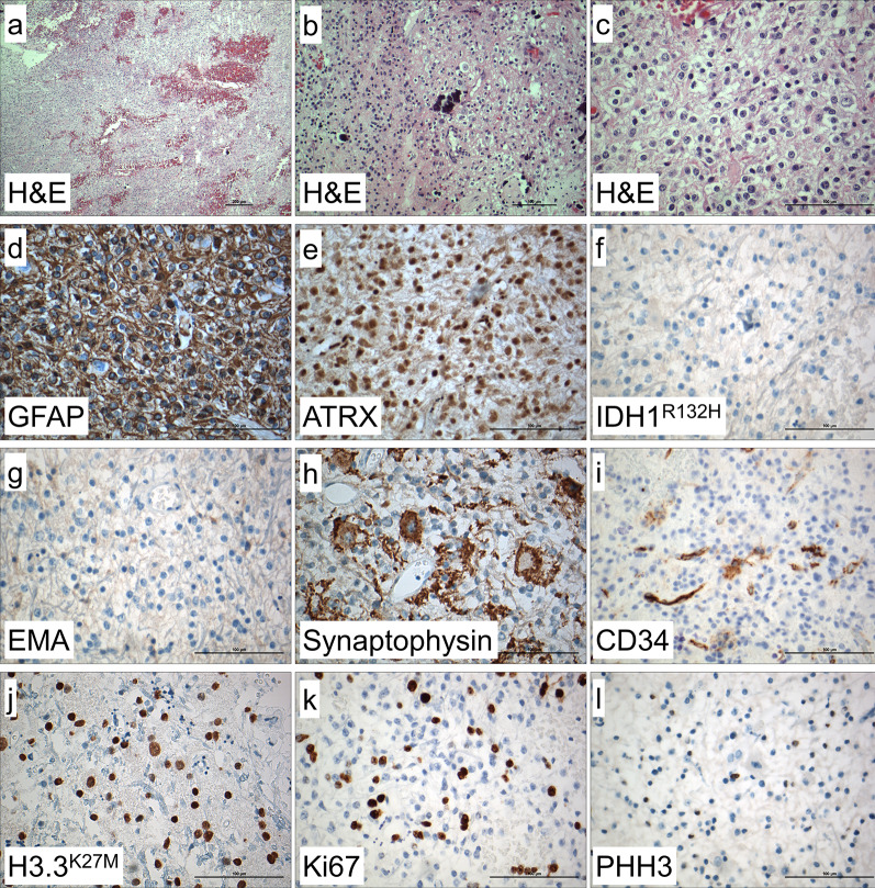
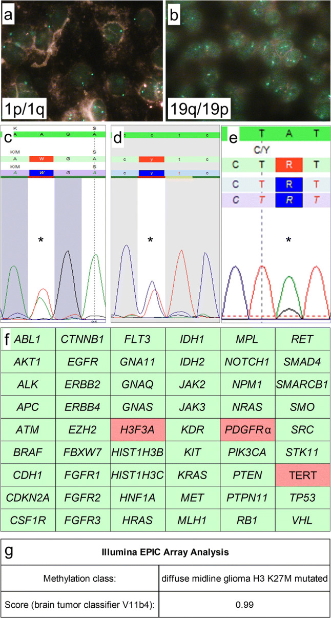
Here, we report on a 28-year old male patient presenting with neck and shoulder pain, dysesthesia of all four limbs and hypesthesia of both hands, without motor deficits. Magnetic resonance imaging showed an intradural, intramedullary mass of the cervical spinal cord of 6.4 cm length and 1.7 cm diameter. The patient underwent surgical resection. Histological and immunohistochemical evaluation showed pleomorphic glial tumor cells, mitoses, calcifications, and atypical ganglioid cells compatible with the morphology of anaplastic ganglioglioma (WHO Grade III). Extensive molecular workup revealed H3F3A K27M, TERT C228T and PDGFRα Y849C mutations indicating poor prognosis. The H3F3A K27M mutation assigned the tumor to the molecular group of diffuse midline glioma (WHO Grade IV). Epigenome-wide methylation profiling confirmed the methylation class of diffuse midline glioma. Thus, this is a very rare case of malignant glioma with H3 K27M genotype phenotypically mimicking anaplastic ganglioglioma. This case emphasizes the importance of comprehensive morphological and molecular workup including methylome profiling for advanced patient care.

Keywords: Anaplastic ganglioglioma; Diffuse midline glioma; H3F3A K27M; Methylome analysis; PDGFRα Y849C; TERT C228T.


Conflict of interest statement

The authors declare that they have no conflicts of interest.

Figures

Fig. 1
Fig. 1
Radiological findings. Sagittal (a) and axial (b) T2 weighted magnetic resonance imaging (MRI) showing an inhomogeneous hyperintense intramedullary tumor in the upper cervical cord with a consecutive syringomyelia below the lesion. Sagittal (c) and axial (d) postcontrast T1 weighted imaging demonstrating a mild and inhomogeneous contrast enhancement in the central parts of the lesion with minimal central hemorrhage (without hemosiderin capping)
Fig. 2
Fig. 2
Histological and immunohistochemical findings. In H&E stained sections, mildly to highly pleomorphic glial tumor cells with glial cell processes and hemorrhages were detected (a). Calcifications and very round tumor cells with only short processes and perinuclear halos (b), intermingled within neuronal cells showing dysmorphic appearance, were also visible (c). Immunohistochemistry showed tumor cells being positive for GFAP (d) with retained ATRX expression (e), and no expression of IDH1 R132H mutant protein (f). Tumor cells were negative for EMA (g). Ganglionic cells were positive for synaptophysin (h) and there were some CD34 positive tumor cells (i). Histone H3.3 K27M mutant protein was strongly expressed in the nuclei of tumor cells (j). Ki67 index showed 20% positive cells (k) and there were some PHH3 (H3S10p) positive cells (l). Magnification: a: 10 ×, b: 20 ×, c–l: 40 ×
Figure. 3
Figure. 3
Molecular genetic findings. FISH-analysis showed retained 1p (a) and 19q (b) expression. Mutation analysis using Sanger sequencing showed a H3F3A K27M mutation (c) and a TERT C228T promoter mutation (d), NGS showed a PDGFRα Y849C mutation (e). Further molecular analysis showed wild-type status of all other 51 genes including BRAF, HIST1H3B, HIST1H3C, IDH1, and IDH2 status (f). Epigenome-wide methylation profiling using the Illumina EPIC Array at the Department of Neuropathology, University Hospital Heidelberg/German Cancer Research Center, allocated the tumor to the methylation class of diffuse midline glioma H3 K27M mutant (g). Magnification: a, b: 100 × oil immersion. c–e: mutations are indicated using “*”. f: wild-type gene status is indicated by green, mutant gene status by red color

References

    1. Louis DN, et al. The 2016 world health organization classification of tumors of the central nervous system: a summary. Acta Neuropathol. 2016;131:803–820. doi: 10.1007/s00401-016-1545-1. - DOI - PubMed
    1. Sturm D, et al. Hotspot mutations in H3F3A and IDH1 define distinct epigenetic and biological subgroups of glioblastoma. Cancer Cell. 2012;22:425–437. doi: 10.1016/j.ccr.2012.08.024. - DOI - PubMed
    1. Halliday GC, et al. The case for DNA methylation based molecular profiling to improve diagnostic accuracy for central nervous system embryonal tumors (not otherwise specified) in adults. J Clin Neurosci. 2018;47:163–167. doi: 10.1016/j.jocn.2017.09.013. - DOI - PubMed
    1. Sahm F, et al. DNA methylation-based classification and grading system for meningioma: a multicentre, retrospective analysis. Lancet Oncol. 2017;18:682–694. doi: 10.1016/S1470-2045(17)30155-9. - DOI - PubMed
    1. Pajtler KW, et al. Molecular classification of ependymal tumors across all CNS compartments, histopathological grades, and age groups. Cancer Cell. 2015;27:728–743. doi: 10.1016/j.ccell.2015.04.002. - DOI - PMC - PubMed

LinkOut - more resources