

doi: 10.1007/s13238-020-00731-9.
Structure of African swine fever virus p15 reveals its dual role for membrane-association and DNA binding
Affiliations
- PMID: 32451720
- PMCID: PMC7381529
- DOI: 10.1007/s13238-020-00731-9
Item in Clipboard
Structure of African swine fever virus p15 reveals its dual role for membrane-association and DNA binding
Protein Cell.
2020 Aug.
No abstract available
Figures
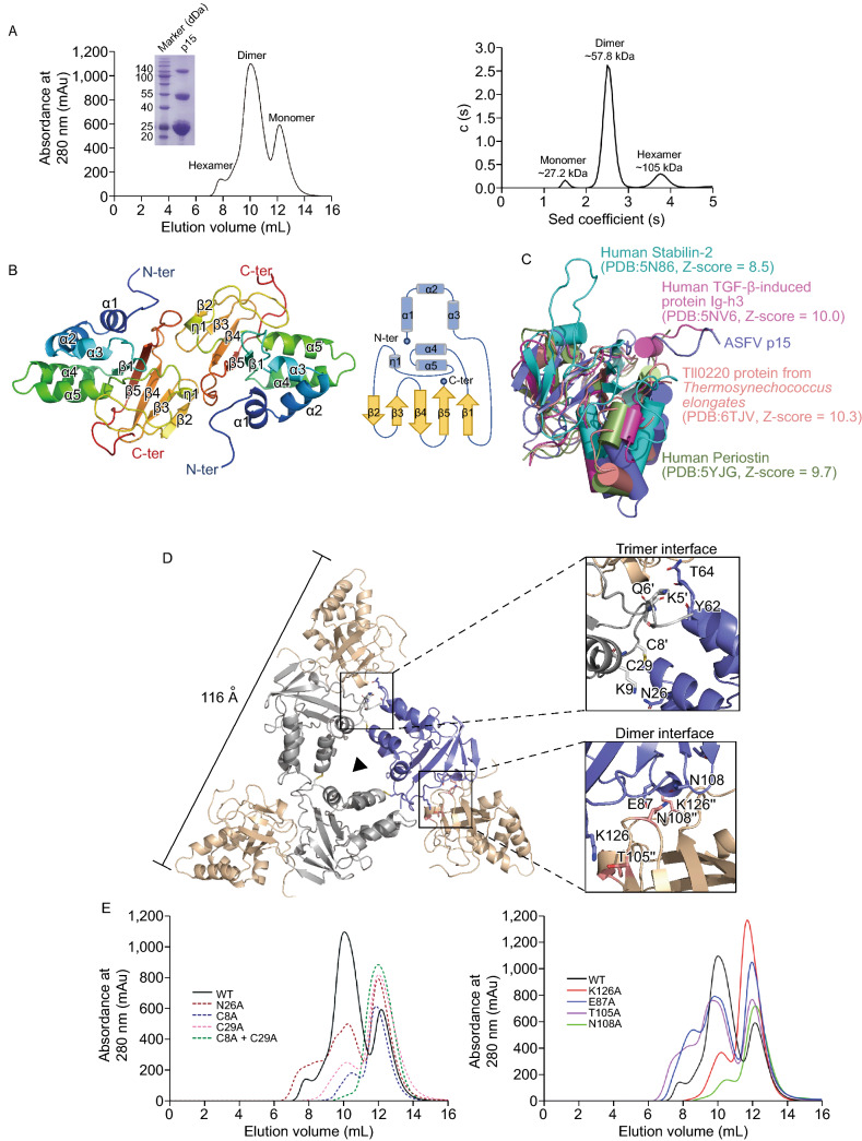
The overall structure of ASFV p15. (A) Size-exclusion chromatogram of ASFV p15 protein. Three distinct p15 peaks corresponding to hexamer, dimer and monomer,respectively. The states of p15 in solution are confirmed by Native-PAGE assay and AUC assay. The calculated molecular weights corresponding to each peak in AUC are labelled above the curve. Sed stands for sedimentation. (B) The crystal structure of ASFV p15 dimer is shown in cartoon together with the topology diagram of the monomer. The secondary structure elements are coloured in rainbow and numbered consecutively. In the topology diagram, β-strands are represented as gold arrows, while α-helices are shown as blue cylinders. (C) Superposition of p15 (Slate) with Human stabilin-2 (Cyan), Human TGF-β-induced protein Ig-h3 (Pink), Tll0220 protein from Thermosynechococcus elongates (Salmon), and Human Periostin (Palegreen). All the structures are shown as ribbon. (D) Top view of the ribbon diagram of the ASFV p15 hexamer. Crystallographic symmetry generates the hexametric “three-blade propeller”- like structure, comprised of three dimers. The side length of the hexamer is labelled. The detailed interactions within the trimer interface and dimer interface are shown at the right panel, respectively. The residues on the interface are labelled and shown as sticks. (E) Mutational analysis of the interface by SEC. The absorbance at UV280 nm of proteins is coloured in different coloured lines
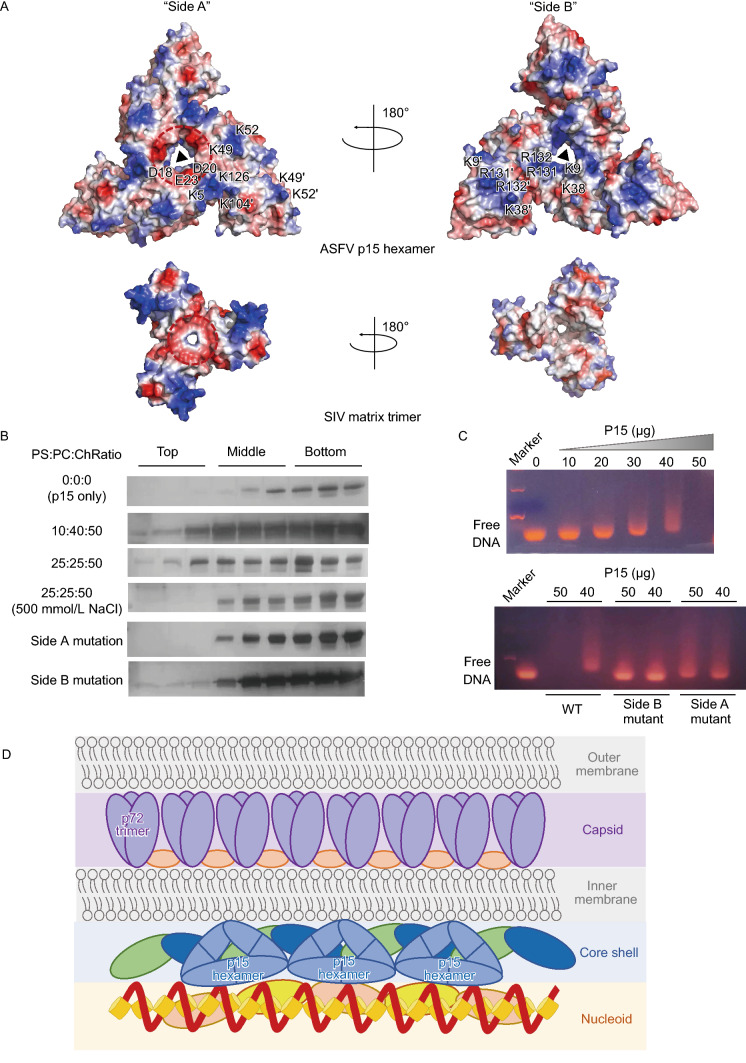
The dual role for membrane-association and DNA binding of ASFV p15. (A) The solvent-accessible surface of ASFV p15 hexamer and SIV matrix trimer are coloured according to electrostatic potential ranging (±5 kT/e) from blue (basic or positively charged) to red (acidic or negatively charged). The front view (“Side A”) and back view (“Side B”) of ASFV p15 hexamer and SIV matrix trimer are displayed. The critical residues on ASFV p15 surface are labelled. The negatively charged regions of ASFV p15 hexamer and SIV matrix trimer are highlighted by a red circle. (B) Liposome flotation experiments of ASFV p15 protein. Interaction of wild-type p15 and mutants with liposomes are tested in flotation experiments against different lipid compositions and NaCl concentrations. Top, Middle, and Bottom fractions of the discontinuous gradient were analyzed by SDS-PAGE and stained with rapid silver staining kit. PS:phosphatidylserine, PC: phosphatidylcholine, Ch: cholesterol. (C) EMSA assay of ASFV p15 wild-type protein and mutants binding to dsDNA. The amount of p15 protein is labelled on the top, unbounded free DNA are labelled on the left side. (D) A possible model illustrating the location and function of p15 hexamer in ASFV core shell structure. The five layers of ASFV are labelled on the right side. The p15 hexamer is shown as a blue triangle. The other proteins are shown as different colour and size shapes. The DNA-genome is shown as a dark red spiral
References
-
- Andres G, Garcia-Escudero R, Vinuela E, Salas ML, Rodriguez JM. African swine fever virus structural protein pE120R is essential for virus transport from assembly sites to plasma membrane but not for infectivity. J Virol. 2001;75:6758–6768. doi: 10.1128/JVI.75.15.6758-6768.2001. - DOI - PMC - PubMed
Publication types
MeSH terms
Substances
LinkOut - more resources
Full Text Sources
