. 2020 Jun;17(3):335-350.
doi: 10.1007/s13770-020-00265-5. Epub 2020 May 26.

Hypoxia Pretreatment Promotes Chondrocyte Differentiation of Human Adipose-Derived Stem Cells via Vascular Endothelial Growth Factor

Affiliations

Hypoxia Pretreatment Promotes Chondrocyte Differentiation of Human Adipose-Derived Stem Cells via Vascular Endothelial Growth Factor

Ok Kyung Hwang et al. Tissue Eng Regen Med. 2020 Jun.

Abstract

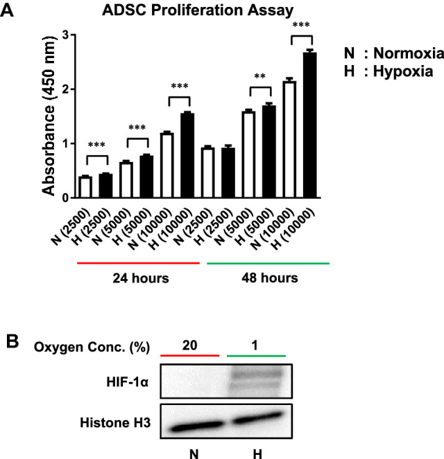
Background: Human adipose tissue-derived stem cells (ADSCs) are attractive multipotent stem cell sources with therapeutic potential in various fields requiring repair and regeneration, such as acute and chronically damaged tissues. ADSC is suitable for cell-based therapy, but its use has been hampered due to poor survival after administration. Potential therapeutic use of ADSC requires mass production of cells through in vitro expansion. Many studies have consistently observed the tendency of senescence by mesenchymal stem cell (MSC) proliferation upon expansion. Hypoxia has been reported to improve stem cell proliferation and survival.

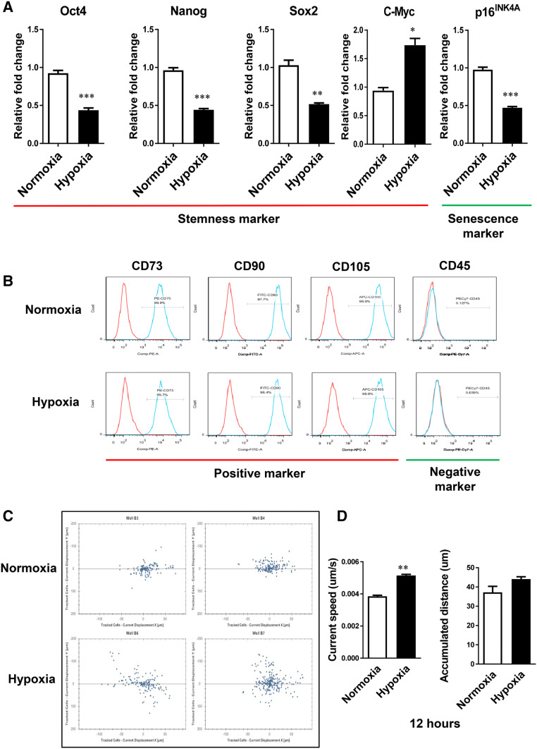
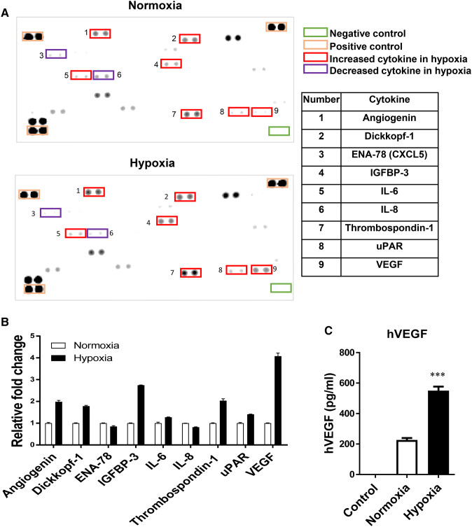
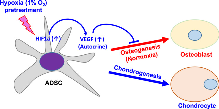
Methods: We investigated the effects of hypoxia pretreatment on ADCS proliferation, migration capacity, differentiation potential and cytokine production. We also analyzed the effects of vascular endothelial growth factor (VEGF) on osteogenic and chondrogenic differentiation of ADSCs by hypoxia pretreatment.

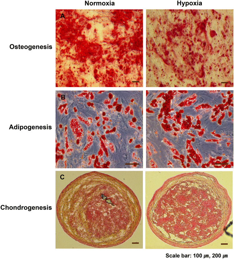
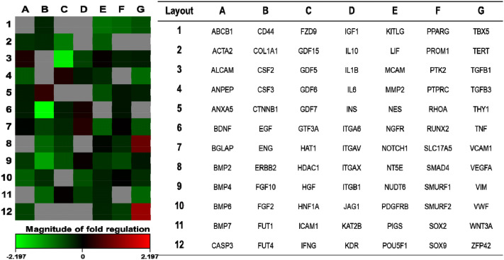
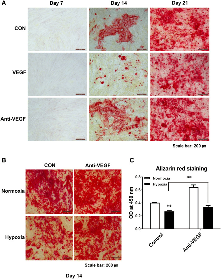
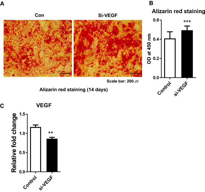
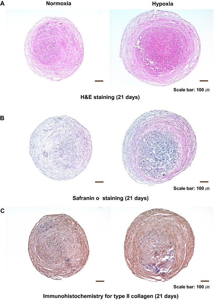
Results: Hypoxia pretreatment increased the proliferation of ADSCs by increasing VEGF levels. Interestingly, hypoxia pretreatment significantly increased chondrogenic differentiation but decreased osteogenic differentiation compared to normoxia. The osteogenic differentiation of ADSC was decreased by the addition of VEGF but increased by the depletion of VEGF. We have shown that hypoxia pretreatment increases the chondrogenic differentiation of ADSCs while reducing osteogenic differentiation in a VEGF-dependent manner.

Conclusion: These results show that hypoxia pretreatment can provide useful information for studies that require selective inhibition of osteogenic differentiation, such as cartilage regeneration.

Keywords: Adipose-derived stem cells (ADSCs); Cartilage regeneration; Differentiation; Hypoxia; VEGF.


Conflict of interest statement

The authors have no conflicts to disclose.

Figures

Fig. 1
Fig. 1
Effects of hypoxia pretreatment on the proliferation of ADSCs. A ADSC proliferation was significantly increased at 24 and 48 h by hypoxia compared to normoxia and measured using the CCK-8 kit. B Nuclear proteins were prepared from ADSCs exposed to hypoxia for 6 h and Western blotting was performed. The expression of HIF-1α was increased by hypoxia. The results are shown as the mean ± SD values. The statistical significance of hypoxia versus normoxia was determined by Student’s t-test (*p < 0.05 and **p < 0.01). N: normoxia (20% O2), H: hypoxia (1% O2)
Fig. 2
Fig. 2
Effect of hypoxia pretreatment on the properties of ADSCs. A The mRNA expression of the indicated gene was analyzed using real-time RT-PCR. The expression of stem cell markers Oct4, Nanog, and Sox2 in hypoxia was significantly lower than in normoxia. The expression of stem cell marker c-Myc in hypoxia was significantly higher than in normoxia. The expression of senescence marker p16INK4a in hypoxia was significantly lower than in normoxia. B In immunophenotype analysis, the expression of mesenchymal surface markers CD73, CD90, and CD105 was not significantly changed by hypoxia compared to normoxia. C The migration ability of ADSCs was increased in hypoxia compared to normoxia. D The current step and accumulated distance of ADSCs cultured in hypoxia were increased compared to normoxia at 12 h. The results are shown as the mean ± SD values. The statistical significance of hypoxia versus normoxia was determined by Student’s t-test (*p < 0.05 and **p < 0.01)
Fig. 3
Fig. 3
Effects of hypoxia pretreatment on the differentiation potential of ADSCs. ADSCs were cultured at 4.5 × 105 cells/100 mm dish in hypoxia or normoxia for 48 h and induced to differentiate along adipogenic, osteogenic, and chondrogenic lines for 21 days. A Hypoxia inhibited the osteogenic differentiation efficiency compared to normoxia as shown by Alizarin Red staining. B No significant difference was observed in adipogenesis between normoxia and hypoxia by Oil Red O staining. C Hypoxia increased the chondrogenic differentiation efficiency compared to normoxia as shown by Safranin O staining
Fig. 4
Fig. 4
Hypoxia reduced the mineralization capacity and the expression of osteogenesis-related genes in ADSCs. A Bone mineralization was reduced by hypoxia compared to normoxia as shown by Alizarin Red staining at 7, 14, and 21 days after the induction of osteogenesis. B The mRNA expression of the indicated genes was analyzed using real-time RT-PCR. The results are shown as the mean ± SD values. The statistical significance of hypoxia versus normoxia was determined by Student’s t-test (*p < 0.05 and **p < 0.01)
Fig. 5
Fig. 5
Effect of hypoxia pretreatment on the differential expression of mesenchymal stem cell-related genes. The heat map shows the changes in mesenchymal stem cell-related genes in ADSCs cultured at 4.5 × 105 cells/100 mm dish for 48 h in hypoxia compared to normoxia. Gradient indicates the magnitude of the change in gene expression
Fig. 6
Fig. 6
Effect of hypoxia on the secretion of cytokines and VEGF by ADSCs. ADSCs were cultured at 4.5 × 105 cells/100 mm dish for 48 h in hypoxia or normoxia. A Cytokines were measured in cell culture supernatants using the Human XL Cytokine Array Kit according to the manufacturer’s protocol. B The expression of angiogenin, Dickkopf-1, IGFBP-3, IL-6, thrombospondin-1, uPAR, and VEGF by ADSCs in hypoxia was higher than in normoxia. C The secretion of VEGF into the medium by ADSCs in hypoxia was significantly increased compared to normoxia. The results are shown as the mean ± SD values. The statistical significance of hypoxia versus normoxia was determined by Student’s t-test (*p < 0.05 and **p < 0.01)
Fig. 7
Fig. 7
Effect of VEGF and anti-VEGF antibody on the differentiation of ADSCs into osteoblasts. A The differentiation of ADSCs into osteoblasts was induced by the addition of VEGF to the medium. The positively stained Alizarin Red area was smaller in cells treated with VEGF compared to the control at 14 and 21 days. B, C The differentiation of ADSCs into osteoblasts in hypoxia was restored by VEGF antibody. The results are shown as the mean ± SD values. The statistical significance of hypoxia versus normoxia was determined by Student’s t-test (*p < 0.05 and **p < 0.01)
Fig. 8
Fig. 8
Effect of VEGF siRNA on the differentiation of ADSCs into osteoblasts. The differentiation of ADSCs to osteoblasts was induced after transfection with VEGF siRNA. A, B The positively stained Alizarin Red area was increased in cells transfected with VEGF siRNA (si-VEGF) compared to control at 14 days. C The mRNA expression of VEGF gene was analyzed using real-time RT-PCR after transfection with VEGF siRNA. The results are shown as the mean ± SD values. The statistical significance of hypoxia versus normoxia was determined by Student’s t-test (*p < 0.05 and **p < 0.01)
Fig. 9
Fig. 9
Effect of hypoxia pretreatment on the chondrogenic differentiation potential of ADSC. A The mass of cells differentiated into chondrocytes was increased by hypoxia compared to normoxia. B, C Hypoxia increased the chondrogenic differentiation efficiency compared to normoxia as shown by Safranin O staining and immunohistochemical analysis for type II collagen
Fig. 10
Fig. 10
Schematic diagram of regulation by hypoxia pretreatment in the differentiation of ADSC into osteoblasts and chondrocytes. Hypoxia (1% O2) pretreatment increases VEGF through increased expression of HIF1a, and increased VEGF inhibits osteogenic differentiation but promotes chondrogenic differentiation

Similar articles

Cited by

References

    1. Dao LT, Park EY, Hwang OK, Cha JY, Jun HS. Differentiation potential and profile of nuclear receptor expression during expanded culture of human adipose tissue-derived stem cells reveals PPARgamma as an important regulator of Oct4 expression. Stem Cells Dev. 2014;23:24–33. - PubMed
    1. Friedenstein AJ, Gorskaja JF, Kulagina NN. Fibroblast precursors in normal and irradiated mouse hematopoietic organs. Exp Hematol. 1976;4:267–274. - PubMed
    1. De Ugarte DA, Morizono K, Elbarbary A, Alfonso Z, Zuk PA, Zhu M, et al. Comparison of multi-lineage cells from human adipose tissue and bone marrow. Cells Tissues Organs. 2003;174:101–109. doi: 10.1159/000071150. - DOI - PubMed
    1. Zhu Y, Liu T, Song K, Fan X, Ma X, Cui Z. Adipose-derived stem cell: a better stem cell than BMSC. Cell Biochem Funct. 2008;26:664–675. - PubMed
    1. Zuk PA, Zhu M, Ashjian P, De Ugarte DA, Huang JI, Mizuno H, et al. Human adipose tissue is a source of multipotent stem cells. Mol Biol Cell. 2002;13:4279–4295. - PMC - PubMed

Publication types

MeSH terms

Substances

LinkOut - more resources