

State of the art in total body PET
- PMID: 32451783
- PMCID: PMC7248164
- DOI: 10.1186/s40658-020-00290-2
State of the art in total body PET
Abstract
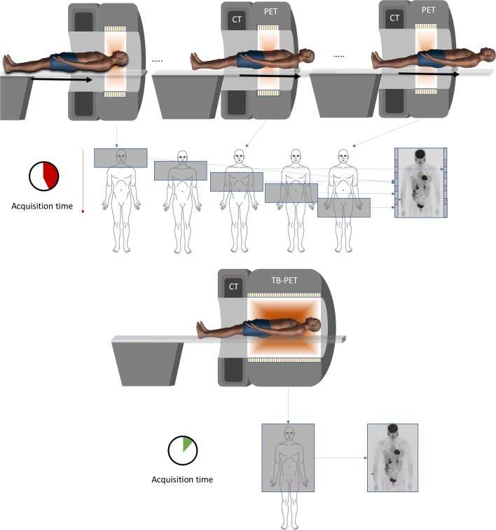
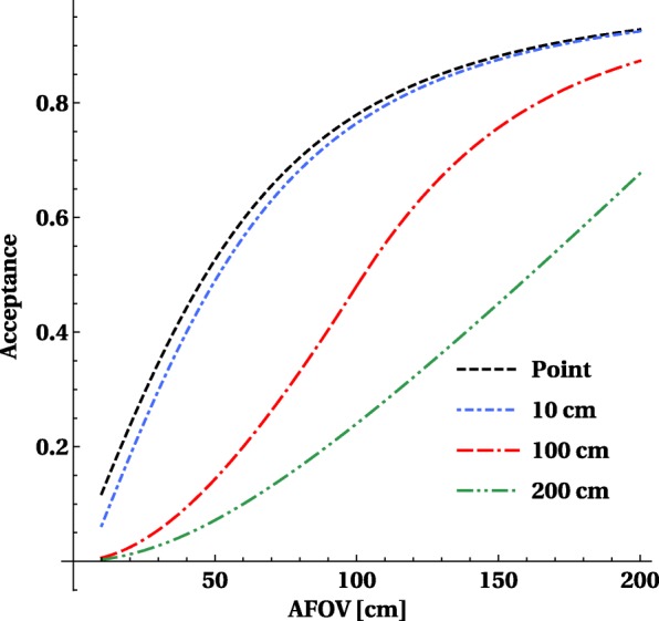
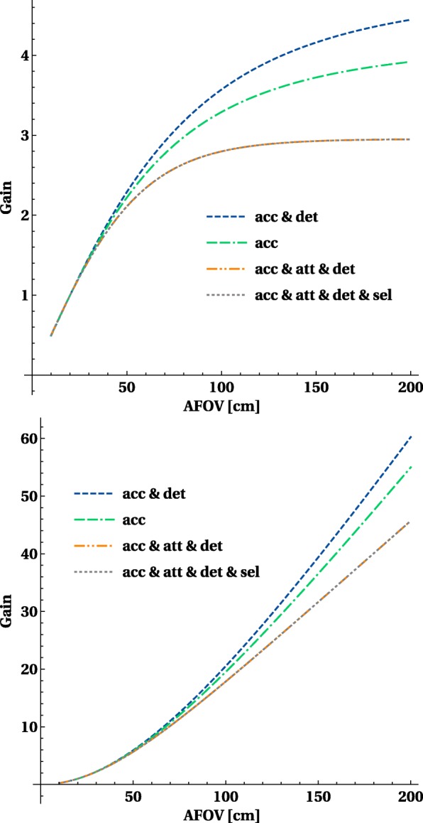
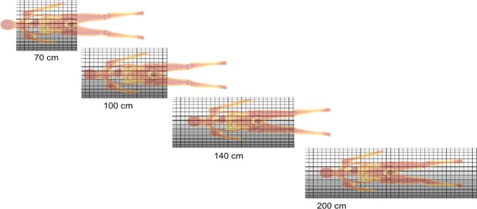
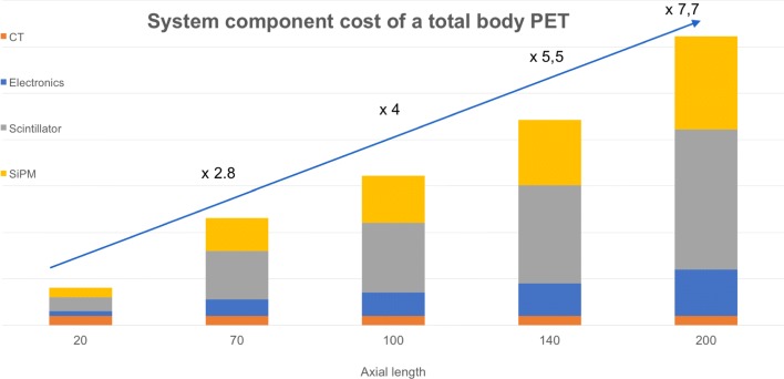
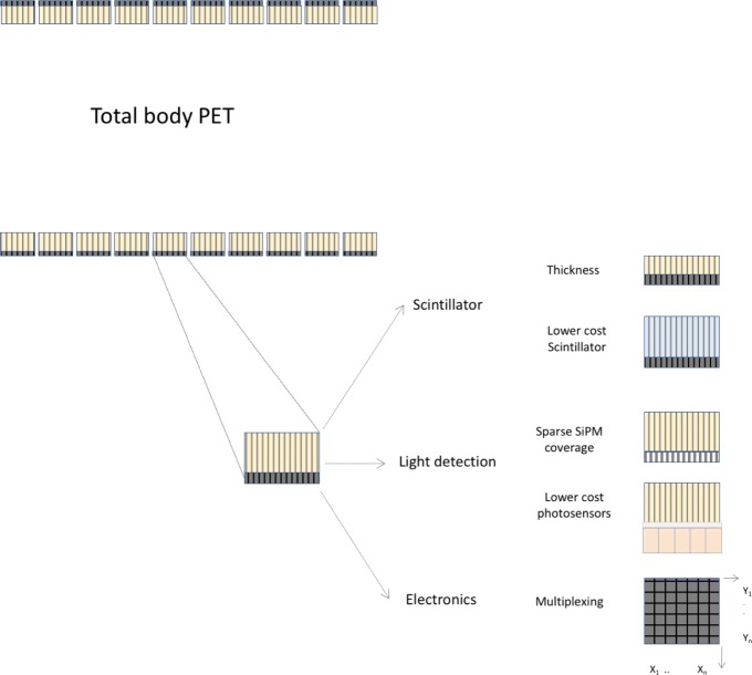
The idea of a very sensitive positron emission tomography (PET) system covering a large portion of the body of a patient already dates back to the early 1990s. In the period 2000-2010, only some prototypes with long axial field of view (FOV) have been built, which never resulted in systems used for clinical research. One of the reasons was the limitations in the available detector technology, which did not yet have sufficient energy resolution, timing resolution or countrate capabilities for fully exploiting the benefits of a long axial FOV design. PET was also not yet as widespread as it is today: the growth in oncology, which has become the major application of PET, appeared only after the introduction of PET-CT (early 2000).The detector technology used in most clinical PET systems today has a combination of good energy and timing resolution with higher countrate capabilities and has now been used since more than a decade to build time-of-flight (TOF) PET systems with fully 3D acquisitions. Based on this technology, one can construct total body PET systems and the remaining challenges (data handling, fast image reconstruction, detector cooling) are mostly related to engineering. The direct benefits of long axial FOV systems are mostly related to the higher sensitivity. For single organ imaging, the gain is close to the point source sensitivity which increases linearly with the axial length until it is limited by solid angle and attenuation of the body. The gains for single organ (compared to a fully 3D PET 20-cm axial FOV) are limited to a factor 3-4. But for long objects (like body scans), it increases quadratically with scanner length and factors of 10-40 × higher sensitivity are predicted for the long axial FOV scanner. This application of PET has seen a major increase (mostly in oncology) during the last 2 decades and is now the main type of study in a PET centre. As the technology is available and the full body concept also seems to match with existing applications, the old concept of a total body PET scanner is seeing a clear revival. Several research groups are working on this concept and after showing the potential via extensive simulations; construction of these systems has started about 2 years ago. In the first phase, two PET systems with long axial FOV suitable for large animal imaging were constructed to explore the potential in more experimental settings. Recently, the first completed total body PET systems for human use, a 70-cm-long system, called PennPET Explorer, and a 2-m-long system, called uExplorer, have become reality and first clinical studies have been shown. These results illustrate the large potential of this concept with regard to low-dose imaging, faster scanning, whole-body dynamic imaging and follow-up of tracers over longer periods. This large range of possible technical improvements seems to have the potential to change the current clinical routine and to expand the number of clinical applications of molecular imaging. The J-PET prototype is a prototype system with a long axial FOV built from axially arranged plastic scintillator strips.This paper gives an overview of the recent technical developments with regard to PET scanners with a long axial FOV covering at least the majority of the body (so called total body PET systems). After explaining the benefits and challenges of total body PET systems, the different total body PET system designs proposed for large animal and clinical imaging are described in detail. The axial length is one of the major factors determining the total cost of the system, but there are also other options in detector technology, design and processing for reducing the cost these systems. The limitations and advantages of different designs for research and clinical use are discussed taking into account potential applications and the increased cost of these systems.
Keywords: PET; PET-CT; Sensitivity.
Conflict of interest statement
The authors declare that they have no competing interests.
Figures


References
-
- Abstracts of the total body PET conference. EJNMMI Phys. 19; 5. 10.1186/s40658-018-0218-7.
-
- Beltrame P, Bolle E, Braem A, Casella C, Chesi E, Clinthorne N, De Leo R, Dissertori G, Djambazov L, Fanti V, et al. The AX-PET demonstrator design, construction and characterization. Nucl Instrum Methods Phys Res Sect A Accel Spectrometers Detectors Assoc Equip. 2011;654:546–59.
Publication types
LinkOut - more resources
Full Text Sources
Other Literature Sources
Research Materials
