. 2020 May 26:9:e58603.
doi: 10.7554/eLife.58603.

SARS-CoV-2 strategically mimics proteolytic activation of human ENaC

Affiliations

SARS-CoV-2 strategically mimics proteolytic activation of human ENaC

Praveen Anand et al. Elife. .

Abstract

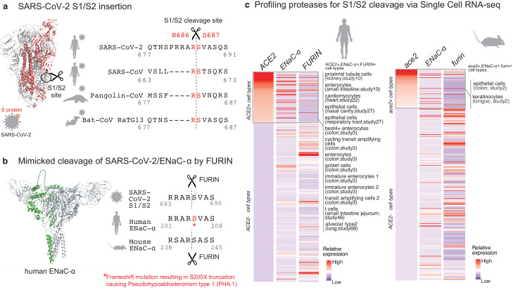
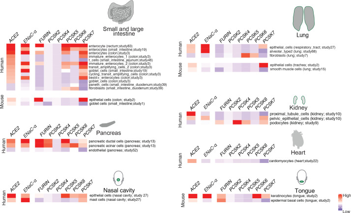
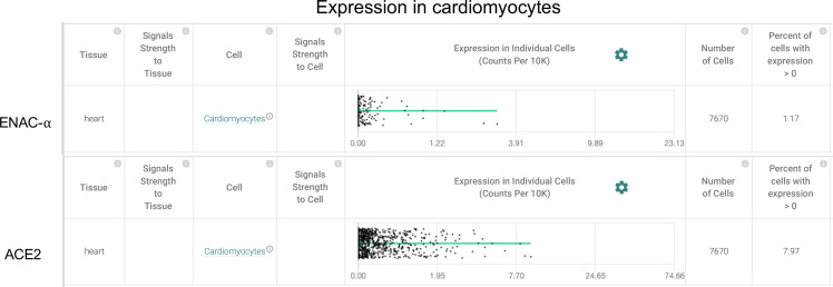
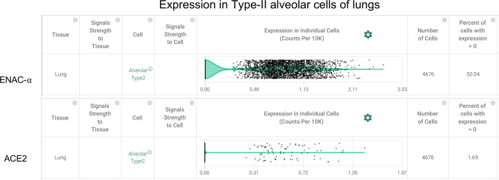
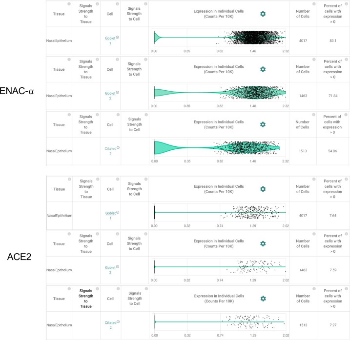
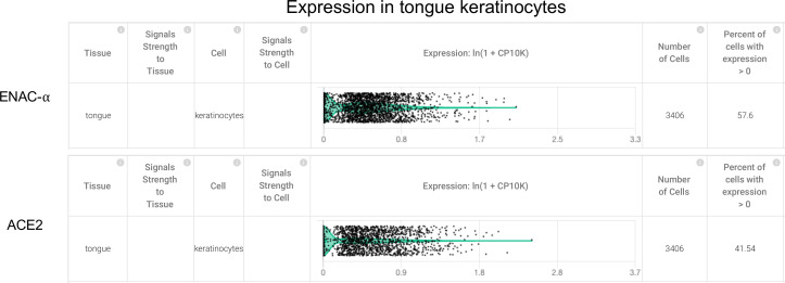
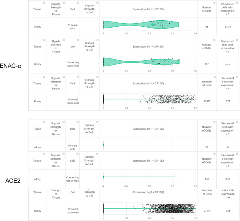
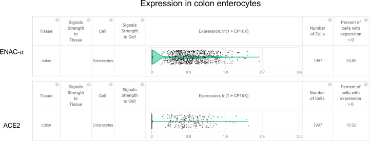
Molecular mimicry is an evolutionary strategy adopted by viruses to exploit the host cellular machinery. We report that SARS-CoV-2 has evolved a unique S1/S2 cleavage site, absent in any previous coronavirus sequenced, resulting in the striking mimicry of an identical FURIN-cleavable peptide on the human epithelial sodium channel α-subunit (ENaC-α). Genetic alteration of ENaC-α causes aldosterone dysregulation in patients, highlighting that the FURIN site is critical for activation of ENaC. Single cell RNA-seq from 66 studies shows significant overlap between expression of ENaC-α and the viral receptor ACE2 in cell types linked to the cardiovascular-renal-pulmonary pathophysiology of COVID-19. Triangulating this cellular characterization with cleavage signatures of 178 proteases highlights proteolytic degeneracy wired into the SARS-CoV-2 lifecycle. Evolution of SARS-CoV-2 into a global pandemic may be driven in part by its targeted mimicry of ENaC-α, a protein critical for the homeostasis of airway surface liquid, whose misregulation is associated with respiratory conditions.

Keywords: COVID-19; ENaC; SARS-CoV-2; acute respiratory distress syndrome; computational biology; coronavirus; human; human biology; medicine; molecular mimicry; mouse; systems biology; virus.

Plain language summary

Viruses hijack the cellular machinery of humans to infect their cells and multiply. The virus causing the global COVID-19 pandemic, SARS-CoV-2, is no exception. Identifying which proteins in human cells the virus co-opts is crucial for developing new ways to diagnose, prevent and treat COVID-19 infections. SARS-CoV-2 is covered in spike-shaped proteins, which the virus uses to gain entry into cells. First, the spikes bind to a protein called ACE2, which is found on the cells that line the respiratory tract and lungs. SARS-CoV-2 then exploits enzymes called proteases to cut, or cleave, its spikes at a specific site which allows the virus to infiltrate the host cell. Proteases identify which proteins to target based on the sequence of amino acids – the building blocks of proteins – at the cleavage site. However, it remained unclear which human proteases SARS-CoV-2 co-opts and whether its cut site is similar to human proteins. Now, Anand et al. show that the spike proteins on SARS-CoV-2 may have the same sequence of amino acids at its cut site as a human epithelial channel protein called ENaC-α. This channel is important for maintaining the balance of salt and water in many organs including the lungs. Further analyses showed that ENaC-α is often found in the same types of human lung and respiratory tract cells as ACE2. This suggests that SARS-CoV-2 may use the same proteases that cut ENaC-α to get inside human respiratory cells. It is possible that by hijacking the cutting mechanism for ENaC-α, SARS-CoV-2 interferes with the balance of salt and water in the lungs of COVID-19 patients. This may help explain why the virus causes severe respiratory symptoms. However, more studies are needed to confirm that the proteases that cut ENaC-α also cut the spike proteins on SARS-CoV-2, and how this affects the respiratory health of COVID-19 patients.


Conflict of interest statement

PA, AP, MA, AV, VS The author is an employee of nference

Figures

Figure 1.
Figure 1.. Targeted molecular mimicry by SARS-CoV-2 of human ENaC-ɑ and profiling ACE2-FURIN-ENaC-ɑ co-expression.
(a) The cartoon representation of the S-protein homotrimer from SARS-CoV-2 is shown (PDB ID: 6VSB). One of the monomers is highlighted in red. The alignment of the S1/S2 cleavage site required for the activation of SARS-CoV-2, SARS-CoV, Pangolin-CoV, and Bat-CoV RaTG13 are shown. The four amino acid insertion evolved by SARS-CoV-2, along with the abutting cleavage site is shown in a box. (b) The cartoon representation of human ENaC protein is depicted (PDB ID: 6BQN; chain in green), highlighting the ENaC-ɑ chain in green. The alignment on the right captures FURIN cleavage at the S1/S2 site of SARS-CoV-2, along with its striking molecular mimicry of the identical peptide from human ENaC-ɑ protein (dotted loop in the cartoon rendering of human ENaC). The alignment further shows the equivalent 8-mer peptide of mouse ENaC-ɑ that is also known to be cleaved by FURIN. One of the known genetic alterations on human ENaC-ɑ is highlighted as well (Welzel et al., 2013). (c) The single cell transcriptomic co-expression of ACE2, ENaC-ɑ, and FURIN is summarized. The heatmap depicts the mean relative expression of each gene across the identified cell populations. The human and mouse single cell RNA-seq are visualized independently. The cell types are ranked based on decreasing expression of ACE2. The box highlights the ACE2 positive cell types in human and mouse samples.
Figure 2.
Figure 2.. Expression profiling of identified proteases.
The heatmap depicts the relative expression of ACE2 and ENaC-ɑ along with a list of proteases that can potentially cleave the S1/S2 site. The relative expression levels are denoted on a scale of blue (low) to red (high). The rows denote proteases and columns denote cell-types.
Figure 2—figure supplement 1.
Figure 2—figure supplement 1.. Cardiomyocytes express ENaC-ɑ (SCNN1A) and ACE2 (Primary data processed from Pubmed ID:31915373 and hosted on https://academia.nferx.com/).
Figure 2—figure supplement 2.
Figure 2—figure supplement 2.. Type-II Alveolar cells of the lungs express ENaC-ɑ (SCNN1A) and ACE2 (Primary data processed from Pubmed ID: 31892341 and hosted on https://academia.nferx.com/).
Figure 2—figure supplement 3.
Figure 2—figure supplement 3.. Goblet cells and Ciliated cells of the nasal epithelial layer express SCNN1A (ENaC-ɑ) and ACE2 (Primary data processed from Pubmed ID: 32327758 and hosted on https://academia.nferx.com/).
Figure 2—figure supplement 4.
Figure 2—figure supplement 4.. Tongue keratinocytes express SCNN1A (ENaC-ɑ) and ACE2 (Primary data processed from Pubmed ID:30283141 and hosted on https://academia.nferx.com/).
Figure 2—figure supplement 5.
Figure 2—figure supplement 5.. Higher expression of SCNN1A was detected in 58% of the principal cells in the collecting duct 47% of the connecting tubule cells from the kidney, but ACE2 expression was not detected in these cell types.
Although only 2.77% of the proximal tubule cells had detectable expression of SCNN1A, a higher percentage (8.46%) of these cells were also observed to express ACE2 (Primary data processed from Pubmed ID: 31604275 and hosted on https://academia.nferx.com/).
Figure 2—figure supplement 6.
Figure 2—figure supplement 6.. Colon enterocytes express SCNN1A (ENaC-ɑ) and ACE2 (Primary data processed from Pubmed ID:31348891 and hosted on https://academia.nferx.com/).

References

    1. Belouzard S, Chu VC, Whittaker GR. Activation of the SARS coronavirus spike protein via sequential proteolytic cleavage at two distinct sites. PNAS. 2009;106:5871–5876. doi: 10.1073/pnas.0809524106. - DOI - PMC - PubMed
    1. Belouzard S, Millet JK, Licitra BN, Whittaker GR. Mechanisms of coronavirus cell entry mediated by the viral spike protein. Viruses. 2012;4:1011–1033. doi: 10.3390/v4061011. - DOI - PMC - PubMed
    1. Butterworth MB. Regulation of the epithelial sodium channel (ENaC) by membrane trafficking. Biochimica Et Biophysica Acta (BBA) - Molecular Basis of Disease. 2010;1802:1166–1177. doi: 10.1016/j.bbadis.2010.03.010. - DOI - PMC - PubMed
    1. Carrillo-Tripp M, Shepherd CM, Borelli IA, Venkataraman S, Lander G, Natarajan P, Johnson JE, Brooks CL, Reddy VS. VIPERdb2: an enhanced and web API enabled relational database for structural virology. Nucleic Acids Research. 2009;37:D436–D442. doi: 10.1093/nar/gkn840. - DOI - PMC - PubMed
    1. Coutard B, Valle C, de Lamballerie X, Canard B, Seidah NG, Decroly E. The spike glycoprotein of the new coronavirus 2019-nCoV contains a furin-like cleavage site absent in CoV of the same clade. Antiviral Research. 2020;176:104742. doi: 10.1016/j.antiviral.2020.104742. - DOI - PMC - PubMed

MeSH terms

LinkOut - more resources