. 2020 May 26;16(5):e1008826.
doi: 10.1371/journal.pgen.1008826. eCollection 2020 May.

Congenital hearing impairment associated with peripheral cochlear nerve dysmyelination in glycosylation-deficient muscular dystrophy

Affiliations

Congenital hearing impairment associated with peripheral cochlear nerve dysmyelination in glycosylation-deficient muscular dystrophy

Shigefumi Morioka et al. PLoS Genet. .

Abstract

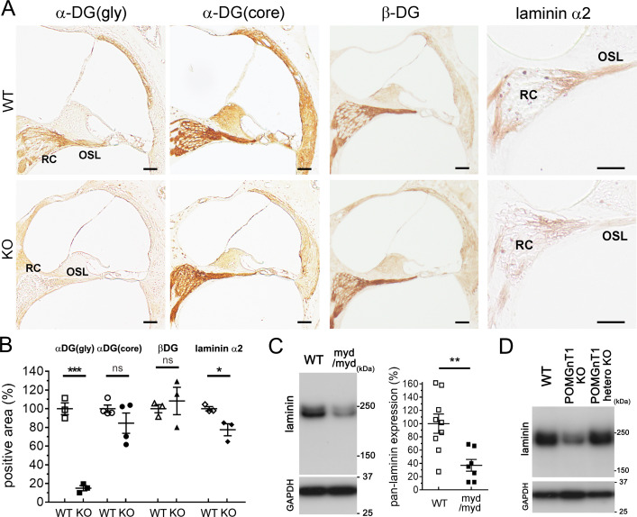
Hearing loss (HL) is one of the most common sensory impairments and etiologically and genetically heterogeneous disorders in humans. Muscular dystrophies (MDs) are neuromuscular disorders characterized by progressive degeneration of skeletal muscle accompanied by non-muscular symptoms. Aberrant glycosylation of α-dystroglycan causes at least eighteen subtypes of MD, now categorized as MD-dystroglycanopathy (MD-DG), with a wide spectrum of non-muscular symptoms. Despite a growing number of MD-DG subtypes and increasing evidence regarding their molecular pathogeneses, no comprehensive study has investigated sensorineural HL (SNHL) in MD-DG. Here, we found that two mouse models of MD-DG, Largemyd/myd and POMGnT1-KO mice, exhibited congenital, non-progressive, and mild-to-moderate SNHL in auditory brainstem response (ABR) accompanied by extended latency of wave I. Profoundly abnormal myelination was found at the peripheral segment of the cochlear nerve, which is rich in the glycosylated α-dystroglycan-laminin complex and demarcated by "the glial dome." In addition, patients with Fukuyama congenital MD, a type of MD-DG, also had latent SNHL with extended latency of wave I in ABR. Collectively, these findings indicate that hearing impairment associated with impaired Schwann cell-mediated myelination at the peripheral segment of the cochlear nerve is a notable symptom of MD-DG.


Conflict of interest statement

The authors have declared that no competing interests exist.

Figures

Fig 1
Fig 1. The dystrophin–dystroglycan–laminin complex and the structure of the α-dystroglycan sugar chain.
A, Illustration showing the dystrophin–dystroglycan–laminin complex in peripheral nerve myelin. Mutations in enzymes modifying the α-dystroglycan (α-DG) sugar chain cause subtypes of muscular dystrophy known as muscular dystrophy-dystroglycanopathy (MD-DG), while dystrophin mutations cause Duchenne muscular dystrophy (Duchenne MD). B, Illustration showing the structure of the α-DG sugar chain and its modifying enzymes. α-DG is modified with O-mannose type glycans, namely CoreM1 and CoreM3. A repeating unit of GlcA-Xyl at the terminal of CoreM3 serves as a laminin-binding moiety. Enzymes referred to in the main text are shown, and their modification sites are indicated by arrows. Man: mannose, GlcNAc: N-acetylglucosamine, Gal: galactose, GalNAc: N-acetylgalacosamine, RboP: ribitol 5-phosphate, Xyl: xylose, GlcA: glucuronic acid.
Fig 2
Fig 2. Moderately impaired hearing in Largemyd/myd mice.
A, ABR thresholds (dB SPL) tested with clicks and pure-tone bursts at 8, 16, and 24 kHz in 2, 3, 5, 8, 10-week-old control (Largewt/wt, n = 3, 6, 5, 10, and 10, respectively), Largemyd/wt (n = 3, 6, 7, 8, and 8, respectively), and Largemyd/myd mice (n = 5, 7, 3, 6, and 6, respectively). Statistical significance was analyzed at all frequencies and ages between control and Largemyd/myd mice, except for 24 kHz frequency at 5 weeks of age. *P < 0.05, **P < 0.01, ***P < 0.001, **** P < 0.0001 using two-way ANOVA with Tukey’s post-hoc test. B, ABR latencies of wave I, wave I-V, and wave III-V and the amplitude of wave I (click stimulation, 90 dB) in 3- and 8-week-old control (n = 5 and 6–7, respectively), Largemyd/wt (n = 5 and 5–6, respectively), and Largemyd/myd mice (n = 6–7 and 6–7, respectively) were graphed. ****P < 0.0001, ***P = 0.0008 in the wave I latency and ****P < 0.0001 in the wave I amplitude (control vs. Largemyd/myd) using two-way ANOVA with Tukey’s post-hoc test. C, Inner ears of 8-week-old control and Largemyd/myd mice were fixed, and the cochleae were morphologically examined by HE staining at the basal turn and Alexa488-conjugated phalloidin staining at the apical and basal turns. No morphological abnormality in inner hair cells (IHCs), outer hair cells (OHCs), or hair cell loss was observed. The results were presented as the mean of three experiments. Scale bars: 50 μm for HE staining and 10 μm for phalloidin staining. RC: Rosenthal’s canal; OSL: osseous spiral lamina; OC: organ of Corti.
Fig 3
Fig 3. Mildly impaired hearing in POMGnT1-KO mice.
A, ABR thresholds (dB SPL) tested with clicks and pure-tone bursts at 8, 16, and 24 kHz in 4, 6, 8, and 10-week-old control (wt/wt; n = 8, 10, 10, and 10, respectively), heterozygous POMGnT1-KO (ko/wt; n = 6, 4, 4, and 4, respectively), and POMGnT1-KO mice (ko/ko; n = 8, 6, 6, and 6, respectively). **P < 0.01, ***P < 0.001, **** P < 0.0001 (control vs. POMGnT1-KO) using two-way ANOVA with Tukey’s post-hoc test. B, ABR latencies of wave I at the click stimulation at 90 dB in 4- and 10-week-old control (n = 6 and 7, respectively), heterozygous POMGnT1-KO (n = 7 and 4, respectively), and POMGnT1-KO (n = 7 and 4, respectively) mice were graphed. *P = 0.0197 (control vs KO) using two-way ANOVA with Tukey’s post-hoc test. C, Inner ears of control and POMGnT1-KO mice were fixed and morphologically examined by HE staining (8-week-old) and Alexa488-conjugated phalloidin staining (12-week-old). No morphological abnormality in inner hair cells (IHCs), outer HC (OHCs), or hair cell loss was observed. The results were presented as the mean of three experiments. Scale bars: 50 μm for HE staining and 10 μm for phalloidin staining. RC: Rosenthal’s canal; OSL: osseous spiral lamina; OC: organ of Corti.
Fig 4
Fig 4. Decreased glycosylated α-DG and laminin levels distal to the glial dome in Largemyd/myd mice.
Inner ears of 8-week-old control and Largemyd/myd mice (A–C) were fixed. A, Immunostaining of glycosylated α-dystroglycan [α-DG(gly)], pan-laminin, and laminin α2 at the basal turn level of the cochlea [glial dome (GD) is indicated by arrowheads]. Scale bars: 200 μm. Magnified immunostainings of β-dystroglycan (β-DG) at the basal turn of the cochlea are also shown (Scale bars: 50 μm). RC: Rosenthal’s canal; OSL: osseous spiral lamina. Asterisks indicate the regions (proximal to the GD) used for TEM analysis (shown in S5 Fig). B, Magnified immunostainings of α-DG(gly) and core α-DG [α-DG(core)] at the basal turn of the cochlea. More magnified immunostained images of α-DG(gly), pan-laminin, and laminin α2 at the RC and OSL of the cochlear basal turn are shown. Scale bars: 50 μm. C, Illustration shows the area including the RC and OSL (surrounded by dots) used for quantitative analysis (Scale bars: 50 μm). Statistical analyses of α-DG(gly), α-DG(core), pan-laminin, laminin α2, and β-DG immunostainings at the RC and OSL. n = 4, 4, 3, 4, and 4, respectively. ****P < 0.0001, P = 0.4587, **P = 0.0093, ****P < 0.0001, and P = 0.9813, respectively (Student’s t-test). ns: not significant.
Fig 5
Fig 5. Decreased levels of glycosylated α-DG and laminin levels distal to the glial dome in POMGnT1-KO and Largemyd/myd mice.
A, Inner ears of 16-week-old control and POMGnT1-KO mice were fixed. Glycosylated α-dystroglycan [α-DG(gly)], core α-DG [α-DG(core)], β-dystroglycan (β-DG), and laminin α2 immunostainings at the basal turn level of the cochlea. RC: Rosenthal’s canal; OSL: osseous spiral lamina. Scale bars: 50 μm. B, Statistical analyses of α-DG(gly), α-DG(core), β-DG, and laminin α2 immunostainings at the RC and OSL. n = 3, 4, 3, and 3, respectively. ***P = 0.0002, P = 0.2274, P = 0.6166, and *P = 0.0294, respectively (Student’s t-test). C and D, Pan-laminin levels in the spiral ganglion (SG) of P5-7 control and Largemyd/myd mice (C) and control, heterozygous POMGnT1-KO, and POMGnT1-KO mice (D) were detected by immunoblotting with GAPDH as loading controls. Statistical analysis was performed in pairs of control (n = 9) and Largemyd/myd mice (n = 7, P = 0.0040 using Student’s t-test).
Fig 6
Fig 6. Decreased MBP levels in Largemyd/myd and POMGnT1-KO mice at the RC and OSL.
Cochlear sections at the basal turn (9-week-old) and lysates from SG (P5-7) were obtained from control and Largemyd/myd mice (A and B). Cochlear sections at the basal turn (16-week-old) and lysates from SG (P5-7) were also obtained from control and POMGnT1-KO mice (C and D). Immunostaining (A and C) and immunoblotting (B and D) were performed using an MBP antibody, and statistical analyses were conducted using Student’s t-test. Comparable loading of proteins was confirmed by immunoblotting of GAPDH. Scale bars: 50 μm. RC: Rosenthal’s canal; OSL: osseous spiral lamina. Asterisks (A) indicate the regions (OSL) used for TEM analysis (shown in Figs 7 and S4). Arrowheads (B and D) indicate MBP variants used for quantitative analysis. A, n = 4 in each group, **P = 0.0046; B, n = 3 in control mice and 4 in Largemyd/myd mice, **P = 0.0011; C, n = 4 in each group, **P = 0.0086; D, n = 4 in each group, **P = 0.0080.
Fig 7
Fig 7. Abnormal myelination of the peripheral cochlear nerve at the OSL in Largemyd/myd and POMGnT1-KO mice.
Inner ears of 2-, 6-, and 10-week-old control and Largemyd/myd mice (A-C) and 10-week-old control and POMGnT1-KO mice (D) were fixed for transmission electron microscopy (TEM). TEM images at the osseous spiral lamina (OSL, indicated by the asterisks in Fig 6A) were obtained. The asterisks, arrowheads, circles, and triangles indicate naked axons, axons with disrupted myelin, and myelinated axons with vacuoles or aggregates, respectively. Scale bars: 500 nm. The percentage (each obtained by analyzing 50 axons) of axons with abnormal myelination was statistically analyzed in 2-, 6-, and 10-week-old control and Largemyd/myd mice (B) and in 10-week-old control and POMGnT1-KO mice (E). (B) ****P < 0.0001 using two-way ANOVA with Bonferroni’s post-hoc test (control vs. Largemyd/myd). *P = 0.0399 (Largemyd/myd mice at 2 weeks vs. 6 weeks), ***P = 0.0003 (Largemyd/myd mice at 6 weeks vs. 10 weeks), and P < 0.0001 (Largemyd/myd mice at 2 weeks vs. 10 weeks) using one-way ANOVA with Tukey’s post-hoc test. n = 3, 5, and 5 in 2-, 6-, and 10-week-old control mice, respectively. n = 3, 6, and 5 in 2-, 6-, and 10-week-old Largemyd/myd mice, respectively. (E) n = 3 in both groups, **P = 0.0051 using Student’s t-test. C, Distribution of the longest diameter of each myelinated axon in the transverse section at the OSL in 6-week-old control and Largemyd/myd mice are graphed and statistically analyzed. n = 100 from 3 cochleae (30, 30, and 40 in control mice and 29, 33, and 38 in Largemyd/myd mice. **P = 0.0063 using Kolmogorov–Smirnov test.
Fig 8
Fig 8. Delayed latency of wave I of ABR in Fukuyama CMD patients.
A, Hearing function of Fukuyama CMD patients (total, n = 17; homozygous SVA-insertion, n = 7; compound heterozygous mutation with a SVA-insertion, n = 10) was analyzed using ABR. Significantly delayed latency of wave I was observed in analysis using all Fukuyama CMD patients (A), homozygous (homo) patients (B), and heterozygous (hetero) patients (C) compared with age- and sex-matched controls (n = 18, 8, and 10, respectively; ****P < 0.0001, ***P = 0.0006, and ***P = 0.0010, respectively, using Mann-Whitney’s U-test). Interpeak latency between wave I and V (A–C) and wave I amplitude (A–C) was not different between controls and Fukuyama CMD patients (P = 0.8005 and P = 0.3902 in all Fukuyama CMD patients, P = 0.3470 and P = 0.2945 in homo patients, and P = 0.7542 and 0.8677 in hetero patients, respectively, using Mann-Whitney’s U-test).

References

    1. Alford RL, Arnos KS, Fox M, Lin JW, Palmer CG, Pandya A, et al. American College of Medical Genetics and Genomics guideline for the clinical evaluation and etiologic diagnosis of hearing loss. Genet Med. 2014;16(4):347–55. 10.1038/gim.2014.2 . - DOI - PubMed
    1. Cabanillas R, Dineiro M, Cifuentes GA, Castillo D, Pruneda PC, Alvarez R, et al. Comprehensive genomic diagnosis of non-syndromic and syndromic hereditary hearing loss in Spanish patients. BMC Med Genomics. 2018;11(1):58 10.1186/s12920-018-0375-5 - DOI - PMC - PubMed
    1. Koffler T, Ushakov K, Avraham KB. Genetics of Hearing Loss: Syndromic. Otolaryngol Clin North Am. 2015;48(6):1041–61. 10.1016/j.otc.2015.07.007 - DOI - PMC - PubMed
    1. Bowl MR, Simon MM, Ingham NJ, Greenaway S, Santos L, Cater H, et al. A large scale hearing loss screen reveals an extensive unexplored genetic landscape for auditory dysfunction. Nat Commun. 2017;8(1):886 10.1038/s41467-017-00595-4 - DOI - PMC - PubMed
    1. Jecmenica J, Bajec-Opancina A, Jecmenica D. Genetic hearing impairment. Childs Nerv Syst. 2015;31(4):515–9. Epub 2015/02/18. 10.1007/s00381-015-2628-3 . - DOI - PubMed

Publication types

MeSH terms