. 2020 May 26;15(5):e0233263.
doi: 10.1371/journal.pone.0233263. eCollection 2020.

Human adipose-derived mesenchymal stem cells for acute and sub-acute TBI

Affiliations

Human adipose-derived mesenchymal stem cells for acute and sub-acute TBI

Katherine A Ruppert et al. PLoS One. .

Erratum in

Abstract

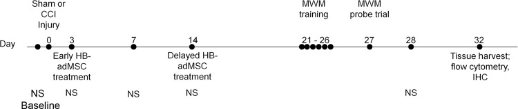
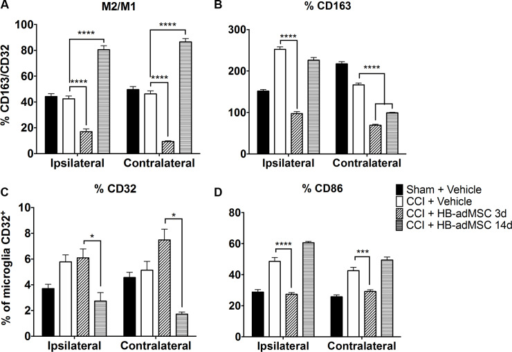
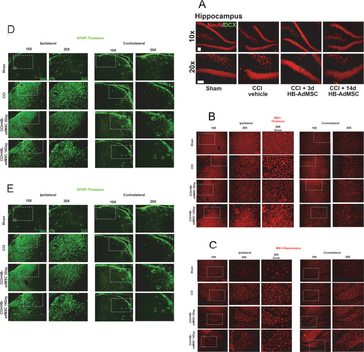
In the U.S., approximately 1.7 million people suffer traumatic brain injury each year, with many enduring long-term consequences and significant medical and rehabilitation costs. The primary injury causes physical damage to neurons, glia, fiber tracts and microvasculature, which is then followed by secondary injury, consisting of pathophysiological mechanisms including an immune response, inflammation, edema, excitotoxicity, oxidative damage, and cell death. Most attempts at intervention focus on protection, repair or regeneration, with regenerative medicine becoming an intensively studied area over the past decade. The use of stem cells has been studied in many disease and injury models, using stem cells from a variety of sources and applications. In this study, human adipose-derived mesenchymal stromal cells (MSCs) were administered at early (3 days) and delayed (14 days) time points after controlled cortical impact (CCI) injury in rats. Animals were routinely assessed for neurological and vestibulomotor deficits, and at 32 days post-injury, brain tissue was processed by flow cytometry and immunohistochemistry to analyze neuroinflammation. Treatment with HB-adMSC at either 3d or 14d after injury resulted in significant improvements in neurocognitive outcome and a change in neuroinflammation one month after injury.


Conflict of interest statement

The authors have read the journal's policy and the authors of this manuscript have the following competing interests: KAR, HP, and AD are paid employees of Hope Bio and have received salary support for their role in this study. SDO and CSC have both received research support from sponsored research agreements between the University of Texas Health Science Center at Houston and Hope Bio. SDO is a Guest Editor on the “Stem Cell Plasticity in Tissue Repair and Regeneration” Call for Papers for PLoS ONE. Hope Bio produces and markets HB-AdMSCs and HB-AdMSC-related products. There are no other patents, products in development or marketed products associated with this research to declare. This does not alter our adherence to PLOS ONE policies on sharing data and materials.

Figures

Fig 1
Fig 1. Experimental timeline.
Animals are tested for baseline behavior measurements prior to injury at Day 0. HB-adMSCs treatment occurs either at Day 3 or Day 14, depending on group. Behavior testing continues on Day 3, 7, 14, 21–26, 27, and 28. Animals are sacrificed and tissues are harvested on Day 32. Neuroscore (NS), Hope Biosciences’ adipose derived mesenchymal stem cells (HB-adMSCs), Morris water maze (MWM), immunohistochemistry (IHC).
Fig 2
Fig 2. Spatial learning and memory.
Animals treated at 3d have significantly shorter latencies compared to CCI on days 2–6. Animals treated at 14d have significantly shorter latencies compared to CCI on days 2, 4, 6. On days 3,4,5 3d treated animals are significantly shorter latency than those treated at 14d. Sham, n = 10, CCI + PBS, n = 13, CCI + HB-adMSCs 3d, n = 7, CCI + HB-adMSCs 14d, n = 3.
Fig 3
Fig 3. Swim distance and probe trials.
Treatment significantly decreased the length of swim on days 2–6 in 3d treated animals. A. Length of swim was significantly decreased on days 2, 3, 5 in 14d treated animals. On days 3, 4, 5 3d treated animals swam significantly shorter distances than 14d treated group. Animals treated at 3d and 14d exhibited decreased distances traveled prior to reaching the location. Treated animals demonstrated significantly shorter swim distances. B. The percent of time spent in the platform quadrant was significantly higher for CCI + HB-adMSCs 14d animals compared to controls. Animals treated at 14d also spent significantly more time in the platform quadrant than those treated at 3d(*, p value<0.05; **, p value<0.01; ****, p value<0.0001). Sham, n = 10, CCI + PBS, n = 13, CCI + HB-adMSCs 3d, n = 7, CCI + HB-adMSCs 14d, n = 3.
Fig 4
Fig 4. Rat brain microglia flow analysis.
The microglia were gated from CD11b/c enriched myeloid cells and defined as CD45+CD11b/c+p2y12+. Microglia were then analyzed for M1 (CD32, CD86) and M2 (CD163) phenotype markers. A. The ratio of CD163+/CD32+ microglia reveals a significant shift toward anti-inflammatory M2 microglia in animals treated at 14 days post-injury, whereas, the 3d treatment group displays a preference toward M1. B. M2 marker CD163 is significantly lower than injured controls for both treatment groups. C. There is a significantly smaller percentage of cells that are positive for M1 marker CD32 in 14d treatment group. D. The same is true for M1 marker CD86 in 3d treatment group. Data represent means ± SEM. Statistical analysis performed by Two-way ANOVA with Tukey’s post hoc test. **, p value<0.01; ****, p value <0.0001. Sham, n = 8, CCI + PBS, n = 8, CCI + HB-adMSCs 3d, n = 4, CCI + HB-adMSCs 14d, n = 2.
Fig 5
Fig 5. Representative localization of neuroinflammation and neurogenesis.
Thin sections from ipsilateral and contralateral hemispheres were immunostained at Day 32. Presented here are portions of the thalamus and hippocampus, specifically the subgranular zone (SGZ). A. Antibodies for NeuN and Doublecortin (DCX) were used to stain for neurogenesis, B, C. IBA-1 for microglial activation and D, E. GFAP for reactive astrocytes. Images are representative of sham, CCI + vehicle, CCI + HB-adMSCs 3d and CCI + HB-adMSCs 14d, at 20x magnification with a 10x inset showing a larger field. Scale bars indicate 200 μm.

References

    1. Taylor C, Bell J, Breiding M, Xu L. Traumatic Brain Injury–Related Emergency Department Visits, Hospitalizations, and Deaths—United States, 2007 and 2013. MMWR Surveill Summ. 2017;66(No. SS-9):1–16. 10.15585/mmwr.ss6609a1 - DOI - PMC - PubMed
    1. Finkelstein E, Corso P, Miller T, associates. The Incidence and Economic Burden of Injuries in the United States. New York (NY): Oxford University Press; 2006.
    1. Kumar A, Loane D. Neuroinflammation after traumatic brain injury: Opportunities for therapeutic intervention. Brain Behav Immun. 2012;26(8):1191–201. 10.1016/j.bbi.2012.06.008 - DOI - PubMed
    1. Liao G, Olson S, Kota D, Hetz R, Smith P, Bedi S, et al. Far-red tracer analysis of traumatic cerebrovascular permeability. J Surg Res. 2014;190(22):628–33. 10.1016/j.jss.2014.05.011 - DOI - PMC - PubMed
    1. Harting MT, Jimenez DF, Xue H, Fischer UM, Baumgartner B, Dash P, et al. Intravenous mesenchymal stem cell therapy for traumatic brain injury. J Neurosurg. 2009;110(6):1189–97. 10.3171/2008.9.JNS08158 - DOI - PMC - PubMed

Publication types