Review
. 2020 Aug;47(8):3752-3771.
doi: 10.1002/mp.14241. Epub 2020 Jun 23.

Multi-energy computed tomography and material quantification: Current barriers and opportunities for advancement

Affiliations
Review

Multi-energy computed tomography and material quantification: Current barriers and opportunities for advancement

Megan C Jacobsen et al. Med Phys. 2020 Aug.

Abstract

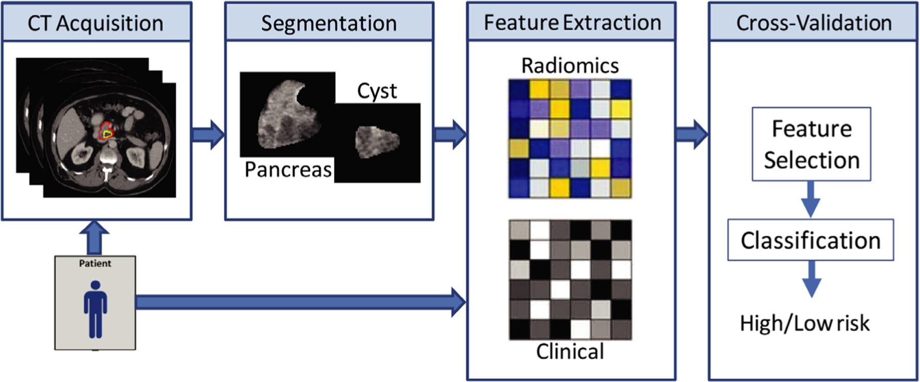
Computed tomography (CT) technology has rapidly evolved since its introduction in the 1970s. It is a highly important diagnostic tool for clinicians as demonstrated by the significant increase in utilization over several decades. However, much of the effort to develop and advance CT applications has been focused on improving visual sensitivity and reducing radiation dose. In comparison to these areas, improvements in quantitative CT have lagged behind. While this could be a consequence of the technological limitations of conventional CT, advanced dual-energy CT (DECT) and photon-counting detector CT (PCD-CT) offer new opportunities for quantitation. Routine use of DECT is becoming more widely available and PCD-CT is rapidly developing. This review covers efforts to address an unmet need for improved quantitative imaging to better characterize disease, identify biomarkers, and evaluate therapeutic response, with an emphasis on multi-energy CT applications. The review will primarily discuss applications that have utilized quantitative metrics using both conventional and DECT, such as bone mineral density measurement, evaluation of renal lesions, and diagnosis of fatty liver disease. Other topics that will be discussed include efforts to improve quantitative CT volumetry and radiomics. Finally, we will address the use of quantitative CT to enhance image-guided techniques for surgery, radiotherapy and interventions and provide unique opportunities for development of new contrast agents.

Keywords: dual-energy computed tomography; photon-counting detector computed tomography; quantitative computed tomography.


Figures

FIG. 1.
FIG. 1.
Example of synchronous scanning of a bone mineral density calibration phantom (white arrow) as it would be positioned under a patient
FIG. 2.
FIG. 2.
Single-energy computed tomography methodology for diagnosis of hepatic steatosis utilizes regions of interest in the liver and spleen, shown here in a 61-yr-old male seen for restaging of a pancreatic neuroendocrine tumor. The liver attenuation in this case is both below 40 HU and more than 10 HU lower than that of the spleen, indicating the presence of diffuse fat within the liver tissue.
FIG. 3.
FIG. 3.
Fatty infiltration of the liver demonstrated on a dual-energy computed tomography fat map in a 75-yr-old woman with pancreatic adenocarcinoma following chemotherapy. The calculated fat fraction for regions of interest in the liver (20.1%) and subcutaneous fat (94.0%) are shown as a percent volume.
FIG. 4.
FIG. 4.
General workflow for development of radiomics-based prediction models. Patients are imaged, and the volume of interest is segmented either manually or automatically. Quantitative radiomics features are extracted from the volume and used along with other clinical or genomics data to develop a statistical model for decision support. Figure reproduced from reference.
FIG. 5.
FIG. 5.
K-edges for iodine (33.2 keV) and tungsten (69.5 keV) allow for K-edge imaging. In energy bin 1 (blue), positioned just below the tungsten K-edge, iodine signal dominates, while in bin 2 (yellow), tungsten signal will be the primary contributor to contrast.
FIG. 6.
FIG. 6.
Representative (a) grayscale computed tomography image slices of a calibration phantom comprising of 0, 5, 10, 15, and 20 mM gadolinium concentrations showing example VOIs (boxes), and (b) cropped grayscale images of VOIs spanning the full range of gadolinium concentrations, for each energy bin of the photon-counting detector, demonstrating the increased attenuation of gadolinium in energy bins greater than the K-edge (50.2 keV). Note that all grayscale intensities were converted to HU. Figure reproduced from Curtis et al.

References

    1. Kim SH, Kamaya A, Willmann JK. CT perfusion of the liver: principles and applications in oncology. Radiology. 2014;272:322–344. - PMC - PubMed
    1. Konstas AA, Goldmakher GV, Lee TY, Lev MH. Theoretic basis and technical implementations of CT perfusion in acute ischemic stroke, part 1: theoretic basis. Am J Neuroradiol. 2009;30:662–668. - PMC - PubMed
    1. Konstas AA, Goldmakher GV, Lee TY, Lev MH. Theoretic basis and technical implementations of CT perfusion in acute ischemic stroke, part 2: technical implementations. Am J Neuroradiol. 2009;30:885–892. - PMC - PubMed
    1. Pullan BR, Fawcitt RA, Isherwood I. Tissue characterization by an analysis of the distribution of attenuation values in computed tomography scans: a preliminary report. J Comput Assist Tomogr. 1978;2:49–54. - PubMed
    1. Norman D, Price D, Boyd D, Fishman R, Newton TH. Quantitative aspects of computed tomography of the blood and cerebrospinal fluid. Radiology. 1977;123:335–338. - PubMed