. 2020 Jun 16;52(6):1022-1038.e7.
doi: 10.1016/j.immuni.2020.04.015. Epub 2020 May 25.

Plasmacytoid Dendritic Cells and Type I Interferon Promote Extrafollicular B Cell Responses to Extracellular Self-DNA

Affiliations

Plasmacytoid Dendritic Cells and Type I Interferon Promote Extrafollicular B Cell Responses to Extracellular Self-DNA

Chetna Soni et al. Immunity. .

Abstract

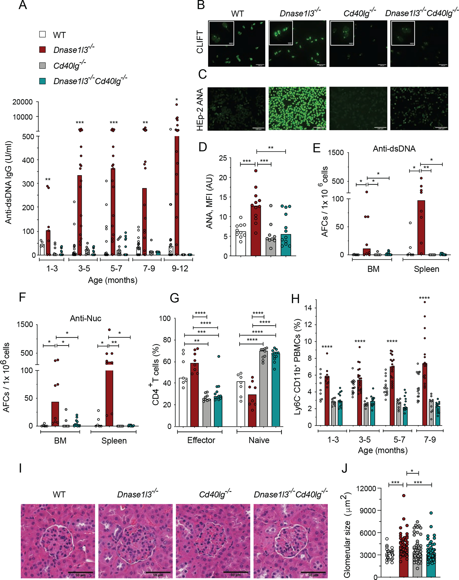
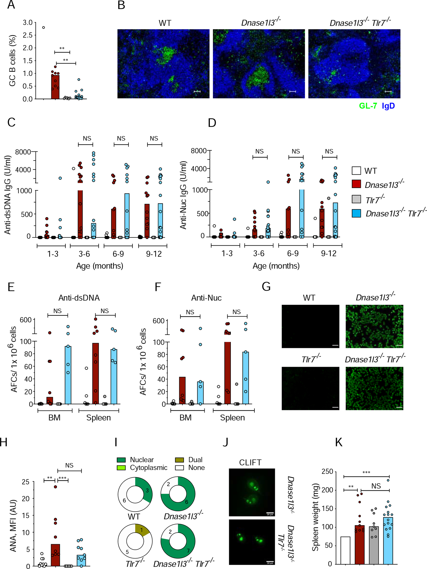
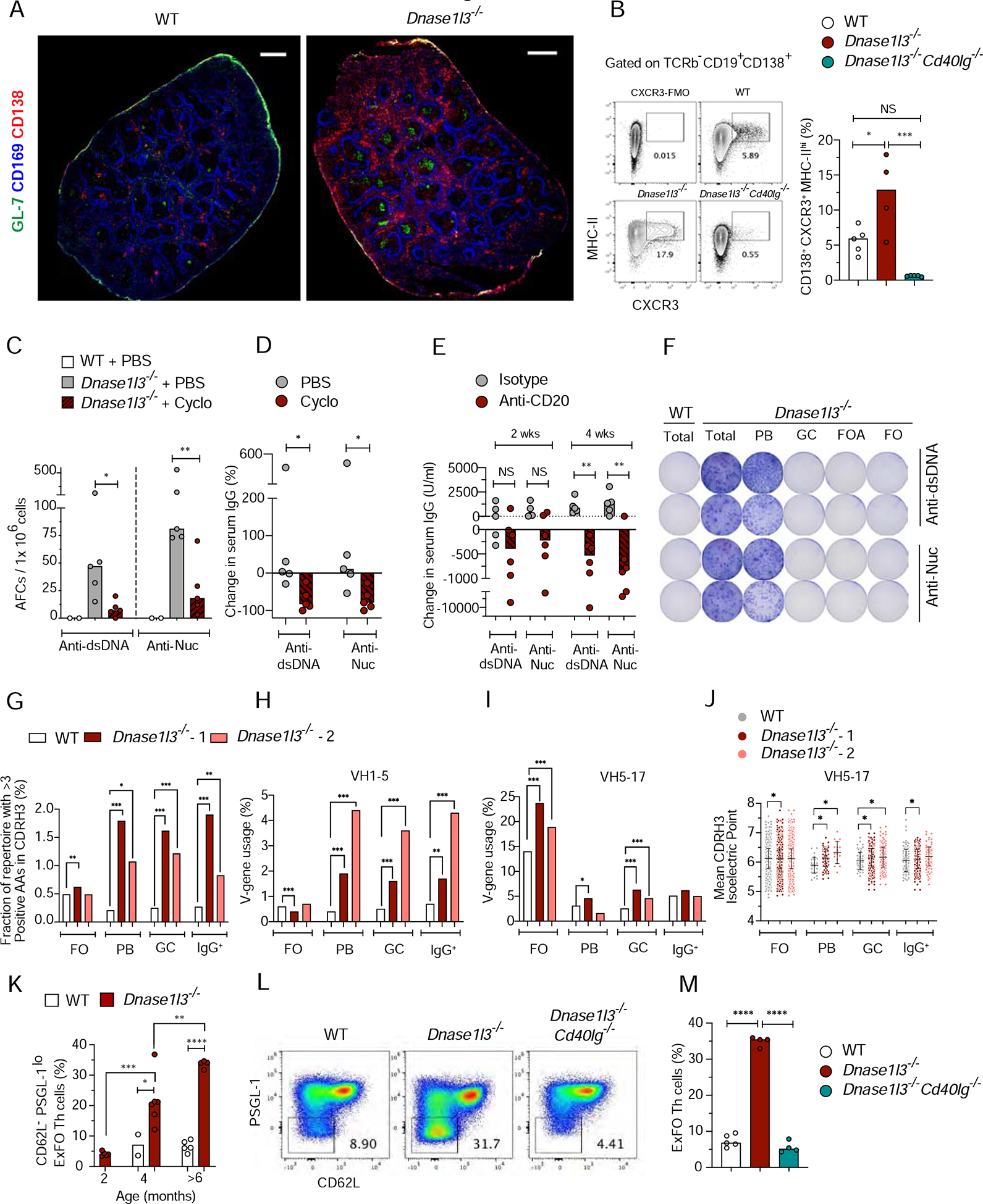
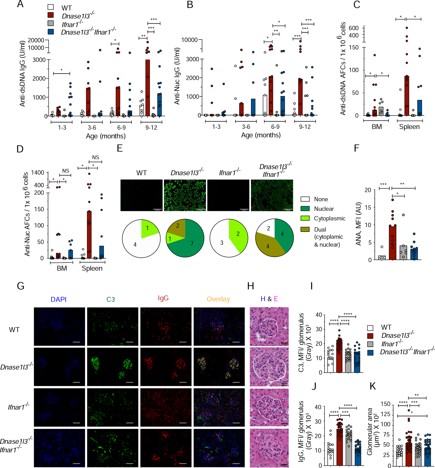
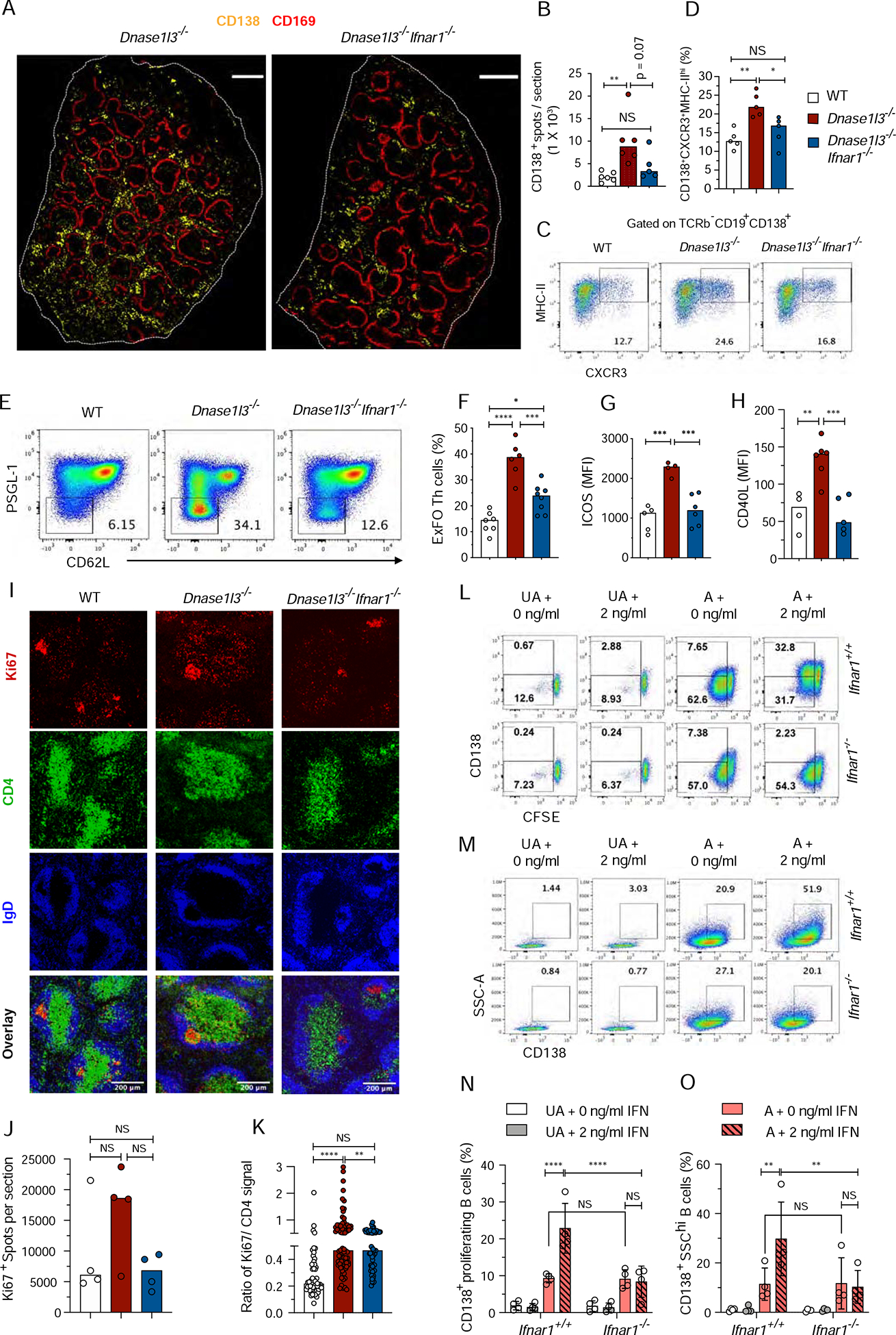
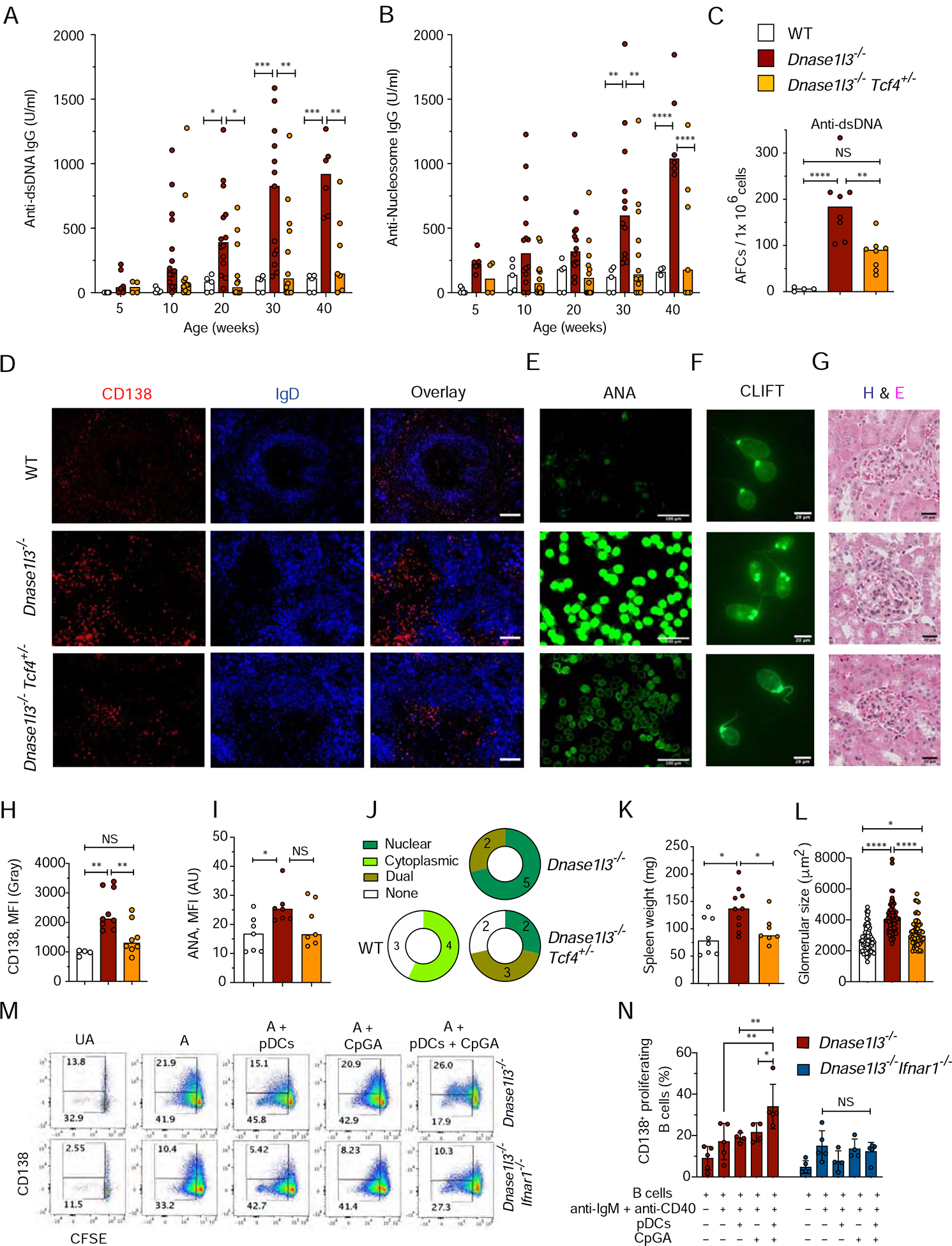
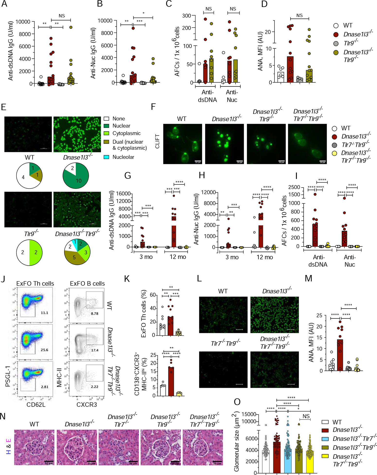
Class-switched antibodies to double-stranded DNA (dsDNA) are prevalent and pathogenic in systemic lupus erythematosus (SLE), yet mechanisms of their development remain poorly understood. Humans and mice lacking secreted DNase DNASE1L3 develop rapid anti-dsDNA antibody responses and SLE-like disease. We report that anti-DNA responses in Dnase1l3-/- mice require CD40L-mediated T cell help, but proceed independently of germinal center formation via short-lived antibody-forming cells (AFCs) localized to extrafollicular regions. Type I interferon (IFN-I) signaling and IFN-I-producing plasmacytoid dendritic cells (pDCs) facilitate the differentiation of DNA-reactive AFCs in vivo and in vitro and are required for downstream manifestations of autoimmunity. Moreover, the endosomal DNA sensor TLR9 promotes anti-dsDNA responses and SLE-like disease in Dnase1l3-/- mice redundantly with another nucleic acid-sensing receptor, TLR7. These results establish extrafollicular B cell differentiation into short-lived AFCs as a key mechanism of anti-DNA autoreactivity and reveal a major contribution of pDCs, endosomal Toll-like receptors (TLRs), and IFN-I to this pathway.

Keywords: DNASE1L3; TLR7; TLR9; anti-DNA antibodies; extrafollicular B cell response; plasmacytoid dendritic cells; systemic lupus erythematosus; type I interferon.


Conflict of interest statement

Declaration of Interests The authors declare no competing interests.

Figures

Figure 1.
Figure 1.. CD40L deficiency in Dnase1l3−/− mice abolishes autoimmunity.
Wild-type (WT), Dnase1l3−/−, Cd40lg−/− and Dnase1l3−/−Cd40lg−/− mice were examined for Abs at the indicated ages or at the 12 month endpoint. (A) Serum anti-dsDNA IgG titers measured by ELISA. (B) CLIFT assay for anti-dsDNA IgG (representative of ≥4 mice per strain). Arrow indicates positive staining of the kinetoplast. Scale bar, 100 μm (insets, 20 μm). (C, D) ANA assay, showing images representative of ≥8 mice per strain (C), and quantitation of fluorescence intensity (D). Scale bars, 100μm. (E, F) Frequency of anti-dsDNA (E) and anti-nucleosome (F) AFCs in the bone marrow (BM) or spleen as determined by ELISpot. (G) Fractions of effector and naive T cells among splenic CD4+ T cells at the endpoint. (H) Fractions of CD11b+Ly6c population among total PBMCs at indicated time points. (I) Glomeruli from H&E-stained kidney sections (representative of ≥3 mice per group). Scale bars, 50 μm. (J) Size of ≥ 20 glomeruli per kidney section from ≥3 mice per group. In A and H, significant differences between Dnase1l3−/− vs Dnase1l3−/−Cd40lg−/−mice are shown. Symbols represent individual mice except in panel J, where they represent individual glomeruli. All bars indicate median. * p ≤ 0.05, ** p ≤ 0.01, ***p ≤ 0.001 and **** p ≤ 0.0001. See also Figure S1.
Figure 2.
Figure 2.. TLR7-deficient Dnase1l3−/− mice do not develop germinal centers but retain anti-DNA reactivity.
Wild-type (WT), Dnase1l3−/−, Tlr7−/− and Dnase1l3−/−Tlr7−/− mice were examined for Abs at the indicated ages or at the 12 month endpoint. (A) Fraction of CD38 GL-7+ GC B cells among splenic B cells at the endpoint. (B) Spleen sections stained for GC B cells (green) and follicular B cells (blue). Representative of ≥3 mice per group. Scale bars, 30 μm. (C, D) Serum anti-dsDNA (C), and anti-Nucleosome (Nuc, D) IgG titers measured by ELISA. (E, F) Frequency of anti-dsDNA (E), and anti-nucleosome (F) AFCs in the bone marrow (BM) or spleen as determined by ELISpot. (G, H) ANA assay, showing images representative of ≥8 mice per strain (G), and quantitation of fluorescence intensity (H). Scale bars, 100μm. (I) Distribution of ANA reactivity patterns in mice from each group. (J) CLIFT assay for anti-dsDNA IgG (representative of ≥4 mice per strain). Scale bar, 20 μm. (K) Spleen weights at the endpoint. Symbols represent individual mice; bars indicate median. NS= not significant, * p ≤ 0.05, ** p ≤ 0.01 and ***p ≤ 0.001 See also Figure S2.
Figure 3.
Figure 3.. DNA-reactive B cells in Dnase1l3−/− mice develop primarily through ExFO differentiation into plasmablasts.
(A) Spleen sections representative of 3–5 WT or Dnase1l3−/− mice stained for CD138 (red), CD169 (blue) and GL-7 (green). Scale bar, 1000μm. (B) Shown are representative flow plots and quantitation of the fraction of CXCR3+MHC-IIhi (ExFO) B cells, pre-gated on splenic TCRβB220+CD19+CD138+ cells from WT (open symbols), Dnase1l3−/− (red) and Dnase1l3−/−Cd40lg−/− (green) mice. CXCR3-FMO (fluorescence minus one) was used for gate allocation. (C, D) Frequency of anti-dsDNA, and anti-nucleosome (Nuc) antibody-forming cells (AFCs), determined by ELISpot (C); and percent change in serum anti-dsDNA or antiNuc IgG titers determined by ELISA (D), in 6-mo-old WT mice (open) or Dnase1l3−/− mice treated with PBS (grey) or cyclophosphamide (cyclo, red). (E) Change in serum titers of anti-dsDNA or anti-Nuc IgG in 6-mo-old Dnase1l3−/− mice after one dose (100μg/ mouse; i.p.) of treatment with IgG2a isotype control (grey) or anti-CD20 Ab (red) at 2 weeks and upon second dose of a similar treatment at 4 weeks. (F) Image of ELISpot plate representative of 2 experiments with two mice per experiment. Shown are two serial dilutions of anti-dsDNA, and anti-nucleosome (Nuc) antibody-forming cells (AFCs) from total splenocytes of >6-mo-old WT and Dnase1l3−/− mice OR from flow sorted – Plasmablasts (PB); Germinal center B cells (GC); Follicular activated B cells (FOA) and Naïve Follicular B cells (FO) from >6 mo old Dnase1l3−/− mice. Sorted GC, FOA and FO cells were activated for 2h with 5 μg/ml anti-CD40 + 1 μg/ml LPS, and plated onto DNA or Nuc coated ELISpot plates. (G) Percentage of clonotypes in each repertoire that contain >3 positively charged amino acids (AA) within the CDR-H3 region, in FO, PB, GC and IgG+ B cells from WT (open), Dnase1l3−/− 1 (red) and Dnase1l3−/− −2 (salmon). (H, I) Percentage of clonotypes that use the V-gene IGHV1–5 (H), and V-gene IGH5–17 (I) in the indicated B cell subsets and mice. (J) Mean isoelectric point of the CDR-H3 region among the clonotypes that use the V-gene IGH5–17. Each point represents one clonotype. Error bars represent the mean and S.D. of all clonotypes in each group. (K) Flow analysis of the fraction of CD62LPSGL-1loExFO Th cells among splenic B220TCRβ+CD4+ T cells of WT and Dnase1l3−/− mice at the indicated ages. (L, M) Representative flow plots of pre-gated splenic B220TCRβ+CD4+ cells with low ExFO Th cells shown within the gate (L); quantitation of the fraction of ExFO Th cells (M) from >6-mo-old WT, Dnase1l3−/− and Dnase1l3−/−Cd40lg−/− mice. For panels A-E, K and M symbols represent individual mice and bars indicate median. * p ≤ 0.05, ** p ≤ 0.01, ***p ≤ 0.001 and **** p ≤ 0.0001. See also Figure S3.
Figure 4.
Figure 4.. Autoreactivity and autoimmune manifestations in Dnase1l3−/− mice are facilitated by type I interferon signaling.
Wild-type (WT), Dnase1l3−/−, Ifnar1−/− and Dnase1l3−/− Ifnar1−/− mice were examined for Abs at the indicated ages or at the 12 month endpoint. (A and B) Serum anti-dsDNA (A) and anti-Nucleosome (B) IgG titers determined by ELISA. (C and D) ELISpot analysis of the number of anti-dsDNA (C), and anti-nucleosome (D) antibody forming cells (AFCs) in bone marrow (BM) or spleen cells. (E) Images of HEp-2 cells stained with sera to detect ANAs (representative of n≥ 5 per strain). Scale bars, 100 μm. Pie charts with numbers indicate distribution of ANA reactivity patterns in mice from each group. (F) Quantitation of ANA fluorescence intensity. (G-K) Images of kidney sections representative of ≥ 3 mice per group stained for IgG, (red) and C3 (green) and DAPI (blue). Scale bars, 40 μm, (G). Images of glomeruli from H&E-stained kidney sections, representative of ≥3 mice per group. Scale bars 20 μm (H). Quantitation of mean fluorescence intensity of C3 (I), and IgG deposits (J), and size of ≥ 20 glomeruli per kidney section (K) from 3–4 mice per strain. In panels A-D and F, symbols represent individual mice. Panels I-K each symbol represents an individual glomerulus. All bars indicate median. NS= not significant, * p ≤ 0.05, ** p ≤ 0.01, ***p ≤ 0.001 and **** p ≤ 0.0001. See also Figure S4.
Figure 5.
Figure 5.. Type I IFN signaling promotes extrafollicular B cell proliferation and differentiation into antibody-forming cells
. (A, B) Confocal images of half spleens representative of >4, 8–9-mo-old WT, Dnase1l3−/− and Dnase1l3−/−Ifnar1−/− mice, stained for CD169 (red) and CD138+ (yellow). Scale bar, 1000 μm (A); Total number of CD138+ spots per half spleen represented in A (B). Each symbol represents half a spleen section (C, D) Staining profiles of gated splenic CD19+CD138+ cells (CXCR3+MHC-IIhi ExFO B cells highlighted) (C), and frequencies of ExFO B cells (D), in >10-mo-old WT, Dnase1l3−/− and Dnase1l3−/−Ifnar1−/− mice. (E-H) Flow cytometric staining profiles of gated splenic CD4+ T cells (PSGL-1loCD62LloExFO Th cells highlighted) (E); Fraction of ExFO Th cells among splenic CD4+ T cells (F), mean fluorescence intensity of ICOS (G), and CD40L (H) expressed in ExFO Th cells of 6–8-mo-old WT, Dnase1l3−/− and Dnase1l3−/−Ifnar1−/− mice. In panels C-H, wherever applicable, symbols represent individual mice and bars indicate median. (I-K) Confocal images of spleen sections representative of three >8-mo-old WT, Dnase1l3−/− and Dnase1l3−/−Ifnar1−/− mice stained for Ki67+ proliferating cells (red), CD4+ T cells (green) and IgD+ B cells (blue). Scale bars, 200 μm (I); Quantitation of total number of Ki67+ spots per half spleen represented in Fig. S5B. Each symbol represents half a spleen section (J); Ratio of mean fluorescence intensity of Ki67 vs CD4 within a region of interest (ROI) marking CD4+ T cell zones excluding GC areas, from spleen sections represented in Fig.S5B. Each symbol represents a single ROI (K). (L-O) Flow cytometric analysis of proliferation and differentiation of B cells upon in-vitro activation with anti-IgM and anti-CD40 in the presence or absence of IFNα. Shown are representative flow plots from 4 independent experiments of purified CFSE labeled unactivated (UA) or activated (A) B cells from Ifnar+/+ or Ifnar1−/− mice cultured for 72h in the absence (0 ng/ml) or presence of 2 ng/ml of IFNα. The cells in the gates indicate CFSElo proliferating B cells, with (upper gate) or without (lower gate) CD138 expression (L); or CD138+SSChi B cells (M), under the indicated conditions. Fraction of CD138+ proliferating (N) and CD138+SSChi blasting (O) B cells. Each symbol represents an independent experiment. Error bars show mean ± SD. Two-way ANOVA followed by Tukey’s multiple-comparison test was used to compare different treatments within a genotype or 2-way ANOVA followed by Sidak’s multiple comparison test was used to compare different treatments between the two genotypes. NS= not significant, * p ≤ 0.05, ** p ≤ 0.01, ***p ≤ 0.001 and **** p ≤ 0.0001. See also Figure S5.
Figure 6.
Figure 6.. Functional impairment of pDCs in Dnase1l3−/− mice ameliorates autoreactivity and disease.
Wild-type (WT), Dnase1l3−/−, and Dnase1l3−/− mice with monoallelic deficiency of Tcf4 (Dnase1l3−/−Tcf4+/−) were examined for Abs at the indicated ages or at the 12 month endpoint. (A-B) Serum anti-dsDNA IgG (A) and anti-Nucleosome (B) IgG titers by ELISA. (C) Frequency of anti-dsDNA AFCs in the spleen by ELISpot. (D) Spleen sections stained for CD138+ cells (red) and follicular B cells (blue). Representative of ≥3 mice per group. Scale bars 40 μm. (E) ANA assay images, representative of 7 mice per group. Scale bar, 100 μm. (F) CLIFT assay for anti-dsDNA IgG (representative of 5 mice per group). Scale bar: 20 μm. (E) Glomeruli from H&E-stained kidney sections (representative of 3 mice per group). Scale bars, 20 μm. (H) Intensity of CD138 fluorescence in ≥ 4 images of spleen sections from the indicated mice. (I, J) Quantitation of ANA fluorescence intensity (I), and distribution of ANA reactivity patterns (J) in 7 mice per indicated group. (K) Spleen weights of indicated mice at the endpoint. (L) Quantitation of the size of ≥ 20 glomeruli per kidney section from 3 mice per group. Each symbol represents an individual glomerulus; bars indicate median. (M-N) Purified CFSE labeled B cells from Dnase1l3−/− or Dnase1l3−/−Ifnar1−/− mice were left unactivated (UA); activated with anti-IgM + anti-CD40 (A); OR activated with anti-IgM + anti-CD40 in the presence of unstimulated pDCs (A+ pDCs); in the presence of 1μM CpGA (A+ CpGA) or in the presence of pDCs stimulated with 1μM CpGA (A+ pDCs+ CpGA) for 72h after which the B cells were analyzed for proliferation and differentiation using flow cytometry. Gating strategy (M) and quantitation (N) of CFSEloCD138+ proliferating B cells. Error bars show mean ± SD from four independent experiments (symbols). NS= not significant, * p ≤ 0.05, ** p ≤ 0.01, ***p ≤ 0.001 and **** p ≤ 0.0001. See also Figure S6.
Figure 7.
Figure 7.. TLR9 and TLR7 are redundantly required for autoreactivity and autoimmunity
Wild-type (WT), Dnase1l3−/−, Tlr9−/−, Tlr7−/− mice or crosses thereof were examined for Abs at the indicated ages or at the 12 month endpoint. (A-B) Serum anti-dsDNA IgG (A) and anti-Nuc (B) IgG titers by ELISA. (C) Frequency of anti-dsDNA and anti-Nuc AFCs in the spleen by ELISpot. (D, E) ANA assay, showing quantitation of fluorescence intensity (D), representative images and the distribution of reactivity patterns in mice from each group (E). (F) CLIFT assay for anti-dsDNA IgG (representative of ≥4 mice per strain). Scale bar: 20 μm. (G-H) Serum anti-dsDNA IgG (A) and anti-Nucleosome (B) IgG titers by ELISA. (I) Frequency of anti-dsDNA and anti-nucleosome AFCs in the spleen as determined by ELISpot. (J) Staining profiles of gated splenic CD4+ T cells (PSGL-1loCD62Llo ExFO Th cells highlighted) and CD19+ CD138+ cells (CXCR3+ MHC-IIhi ExFO B cells highlighted). (K) Frequencies of populationss highlighted in J at the endpoint. (L-M) ANA assay, showing images representative of ≥8 mice per strain (L), and quantitation of fluorescence intensity (M). Scale bars, 100μm. (N) Glomeruli from H&E-stained kidney sections (representative of 4 mice per group). Scale bars, 20 μm. (O) Size of ≥ 20 glomeruli per kidney section from 4 mice per group. Symbols represent individual mice except in panel O, where they represent individual glomeruli. All bars indicate median. NS= not significant, * p ≤ 0.05, ** p ≤ 0.01, ***p ≤ 0.001 and **** p ≤ 0.0001. See also Figure S7.

References

    1. Agrawal H, Jacob N, Carreras E, Bajana S, Putterman C, Turner S, Neas B, Mathian A, Koss MN, Stohl W, et al. (2009). Deficiency of type I IFN receptor in lupus-prone New Zealand mixed 2328 mice decreases dendritic cell numbers and activation and protects from disease. J Immunol 183, 6021–6029. - PMC - PubMed
    1. Al-Mayouf SM, Sunker A, Abdwani R, Abrawi SA, Almurshedi F, Alhashmi N, Al Sonbul A, Sewairi W, Qari A, Abdallah E, et al. (2011). Loss-of-function variant in DNASE1L3 causes a familial form of systemic lupus erythematosus. Nat Genet 43, 1186–1188. - PubMed
    1. Barrat FJ, Meeker T, Gregorio J, Chan JH, Uematsu S, Akira S, Chang B, Duramad O, and Coffman RL (2005). Nucleic acids of mammalian origin can act as endogenous ligands for Toll-like receptors and may promote systemic lupus erythematosus. J Exp Med 202, 1131–1139. - PMC - PubMed
    1. Barrat FJ, and Su L (2019). A pathogenic role of plasmacytoid dendritic cells in autoimmunity and chronic viral infection. J Exp Med. - PMC - PubMed
    1. Bennett L, Palucka AK, Arce E, Cantrell V, Borvak J, Banchereau J, and Pascual V (2003). Interferon and granulopoiesis signatures in systemic lupus erythematosus blood. J Exp Med 197, 711–723. - PMC - PubMed

Publication types

MeSH terms