Review
. 2020 Jul:144:A3-A13.
doi: 10.1016/j.yjmcc.2020.05.012. Epub 2020 May 23.

A 20/20 view of ANT function in mitochondrial biology and necrotic cell death

Affiliations
Review

A 20/20 view of ANT function in mitochondrial biology and necrotic cell death

Michael J Bround et al. J Mol Cell Cardiol. 2020 Jul.

Abstract

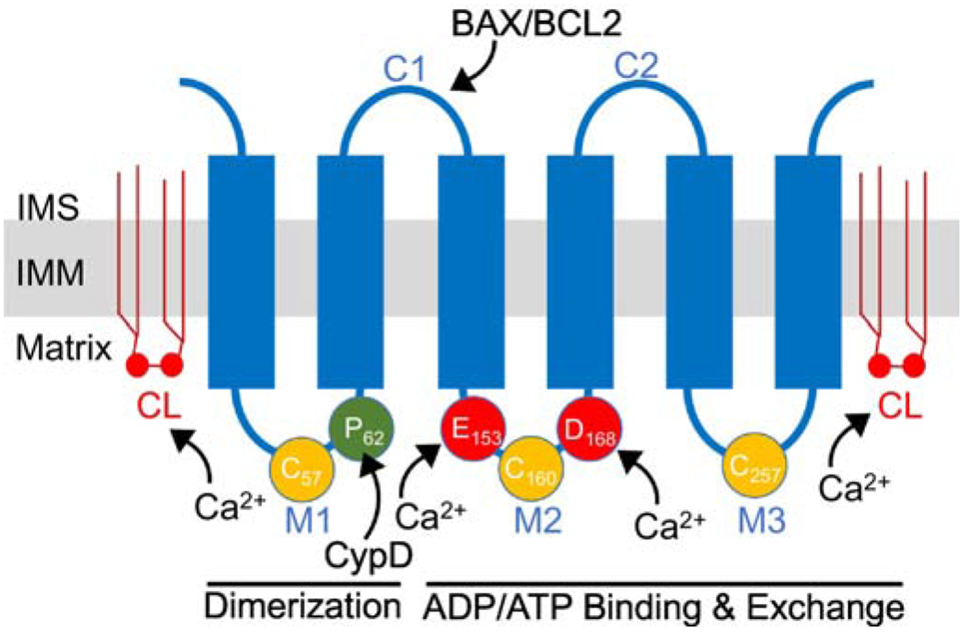
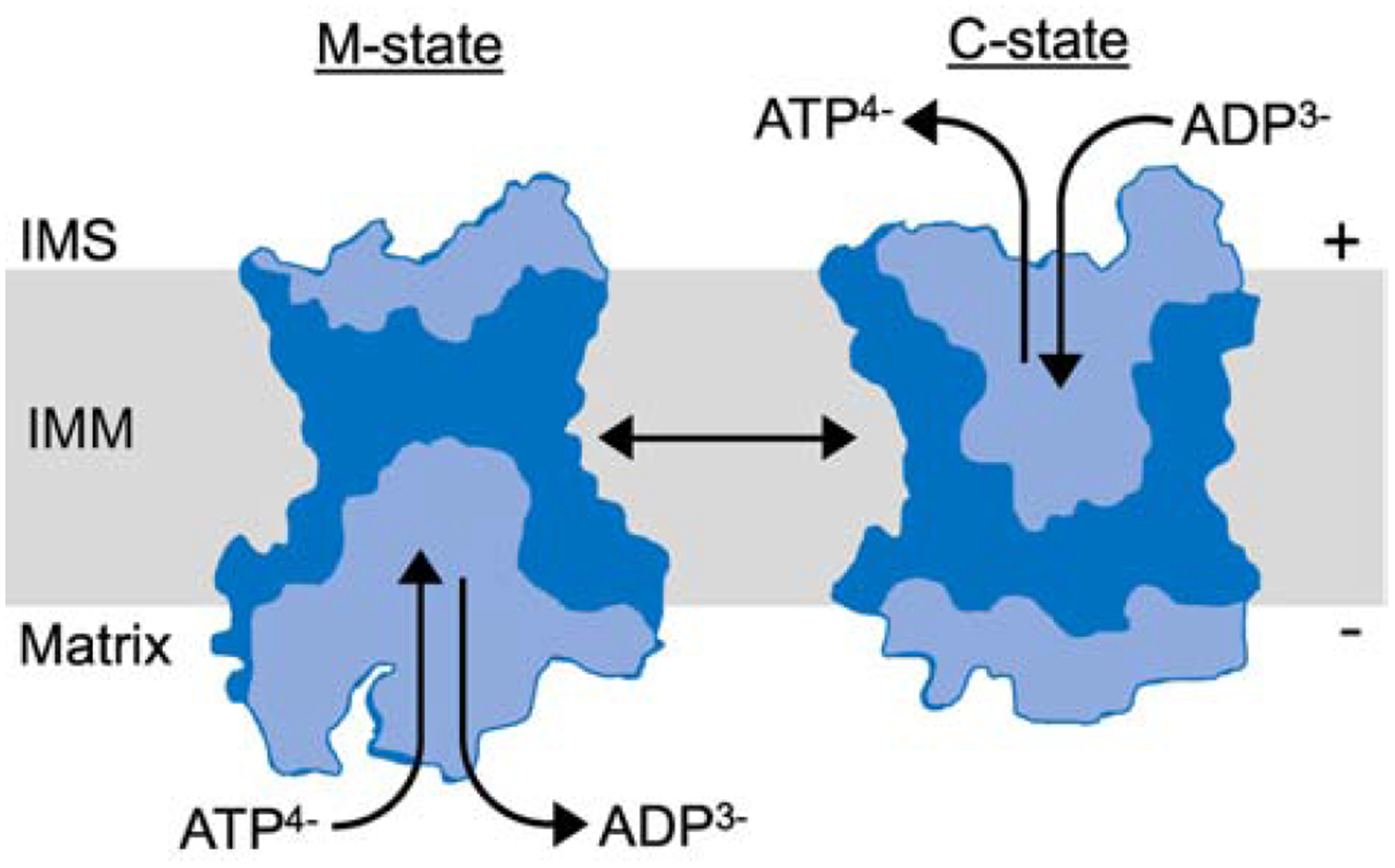
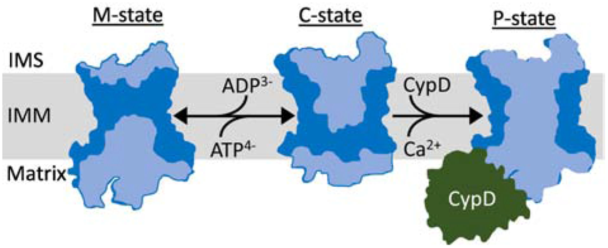
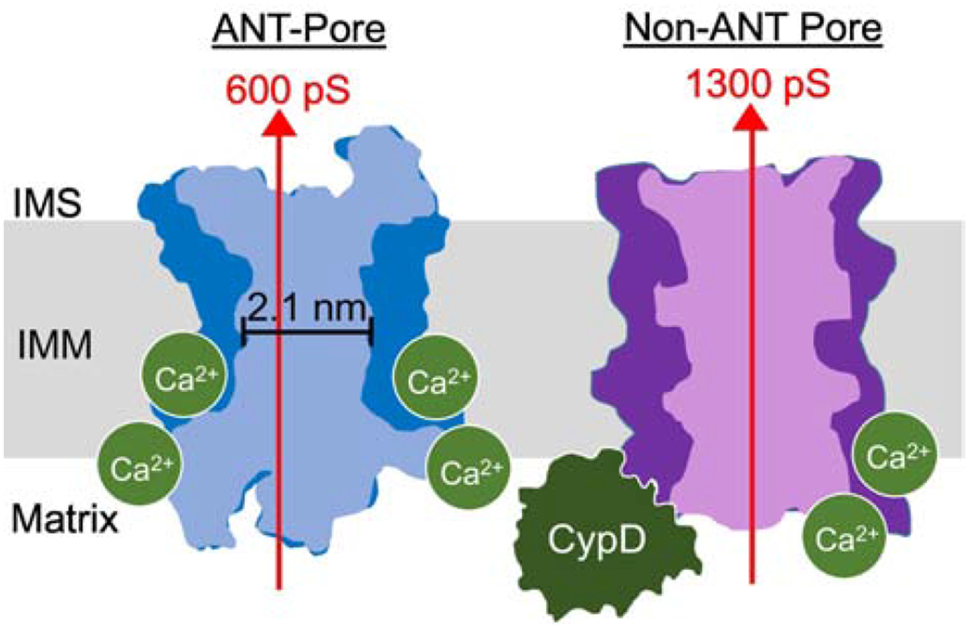
The adenosine nucleotide translocase (ANT) family of proteins are inner mitochondrial membrane proteins involved in energy homeostasis and cell death. The primary function of ANT proteins is to exchange cytosolic ADP with matrix ATP, facilitating the export of newly synthesized ATP to the cell while providing new ADP substrate to the mitochondria. As such, the ANT proteins are central to maintaining energy homeostasis in all eukaryotic cells. Evidence also suggests that the ANTs constitute a pore-forming component of the mitochondrial permeability transition pore (MPTP), a structure that forms in the inner mitochondrial membrane that is thought to underlie regulated necrotic cell death. Additionally, emerging studies suggest that ANT proteins are also critical for mitochondrial uncoupling and for promoting mitophagy. Thus, the ANTs are multifunctional proteins that are poised to participate in several aspects of mitochondrial biology and the greater regulation of cell death, which will be discussed here.

Keywords: ANT; ATP; Heart; Mitochondria; Necrotic cell death.


Conflict of interest statement

Declaration of Competing Interest All authors confirm no conflict of interest with the current study.

Figures

Figure 1:
Figure 1:. ANT protein schematic.
Schematic of ANT protein monomer topology in the inner mitochondrial membrane (IMM). ANT proteins have six transmembrane domains and two mitochondrial intermembrane (IMS) space facing domains (C1-C2). C1 contains a BAX/BCL2 binding motif. In addition, there are three mitochondrial matrix facing loops (M1-M3). The M1 loop is thought to be involved in dimerization and it contains Proline-62 (P62) that is thought to interact with CypD. This M1 loop also contains Cysteine-57 (C57) that is thought to play a role in ANT dimerization. The M2 and M3 loops are involved in ADP/ATP binding and nucleotide exchange. M2 and M3 contain two modifiable cysteines (C160 and C257) that are thought to be sensitive to oxidative stress. In addition, M2 contains two acidic amino acids, Gultamate-153 (E153) and Asparatate-168 (D168), important to ADP binding that may also be involved in Ca2+ sensing and regulation. In addition, ANT proteins are associated with six molecules of cardiolipin (depicted by CL) in the inner membrane that is required for protein folding and sensitivity to Ca2+ regulation. Amino acid residue numbers are taken from human ANT1.
Figure 2:
Figure 2:. ANT model of ADP/ATP exchange.
ANT interconverts between a mitochondrial matrix facing m-state conformation and intermembrane space (IMS) facing c-state. In charged mitochondria ATP is exported and ADP is imported, causing the conformational change. The ANT m-state and c-state diagrams were adapted from the ANT crystal structures [7]. Light blue areas depict ANT solute accessible cavities.
Figure 3:
Figure 3:. ANT model of MPTP.
ANT is hypothesized to form a non-selective pore with properties similar to the MPTP. The ANT interconverts between the m- and c-states during ADP/ATP exchange. The m-state is protected and cannot transition directly into the MPTP-like pore, the ‘p-state’. The ANT c-state can convert into the p-state in response to a variety of activating stresses including binding CypD and high matrix Ca2+ levels. According to this model the MPTP is produced when ANT monomers become simultaneously open to both the matrix and intermembrane space (IMS), forming a pore. The ANT m-state and c-state diagrams were adapted from ANT crystal structures [7], while the hypothetical p-state diagram was generated by superimposing the m- and c-states crystal structures to create an ANT-species open to both the matrix and IMS side of the inner mitochondrial membrane (IMM). Light blue areas depict solute accessible ANT cavities. CypD is not drawn to scale and is included for illustration only.
Figure 4:
Figure 4:. The “Multiple Pore” model of MPTP.
The MPTP is hypothesized to be comprised of multiple protein species, one of which is the ANT. In our model, ANT generates a pore with a conductance of up to 300–600 pS [123, 151] and with a theoretical diameter of 2.1 nm (based on the narrowest channel opening in ANT crystal structures [7]). ANT-pore activity is regulated by CypD but can also activate in the absence of CypD in response to very high levels of matrix Ca2+. It is currently unclear if Ca2+ binds to ANT directly to regulate its MPTP-activity, however we favor a model where Ca2+ directly activates the channel since Ca2+ treatment can activate channel behavior in vesicles containing purified ANT [–123]. There is at least one non-ANT pore forming species. This non-ANT pore responds to matrix Ca2+ but absolutely requires CypD to activate. This non-ANT pore may generate a conductance of 1300 pS [151]. The ANT pore diagram was generated from superimposing the m-state and c-state crystal structures as in Figure 3 [7].

References

    1. Aquila H, Link TA, Klingenberg M. Solute carriers involved in energy transfer of mitochondria form a homologous protein family. FEBS Lett 1987; 212: 1–9. - PubMed
    1. Monne M, Palmieri F. Antiporters of the mitochondrial carrier family. Curr Top Membr 2014; 73: 289–320. - PubMed
    1. Klingenberg M. The ADP and ATP transport in mitochondria and its carrier. Biochim Biophys Acta 2008; 1778: 1978–2021. - PubMed
    1. Pebay-Peyroula E, Dahout-Gonzalez C, Kahn R, Trezeguet V, Lauquin GJ, Brandolin G. Structure of mitochondrial ADP/ATP carrier in complex with carboxyatractyloside. Nature 2003; 426: 39–44. - PubMed
    1. Aquila H, Misra D, Eulitz M, Klingenberg M. Complete amino acid sequence of the ADP/ATP carrier from beef heart mitochondria. Hoppe Seylers Z Physiol Chem 1982; 363: 345–9. - PubMed

Publication types

MeSH terms

Substances