

Reflectance confocal microscopy terminology glossary for melanocytic skin lesions: A systematic review
- PMID: 32454102
- PMCID: PMC8387955
- DOI: 10.1016/j.jaad.2020.05.097
Reflectance confocal microscopy terminology glossary for melanocytic skin lesions: A systematic review
Abstract
Background: There is lack of uniformity in the reflectance confocal microscopy (RCM) terminology for melanocytic lesions.
Objective: To review published RCM terms for melanocytic lesions and identify redundant, synonymous terms.
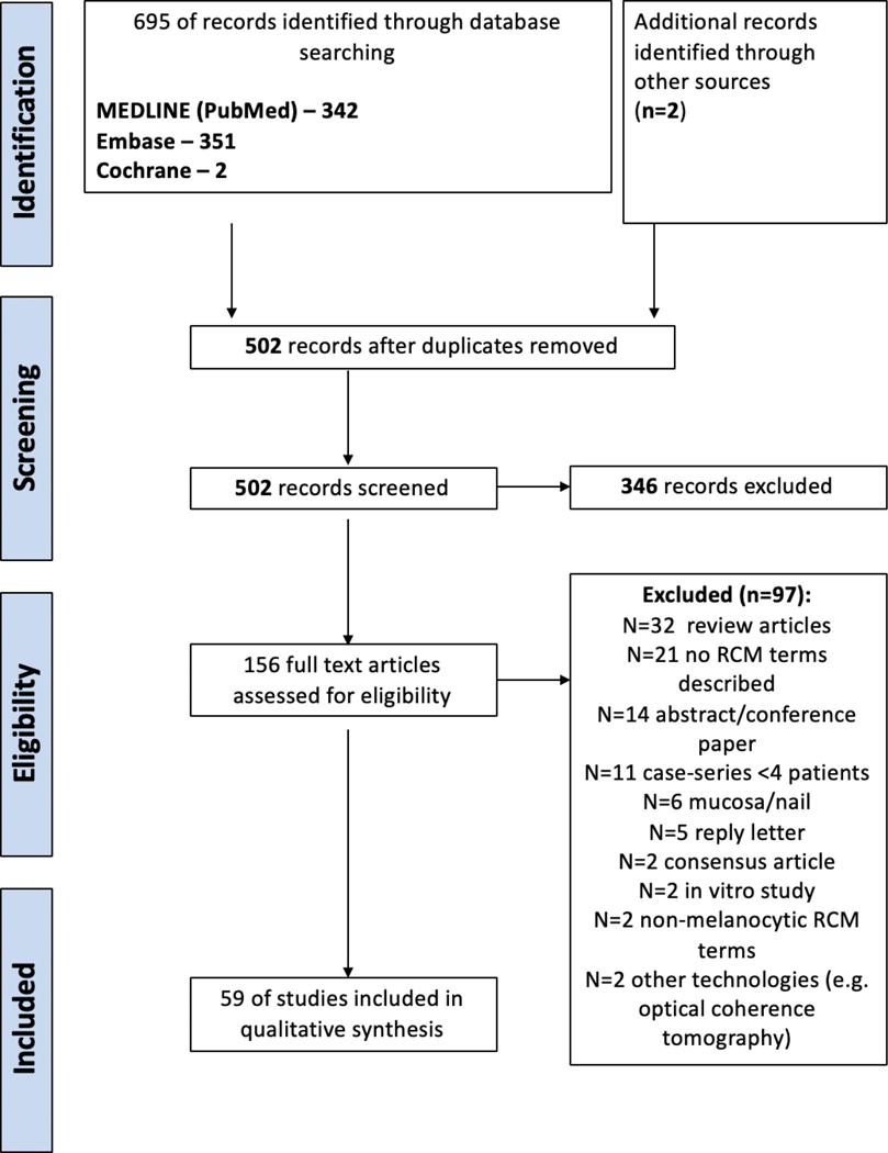
Methods: A systematic review of original research articles adhering to Preferred Reporting Items for Systematic Reviews and Meta-analyses (PRISMA) guidelines was conducted until August 15, 2018. Two investigators gathered all published RCM terms used to describe melanoma and melanocytic nevi. Synonymous terms were grouped based on similarity in definition and in histopathologic correlation.
Results: Out of 156 full-text screened articles, 59 studies met the inclusion criteria. We identified 209 terms; 191 (91.4%) corresponding to high-magnification/cellular-level terms and 18 (8.6%) corresponding to low-magnification/architectural patterns terms. The overall average use frequency of RCM terms was 3.1 times (range, 1-31). By grouping of individual RCM terms based on likely synonymous definitions and by eliminating terms lacking clear definition, the total number of RCM terms could be potentially reduced from 209 to 40 terms (80.8% reduction).
Limitations: Non-English and non-peer-reviewed articles were excluded.
Conclusions: This systematic review of published RCM terms identified significant terminology redundancy. It provides the basis for subsequent terminology consensus on melanocytic neoplasms.
Keywords: diagnosis; melanocytic; melanoma; nevus; noninvasive; reflectance confocal microscopy; systematic review.
Copyright © 2020 American Academy of Dermatology, Inc. Published by Elsevier Inc. All rights reserved.
Conflict of interest statement
Figures
Similar articles
-
Reflectance confocal microscopy terminology glossary for nonmelanocytic skin lesions: A systematic review.J Am Acad Dermatol. 2019 May;80(5):1414-1427.e3. doi: 10.1016/j.jaad.2018.12.007. Epub 2018 Dec 8. J Am Acad Dermatol. 2019. PMID: 30529706 Free PMC article.
-
Role of In Vivo Reflectance Confocal Microscopy in the Analysis of Melanocytic Lesions.Acta Dermatovenerol Croat. 2018 Apr;26(1):64-67. Acta Dermatovenerol Croat. 2018. PMID: 29782304 Review.
-
In vivo reflectance confocal microscopy imaging of melanocytic skin lesions: consensus terminology glossary and illustrative images.J Am Acad Dermatol. 2007 Oct;57(4):644-58. doi: 10.1016/j.jaad.2007.05.044. Epub 2007 Jul 16. J Am Acad Dermatol. 2007. PMID: 17630045
-
Nevomelanocytic atypia detection by in vivo reflectance confocal microscopy.Medicina (Kaunas). 2014;50(4):209-15. doi: 10.1016/j.medici.2014.09.008. Epub 2014 Oct 1. Medicina (Kaunas). 2014. PMID: 25458957
-
[Translated article] Reflectance Confocal Microscopy Terminology in Spanish: A Delphi Consensus Study.Actas Dermosifiliogr. 2024 Mar;115(3):T258-T264. doi: 10.1016/j.ad.2024.01.018. Epub 2024 Jan 19. Actas Dermosifiliogr. 2024. PMID: 38244840 English, Spanish.
Cited by
-
Confocal Microscopy for Diagnosis and Management of Cutaneous Malignancies: Clinical Impacts and Innovation.Diagnostics (Basel). 2023 Feb 23;13(5):854. doi: 10.3390/diagnostics13050854. Diagnostics (Basel). 2023. PMID: 36899999 Free PMC article. Review.
-
Editorial: Methods in skin cancer.Front Oncol. 2023 Mar 3;13:1150450. doi: 10.3389/fonc.2023.1150450. eCollection 2023. Front Oncol. 2023. PMID: 36937449 Free PMC article. No abstract available.
-
Optical imaging guided- 'precision' biopsy of skin tumors: a novel approach for targeted sampling and histopathologic correlation.Arch Dermatol Res. 2021 Sep;313(7):517-529. doi: 10.1007/s00403-020-02126-6. Epub 2020 Aug 25. Arch Dermatol Res. 2021. PMID: 32844312 Free PMC article.
-
Non-Invasive Imaging Including Line-Field Confocal Optical Coherence Tomography (LC-OCT) for Diagnosis of Cutaneous Lymphomas.Cancers (Basel). 2024 Oct 25;16(21):3608. doi: 10.3390/cancers16213608. Cancers (Basel). 2024. PMID: 39518050 Free PMC article.
-
Reflectance confocal microscopy - Consensus terminology glossary in Brazilian Portuguese for normal skin, melanocytic and non-melanocytic lesions.An Bras Dermatol. 2024 Jan-Feb;99(1):100-110. doi: 10.1016/j.abd.2023.05.001. Epub 2023 Sep 28. An Bras Dermatol. 2024. PMID: 37777382 Free PMC article.
References
-
- Rajadhyaksha M, Grossman M, Esterowitz D, Webb RH, Anderson RR. In vivo confocal scanning laser microscopy of human skin: melanin provides strong contrast. J Invest Dermatol 1995;104:946–52. - PubMed
-
- Rajadhyaksha M, Gonzalez S, Zavislan JM, Anderson RR, Webb RH. In vivo confocal scanning laser microscopy of human skin II: advances in instrumentation and comparison with histology. J Invest Dermatol 1999;113:293–303. - PubMed
-
- Guitera P, Pellacani G, Crotty KA, Scolyer RA, Li LX, Bassoli S et al.The impact of in vivo reflectance confocal microscopy on the diagnostic accuracy of lentigo maligna and equivocal pigmented and nonpigmented macules of the face. J Invest Dermatol 2010;130:2080–91. - PubMed
-
- Pellacani G, De Pace B, Reggiani C, Cesinaro AM, Argenziano G, Zalaudek I et al.Distinct melanoma types based on reflectance confocal microscopy. Exp Dermatol 2014;23:414–8. - PubMed
Publication types
MeSH terms
Grants and funding
LinkOut - more resources
Full Text Sources
Other Literature Sources
Medical
Miscellaneous
