

Type I astrocytes and microglia induce a cytokine response in an encephalitic murine coronavirus infection
- PMID: 32454103
- PMCID: PMC7245307
- DOI: 10.1016/j.yexmp.2020.104474
Type I astrocytes and microglia induce a cytokine response in an encephalitic murine coronavirus infection
Abstract
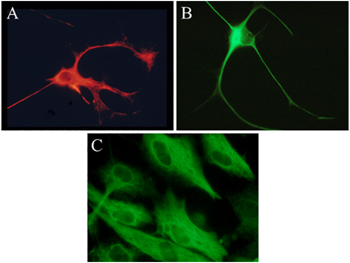
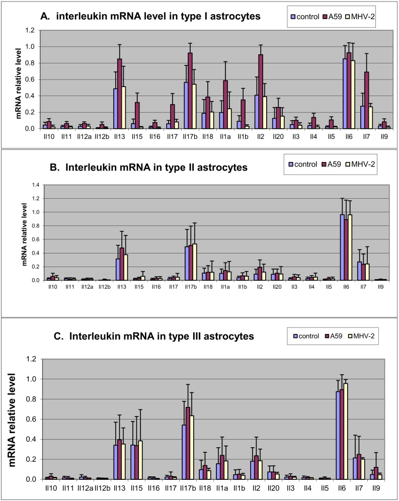
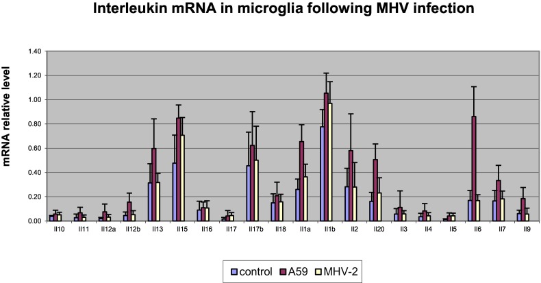
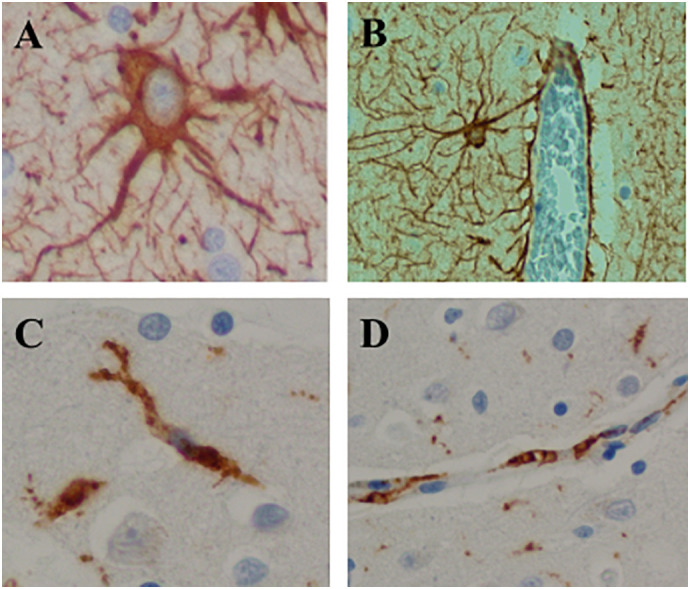
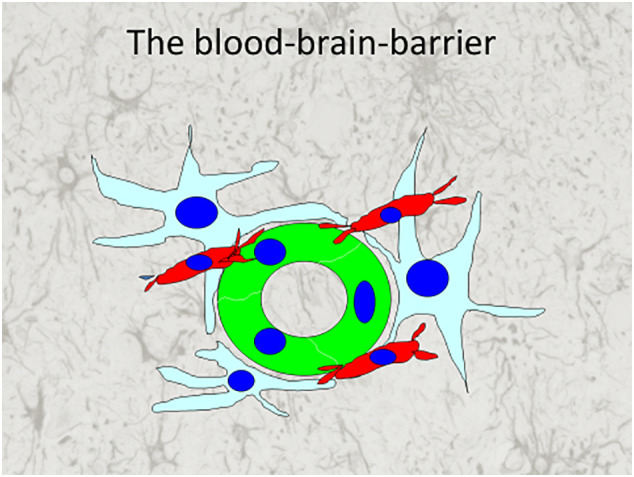
The pathogenesis of viral infections involves an immune response by cytokines, causing a deleterious effect on organ function, in addition to tissue destruction due to viral replication. Clinical symptoms and laboratory findings of the human coronavirus disease COVID-19, caused by the novel coronavirus SARS CoV-2, indicate cytokine involvement. Our laboratory showed that an experimental murine coronavirus (MHV-A59) can be transmitted into the brain by intranasal or intracerebral exposure and that neurovirulence is mediated by cytokine secretion. In this study we investigated which cells in the brain produce cytokines, thus functioning as the brain's innate immune system. Using tissue cultures of microglia, and clonal populations of astrocytes, we found that microglia and type I astrocytes (but not types II and III), produced pro-inflammatory cytokines in response to MHV-A59 infection. A molecularly closely related, non-encephalitic strain of the virus (MHV-2) caused in vitro infection, but without cytokine induction. Furthermore, immunofluorescence and immunohistochemistry revealed that type I astrocytes and microglia have perivascular foot processes necessary for the formation of the perivascular glymphatic system, the anatomical site of the brain's innate immune system. Cytokine secretion by type I astrocytes and microglia, as part of the brain's glymphatic and innate immune system, contributes to the pathogenesis of an encephalitic coronavirus infection, and indicates the rationale for anti-cytokine therapies for COVID-19.
Keywords: Astrocytes; COVID-19; Coronavirus; Cytokines; Microglia; Mouse hepatitis virus.
Copyright © 2020 Elsevier Inc. All rights reserved.
Conflict of interest statement
Declaration of Competing Interest The authors declare that there is no conflict of interest in the publication of this work.
Figures
Similar articles
-
Of Mice and Men: The Coronavirus MHV and Mouse Models as a Translational Approach to Understand SARS-CoV-2.Viruses. 2020 Aug 12;12(8):880. doi: 10.3390/v12080880. Viruses. 2020. PMID: 32806708 Free PMC article. Review.
-
Coronavirus induction of class I major histocompatibility complex expression in murine astrocytes is virus strain specific.J Exp Med. 1994 Sep 1;180(3):1013-23. doi: 10.1084/jem.180.3.1013. J Exp Med. 1994. PMID: 8064222 Free PMC article.
-
Alpha/Beta Interferon (IFN-α/β) Signaling in Astrocytes Mediates Protection against Viral Encephalomyelitis and Regulates IFN-γ-Dependent Responses.J Virol. 2018 Apr 27;92(10):e01901-17. doi: 10.1128/JVI.01901-17. Print 2018 May 15. J Virol. 2018. PMID: 29491163 Free PMC article.
-
A Biosafety Level 2 Mouse Model for Studying Betacoronavirus-Induced Acute Lung Damage and Systemic Manifestations.J Virol. 2021 Oct 27;95(22):e0127621. doi: 10.1128/JVI.01276-21. Epub 2021 Sep 8. J Virol. 2021. PMID: 34495692 Free PMC article.
-
Remodeling of the Immune Response With Aging: Immunosenescence and Its Potential Impact on COVID-19 Immune Response.Front Immunol. 2020 Aug 7;11:1748. doi: 10.3389/fimmu.2020.01748. eCollection 2020. Front Immunol. 2020. PMID: 32849623 Free PMC article. Review.
Cited by
-
Microglia Fighting for Neurological and Mental Health: On the Central Nervous System Frontline of COVID-19 Pandemic.Front Cell Neurosci. 2021 Feb 18;15:647378. doi: 10.3389/fncel.2021.647378. eCollection 2021. Front Cell Neurosci. 2021. PMID: 33737867 Free PMC article. Review.
-
Neuroinvasion and Neurotropism by SARS-CoV-2 Variants in the K18-hACE2 Mouse.Viruses. 2022 May 11;14(5):1020. doi: 10.3390/v14051020. Viruses. 2022. PMID: 35632761 Free PMC article.
-
Parallel electrophysiological abnormalities due to COVID-19 infection and to Alzheimer's disease and related dementia.Alzheimers Dement. 2024 Oct;20(10):7296-7319. doi: 10.1002/alz.14089. Epub 2024 Aug 29. Alzheimers Dement. 2024. PMID: 39206795 Free PMC article.
-
Immune Functions of Astrocytes in Viral Neuroinfections.Int J Mol Sci. 2023 Feb 9;24(4):3514. doi: 10.3390/ijms24043514. Int J Mol Sci. 2023. PMID: 36834929 Free PMC article. Review.
-
Severe Acute Respiratory Syndrome Coronavirus 2 Impact on the Central Nervous System: Are Astrocytes and Microglia Main Players or Merely Bystanders?ASN Neuro. 2020 Jan-Dec;12:1759091420954960. doi: 10.1177/1759091420954960. ASN Neuro. 2020. PMID: 32878468 Free PMC article. Review.
References
MeSH terms
Substances
LinkOut - more resources
Full Text Sources
Medical
Miscellaneous
