. 2020 May 8:2020:4260204.
doi: 10.1155/2020/4260204. eCollection 2020.

Exposure to Air Pollution Exacerbates Inflammation in Rats with Preexisting COPD

Affiliations

Exposure to Air Pollution Exacerbates Inflammation in Rats with Preexisting COPD

Jing Wang et al. Mediators Inflamm. .

Abstract

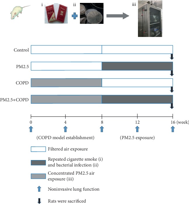
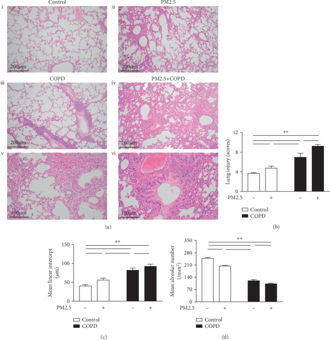
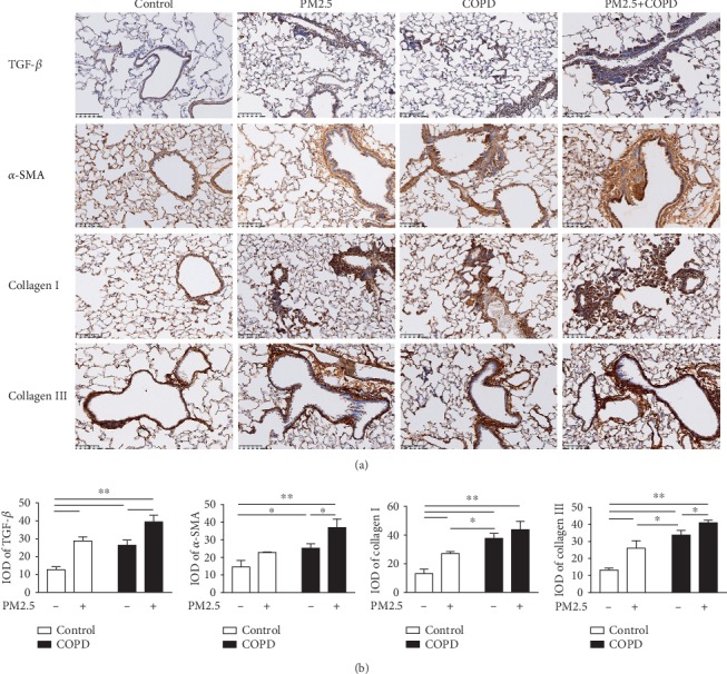
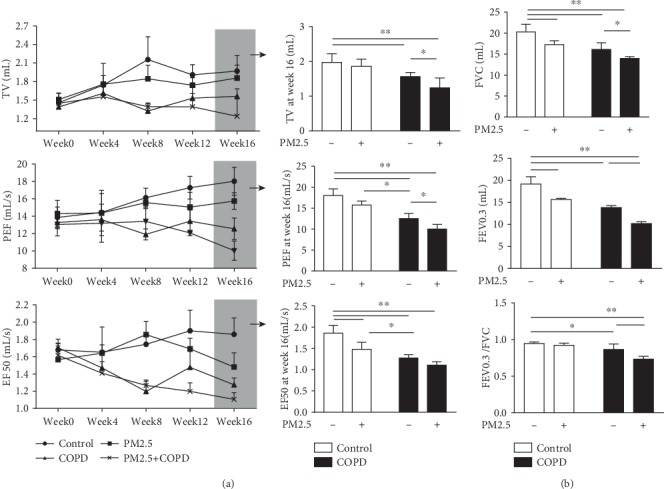
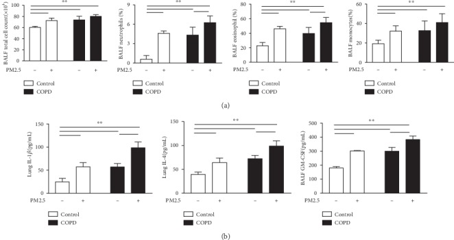
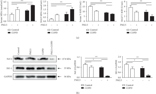
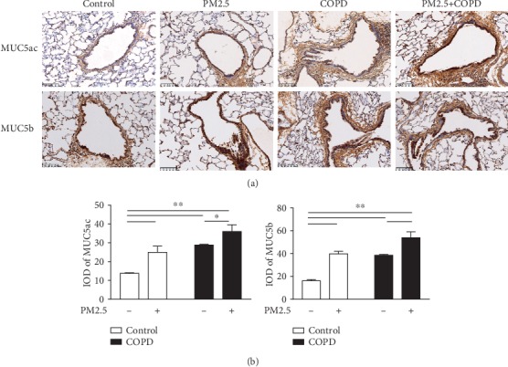
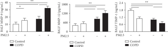
Particulate matter with an aerodynamic diameter equal or less than 2.5 micrometers (PM2.5) is associated with the development of chronic obstructive pulmonary disease (COPD). The mechanisms by which PM2.5 accelerates disease progression in COPD are poorly understood. In this study, we aimed to investigate the effect of PM2.5 on lung injury in rats with hallmark features of COPD. Cardinal features of human COPD were induced in a rat model by repeated cigarette smoke inhalation and bacterial infection for 8 weeks. Then, from week 9 to week 16, some of these rats with COPD were subjected to real-time concentrated atmospheric PM2.5. Lung function, pathology, inflammatory cytokines, oxidative stress, and mucus and collagen production were measured. As expected, the COPD rats had developed emphysema, inflammation, and deterioration in lung function. PM2.5 exposure resulted in greater lung function decline and histopathological changes, as reflected by increased Mucin (MUC) 5ac, MUC5b, Collagen I, Collagen III, and the profibrotic cytokine α-smooth muscle-actin (SMA), transforming growth factor- (TGF-) β1 in lung tissues. PM2.5 also aggravated inflammation, increasing neutrophils and eosinophils in bronchoalveolar lavage fluid (BALF) and cytokines including Interleukin- (IL-) 1β, granulocyte-macrophage colony-stimulating factor (GM-CSF), and IL-4. The likely mechanism is through oxidative stress as antioxidants levels were decreased, whereas oxidants were increased, indicating a detrimental shift in the oxidant-antioxidant balance. Altogether, these results suggest that PM2.5 exposure could promote the development of COPD by impairing lung function and exacerbating pulmonary injury, and the potential mechanisms are related to inflammatory response and oxidative stress.


Conflict of interest statement

The authors report no conflicts of interest in this work.

Figures

Figure 1
Figure 1
Experimental protocol. SD rats were divided into control, PM2.5, COPD, and PM2.5+COPD groups. During weeks 0~8, for the COPD and PM2.5+COPD groups, the COPD rat model was prepared by using repeated cigarette smoke inhalation and bacterial infection. During weeks 8~16, rats in the PM2.5 and PM2.5+COPD groups were exposed to concentrated PM2.5 atmosphere in the whole-body exposure chamber, 4 hours per day. Rats in the control group were exposed to filtered air all the time. At weeks 0, 4, 8, 12, and 16, the noninvasive lung function (TV, PEF, and EF50) were measured. All rats were sacrificed within 24 h of the last PM2.5 exposure. And lung function, pulmonary histopathology, inflammatory cytokines, oxidative stress, mucus secretion, and proteinases/antiproteinases were detected. Control: healthy rats; PM2.5: particulate matter 2.5 treatment alone rats; COPD: chronic obstructive pulmonary disease rats; PM2.5+COPD: COPD rats exposed to prolonged PM2.5 for 8 weeks. TV: tidal volume; PEF: peak expiratory flow; EF50: expiratory flow 50%.
Figure 2
Figure 2
The effect of PM2.5 on lung histological injury in rats with COPD. (a) Lung tissue HE staining of different groups of rats. (i) Control, an intact structure of pulmonary alveoli and airway (magnification, ×100); (ii) PM2.5, mild infiltration of inflammatory cells and thickening of airway wall (magnification, ×100); (iii) COPD, alveolar cavity enlargement and vascular wall thickening (magnification, ×100); (iv) PM2.5+COPD, chronic obstructive bronchiolitis with thickening of the airway wall and infiltration with inflammatory cells (magnification, ×100); (v) COPD, airway wall thickening and inflammatory cell infiltration (magnification, ×200); (vi) PM2.5+COPD, increased thickening of airway wall and infiltration of inflammatory cells compared to COPD (magnification, ×200). (b–d) Quantitative analysis of lung injury, that is, the level of (b) LIS, (c) MLI, and (d) MAN of the lungs in each group. LIS: lung injury scores; MLI: mean linear intercept; MAN: mean alveolar number. The data are expressed as the means ± SD (n = 6~7). ∗∗p < 0.01, p < 0.05.
Figure 3
Figure 3
The effect of PM2.5 on airway remodeling in rats with COPD. (a) Immunohistochemical staining of TGF-β, α-SMA, Collagen I, and Collagen III of lung sections in each group (magnification, ×200). (b) Quantitative analysis of TGF-β, α-SMA, Collagen I, and Collagen III using Image-ProPlus 6.0 software. TGF-β1: transforming growth factor-beta 1; α-SMA: α-smooth muscle-actin; IOD: integral optical density. The data are expressed as the means ± SD (n = 6). ∗∗p < 0.01, p < 0.05.
Figure 4
Figure 4
The effect of PM2.5 on pulmonary function in rats with COPD. (a) The change of noninvasive lung function parameters TV, PEF, and EF50 of rats in each group from week 0 to week 16, as well as at week 16. (b) The change of invasive lung function parameters FVC, FEV0.3, and FEV0.3/FVC of rats in each group at week 16. TV: tidal volume; PEF: peak expiratory flow; EF50: expiratory flow 50%; FVC: forced vital capacity; FEV0.3: forced expiratory volume at 0.3 s; FEV0.3/FVC: forced expiratory volume at 0.3 s/forced vital capacity. The data are expressed as the means ± SD (n = 7). ∗∗p < 0.01, p < 0.05.
Figure 5
Figure 5
The effect of PM2.5 on inflammatory response in rats with COPD. (a) The total cell count and percentage of neutrophils, eosinophils, and macrophages in the BALF of rats in each group. (b) Level of IL-1β and IL-4 in the lung and GM-CSF in the BALF of rats in each group. IL-1β: Interleukin-1β; IL-4: Interleukin-4; GM-CSF: granulocyte-macrophage colony-stimulating factor. The data are expressed as the means ± SD (n = 7). ∗∗p < 0.01, p < 0.05.
Figure 6
Figure 6
The effect of PM2.5 on oxidative stress in rats with COPD. (a) The levels of MDA and T-SOD in the serum and BALF of rats in each group. (b) Protein expression levels of Nrf-2 and HO-1 in the lung measured by Western blotting. MDA: malondialdehyde; T-SOD: total superoxide dismutase; Nrf-2: nuclear factor erythroid 2-related factor 2; HO-1: heme oxygenase-1. The data are expressed as the means ± SD (n = 3~7). ∗∗p < 0.01, p < 0.05.
Figure 7
Figure 7
The effect of PM2.5 exposure on mucus hypersecretion in rats with COPD. (a) Immunohistochemical staining of MUC5ac and MUC5b in the lung sections of each group (magnification, ×200). (b) Quantitative analysis of MUC5ac and MUC5b using Image-ProPlus 6.0 software. MUC5ac: Mucin5ac; MUC5b: Mucin5b; IOD: integral optical density. The data are expressed as the means ± SD (n = 6). ∗∗p < 0.01, p < 0.05.
Figure 8
Figure 8
The effect of PM2.5 on protease-antiprotease imbalance in rats with COPD. Level of MMP-9, MMP-12, and TIMP-1 in the BALF of rats in each group. MMP-9: matrix mettaloproteinase-9; TIMP-1: tissue inhibitors of metalloproteases-1. The data are expressed as the means ± SD (n = 7). ∗∗p < 0.01, p < 0.05.

References

    1. GBD 2017 Disease and Injury Incidence and Prevalence Collaborators. Global, regional, and national incidence, prevalence, and years lived with disability for 354 diseases and injuries for 195 countries and territories, 1990-2017: a systematic analysis for the Global Burden of Disease Study 2017. Lancet. 2018;392:1789–1858. - PMC - PubMed
    1. Fang L., Gao P., Bao H., et al. Chronic obstructive pulmonary disease in China: a nationwide prevalence study. The Lancet Respiratory Medicine. 2018;6(6):421–430. doi: 10.1016/S2213-2600(18)30103-6. - DOI - PMC - PubMed
    1. Singh D., Agusti A., Anzueto A., et al. Global strategy for the diagnosis, management, and prevention of chronic obstructive lung disease: the GOLD science committee report 2019. The European Respiratory Journal. 2019;53(5, article 1900164) doi: 10.1183/13993003.00164-2019. - DOI - PubMed
    1. Eisner M. D., Anthonisen N., Coultas D., et al. An official American Thoracic Society public policy statement: novel risk factors and the global burden of chronic obstructive pulmonary disease. American Journal of Respiratory and Critical Care Medicine. 2010;182(5):693–718. doi: 10.1164/rccm.200811-1757ST. - DOI - PubMed
    1. Denguezli M., Daldoul H., Harrabi I., et al. COPD in nonsmokers: reports from the Tunisian population-based burden of obstructive lung disease study. PLoS One. 2016;11(3, article e0151981) doi: 10.1371/journal.pone.0151981. - DOI - PMC - PubMed