. 2020 May 21;10(5):984.
doi: 10.3390/nano10050984.

Detection of Human p53 In-Vitro Expressed in a Transcription-Translation Cell-Free System by a Novel Conjugate Based on Cadmium Sulphide Nanoparticles

Affiliations

Detection of Human p53 In-Vitro Expressed in a Transcription-Translation Cell-Free System by a Novel Conjugate Based on Cadmium Sulphide Nanoparticles

Víctor Barba-Vicente et al. Nanomaterials (Basel). .

Abstract

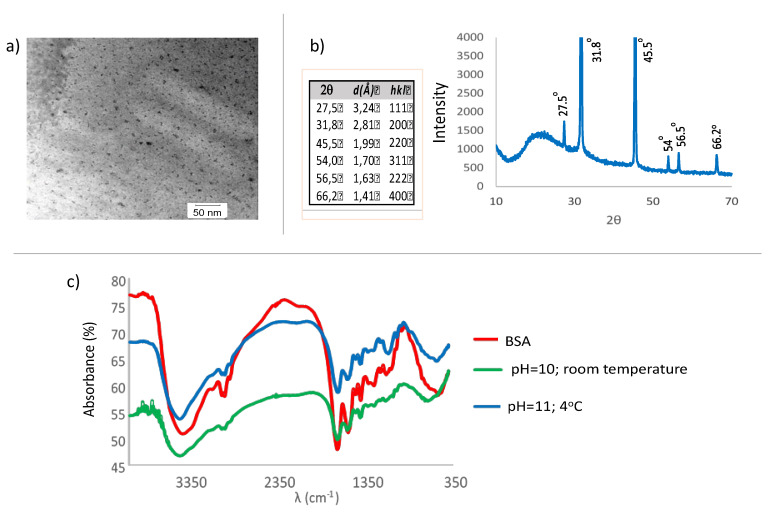
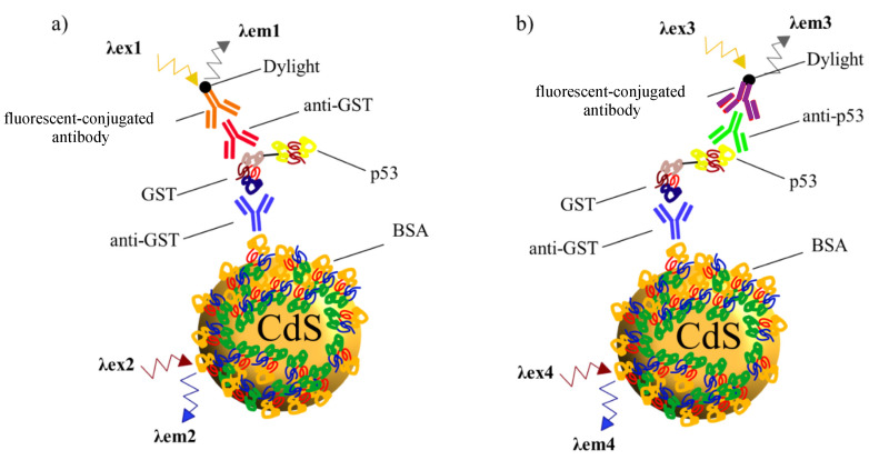
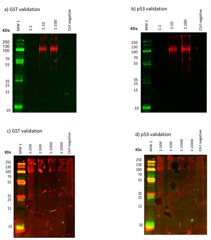
Here, cadmium sulphide quantum dots (CdS QDs) have been synthetized and functionalized with Bovine Serum Albumin (BSA) in a colloidal aqueous solution with a stability of over 3 months. Specific synthesis conditions, in homogeneous phase and at low temperature, have allowed limitation of S2- concentration, hence, as a consequence, there is restricted growth of the nanoparticles (NPs). This fact allows binding with BSA in the most favorable manner for the biomolecule. The presence of Cd2+ ions on the surface of the CdS nanoparticle is counteracted by the negatively charged domains of the BSA, resulting in the formation of small NPs, with little tendency for aggregation. Temperature and pH have great influence on the fluorescence characteristics of the synthetized nanoparticles. Working at low temperatures (4 °C) and pH 10-11 have proven the best result as shown by hydrolysis kinetic control of the thioacetamide precursor of S2- ion. Biological activity of the coupled BSA is maintained allowing subsequent bioconjugation with other biomolecules such as antibodies. The chemical conjugation with anti-Glutathione S-transferase (α-GST) antibody, a common tag employed in human recombinant fusion proteins, produces a strong quenching of fluorescence that proves the possibilities of its use in biological labelling. Finally, p53, onco-human recombinant protein (GST tagged in COOH terminus), has been in situ IVTT (in vitro transcription-translation) expressed and efficiently captured by the α-GST-CdS QD conjugate as a proof of the biocompatibility on IVTT systems and the functionality of conjugated antibody.

Keywords: CdS-BSA quantum dots; IVTT protein expression; bovine serum albumin; fluorescent immunoassays; nanoparticles; protein detection; synthesis.


Conflict of interest statement

The authors declare no conflict of interest.

Figures

Figure 1
Figure 1
Absorption spectrum of the cadmium sulphide-Bovine Serum Albumin (BSA) quantum dots (CdS BSA QDs) 3 days after the synthesis process: pH = 10; Temperature: 22 °C (“A” is absorbance and “d” is diameter).
Figure 2
Figure 2
Photoluminescence behavior of the CdS-BSA QDs 1 day after synthesis under the general conditions described. Excitation and emission slit widths Rex= Rem = 10 nm. (a) Excitation spectrum at λem 496.0 nm. (b) Emission spectrum at λex 369.8 nm.
Figure 3
Figure 3
Influence of the change in pH during synthesis. (a) Evolution of the maximum absorbance values at different times for different pH values. (b) Temporal evolution of the Photoluminescence intensity at different pH values. Under operating conditions the final concentration and their molar ratios were as follows: [Cd2+]F = 7.477 × 10⁻⁴ mol/L; [BSA]F = 3.738 × 10⁻⁴ mol/L; [thioacetamide]F = 3.738 × 10⁻⁴ mol/L; [Cd2+]F/[thioacetamide]F = 2.0; [Cd2+]F/[BSA]F = 2.0, Room temperature.
Figure 4
Figure 4
Influence of the change in pH values during synthesis at low temperatures. (a) Evolution of the absorbance values of the nanoparticles at different times for different pH values. (b) Temporal evolution of the Photoluminescence intensity at different pH values. Under operating conditions the final concentration and their molar ratios were as follows: [Cd2+]F = 7.477 × 10⁻⁴ mol/L; [BSA]F = 3.738 × 10⁻⁴ mol/L; [thioacetamide]F = 3.738 × 10⁻⁴ mol/L; [Cd2+]F/[thioacetamide]F = 2.0; [Cd2+]F/[BSA]F = 2.0, 4 °C.
Figure 5
Figure 5
Influence of [Cd2+]F/[thioacetamide]F at 4 °C. Temporal evolution of Photoluminescence intensity of CdS QDs solutions with different [Cd2+]F/[thioacetamide]F ratios; (a) 4.0, (b) 2.0, (c) 0.8, (d) 0.4.
Figure 6
Figure 6
Influence of [Cd2+]F/[BSA]F at 4 °C. Temporal evolution of Photoluminescence intensity of CdS QDs solutions with different [Cd2+]F/[BSA]F ratios.
Figure 7
Figure 7
(a) Transmission electron microscopy (TEM) image of the CdS nanoparticles; (b) 1 indices hkl for the zinc blende structure for the CdS. Powder X-ray diffraction (XRD) pattern of the CdS particles; (c) Infrared spectra of the CdS nanoparticles. FT-IR spectra (Fourier Transform InfraRed spectra) of BSA and of CdS QDs prepared at pH 10 (room temperature) and at pH 11 (4 °C).
Figure 8
Figure 8
CdS-BSA-α-GST interaction. Variation in fluorescence for the different antibody concentrations studied. Concentration of CdS-BSA: 2.7 × 10⁻⁶ mol/L. Concentration of α-GST: Range from 7.7 × 10⁻⁸ mol/L to 3.1 × 10⁻⁷ mol/L.
Figure 9
Figure 9
Scheme of detection of in vitro human cell- free transcription-translation (IVTT) expressed human recombinant p53 GST tagged protein. Western Blot detection carried in our study based on novel NPs (CdS-BSA). In both cases, antibody against GST conjugated with the novel QDs (blue), catch the GST domain of the IVTT protein. (a) When the goal is detecting the GST domain, another α-GST antibody is used (red) and final detection with a fluorescence dye labeled antibody (orange). (b) For specific recognition of the p53 human recombinant protein, an α-p53 antibody is used (green) and detected with a fluorescence dye labeled antibody (purple).
Figure 10
Figure 10
Detection of the GST protein label and the protein under study, p53, after expression of the recombinant protein p53-GST in an in vitro (cell-free) system in the presence of QDs-BSA-α-GST by Western Blot. (In each gel image, lane 1 corresponds to the molecular weight markers, indicating the values (KDa) in the left column; meanwhile the last lane corresponds to negative control). (a) Western Blot analysis of IVTT mix with cDNA 1 µg and 5 µL of IVTT reagents (dilution 1:1 at 1:100 v/v) for the detection of GST at 480 nm. (b) Western Blot analysis IVTT mix with cDNA 1 µg and 5 µL of IVTT reagents (dilution 1:1 at 1:100 v/v) for detection of p53 at 480 nm. (c) Western Blot analysis IVTT mix with cDNA 1 µg and 25 µL of IVTT reagents (dilution 1:200 to 1:2.000 v/v) for the detection of GST at 680 nm. (d) Western Blot analysis IVTT mix with cDNA 1 µg and 25 µL of IVTT reagents (dilution 1:200 to 1:2000 v/v) for the detection of p53 at 680 nm.
Figure 11
Figure 11
Schematic representation of super-paramagnetic microsphere (black) interaction by a glutathione group (brown), with the active site of the GST enzyme, expressed by IVTT system. New synthetized protein (GST-tagged) is caught by the α-GST antibody (blue) previously conjugated to the novel QD.
Figure 12
Figure 12
Fluorescence emission spectra at λex = 370 nm, (a) microspheres (b) QDs-BSA-α-GST and microspheres, (c) QDs-BSA-α-GST with expression of p53 and microspheres.

References

    1. Kango S., Kalia S., Celli A., Njuguna J., Habibi Y., Kumar R. Surface modification of inorganic nanoparticles for development of organic–inorganic nanocomposites—A review. Prog. Polym. Sci. 2013;38:1232–1261. doi: 10.1016/j.progpolymsci.2013.02.003. - DOI
    1. Katz E., Willner I. Integrated nanoparticle—Biomolecule hybrid systems: Synthesis, properties, and applications. Angew. Chem. Int. Ed. 2004;43:6042–6108. doi: 10.1002/anie.200400651. - DOI - PubMed
    1. Karakoti A.S., Shukla R., Shanker R., Singh S. Surface functionalization of quantum dots for biological applications. Adv. Colloid Interface Sci. 2015;215:28–45. doi: 10.1016/j.cis.2014.11.004. - DOI - PubMed
    1. Bruchez M., Moronne M., Gin P., Weiss S., Alivisatos A.P. Semiconductor nanocrystals as fluorescent biological labels. Science. 1998;281:2013–2016. doi: 10.1126/science.281.5385.2013. - DOI - PubMed
    1. Chan W.C., Nie S. Quantum dot bioconjugates for ultrasensitive nonisotopic detection. Science. 1998;281:2016–2018. doi: 10.1126/science.281.5385.2016. - DOI - PubMed