. 2020 May 21;25(10):2396.
doi: 10.3390/molecules25102396.

Pretreatment of Anthocyanin from the Fruit of Vitis coignetiae Pulliat Acts as a Potent Inhibitor of TNF-α Effect by Inhibiting NF-κB-Regulated Genes in Human Breast Cancer Cells

Affiliations

Pretreatment of Anthocyanin from the Fruit of Vitis coignetiae Pulliat Acts as a Potent Inhibitor of TNF-α Effect by Inhibiting NF-κB-Regulated Genes in Human Breast Cancer Cells

Anjugam Paramanantham et al. Molecules. .

Abstract

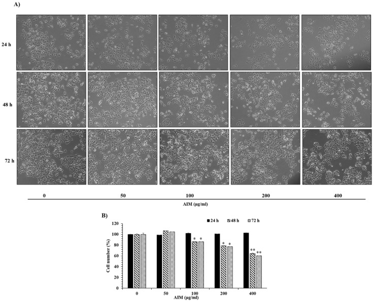
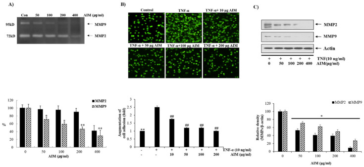
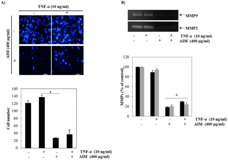
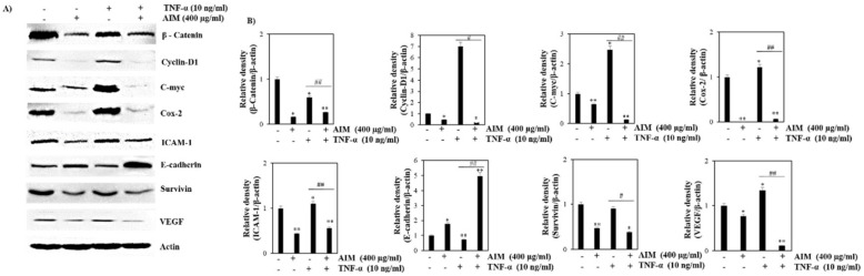
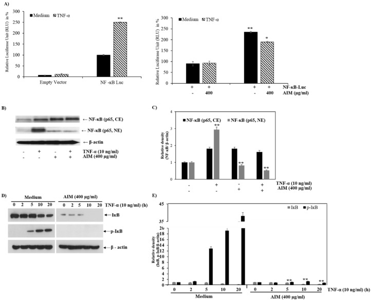
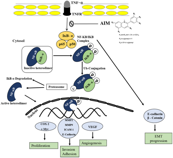
Vitis coignetiae Pulliat (Meoru in Korea) has been used in Korean folk medicine for the treatment of inflammatory diseases and cancers. Evidence suggests that NF-κB activation is mainly involved in cancer cell proliferation, invasion, angiogenesis, and metastasis. TNF-α also enhances the inflammatory process in tumor development. Recently, flavonoids from plants have been reported to have inhibitory effects on NF-κB activities. We investigated the effects of anthocyanins extracted from the fruits of Vitis coignetiae Pulliat (AIM, anthocyanins isolated from Meoru (AIM)) on TNF-α-induced NF-κB activities in MCF-7 human breast cancer cells and the molecules involved in AIM-induced anti-cancer effects, especially on cancer metastasis. We performed cell viability assay, gelatin zymography, invasion assay, and western blot analysis to unravel the anti-NF-κB activity of AIMs on MCF-7 cells. AIM suppressed the TNF-α effects on the NF-κB-regulated proteins involved in cancer cell proliferation (COX-2, C-myc), invasion, and angiogenesis (MMP-2, MMP9, ICAM-1, and VEGF). AIM also increased the expression of E-cadherin, which is one of the hallmarks of the epithelial-mesenchymal transition (EMT) process. In conclusion, this study demonstrates that the anthocyanins isolated from the fruits of Vitis coignetiae Pulliat acts as an inhibitor of TNF-α induced NF-κB activation, and subsequent downstream molecules involved in cancer proliferation, invasion, adhesion, angiogenesis, and thus have anti-metastatic activities in MCF-7 breast cancer cells.

Keywords: NF-κB; Vitis coignetiae Pulliat; anthocyanins; breast cancer.


Conflict of interest statement

The authors declare no conflicts of interest.

Figures

Figure 1
Figure 1
The inhibitory effects of anthocyanins isolated from Meoru (AIM) on cancer cell proliferation of MCF-7 breast cancer cells. (A) Morphological representation of MCF-7 cells with AIM treatment at different concentrations (0, 50, 100, 200, and 400 μg/mL) and time points (24 h, 48 h, and 72 h) were observed under light microscope (magnification, ×200; the length of scale bar, 50 μm). (B) Dose-dependent inhibitory effects of AIM on cell proliferation were assessed by the MTT assay. Cells were treated with indicated concentrations of AIM (0, 50, 100, 200, and 400 μg/mL) for 24 h, 48 h, and 72 h. Data are mean ±SD values from three independent experiments. * p < 0.05, ** p < 0.01 versus control.
Figure 2
Figure 2
Inhibitory effects of AIM on cancer cell adhesion of MCF-7 Cells. (A) The cells were grown in 6-well plates with the seeding density of 5 × 10⁴ cells/well. MMP-2 and MMP-9 levels in gelatin zymography were assessed by densitometry. (Upper) The cells were treated with and without AIM for 24 h and the conditioned medium was taken to perform gelatin zymography. (Lower) MMP-2 and MMP-9 are expressed as a percentage of the activities against untreated cells. (B) The cells were seeded at the density of 5 × 10⁴ cells/mL. The cells were treated with indicated concentrations of AIM (0, 50, 100, and 200 μg/mL) for 24 h and the AIM effect on cancer cell invasion was assessed. For the group treated with AIM and TNF-α, cells were pre-treated with AIM (0, 10, 50, 100, and 200 μg/mL) for 1 h and then treated with TNF-α (10 ng/mL). ** p < 0.01 vs. the control group, ## p < 0.01 vs. the TNF-treated group. (C) Cells (5 × 10⁴ cells), either left untreated or pre-treated with AIM for 1 h, were exposed to TNF-α (10 ng/mL) for 24 h. (Upper) 30 μg of whole cell protein lysate were used for western blot analysis using antibodies of MMP-2 and MMP9. (Lower) Densitometry analysis of the data in western blot analysis by ImageJ software. The values were normalized against β-actin. ** p < 0.01 vs. the control group.
Figure 3
Figure 3
Inhibitory effects of AIM on the expression of TNF-treated MMP-2 and MMP-9 in MCF-7 cells. (A) AIM greatly influenced TNF-α -stimulated cell invasion of MCF-7 cells. (Upper) The Transwell invasion assay was performed using a Boyden chamber treated with or without TNF-α in the presence of AIM (400 μg/mL) at 37 °C for 24 h in a CO2 incubator. (Lower) Graphical representation of the number of invasive cells present were expressed in percentage. (B) MCF-7 cells were treated with TNF-α. (Upper) MMP-2 and MMP-9 protein secreted in the treated medium was used for the gelatin zymography analysis. (Lower) MMP-2 and MMP-9 enzyme activities were expressed in percentage and compared against the untreated cells. Data are the mean ± SD values of three independent experiments. * p < 0.05 versus the control.
Figure 4
Figure 4
Effects of AIM and TNF-α induced AIM on NF-κB-regulated proteins involved in proliferation, invasion, and angiogenesis. MCF-7 cells were seeded with the seeding density of 5 × 10⁴ cells and pretreated AIM (400 μg/ml) for 1 h, followed by the treatment of TNF-α (10 ng/mL) for 24 h. The control cells were left untreated. The whole cell protein lysate was prepared and 30 μg of proteins were resolved in SDS-Polyacrylamide gels. (A) Western blot analysis of various NF-κB related proteins involved in cancer cell proliferation, invasion, and angiogenesis, (B) Densitometry analysis of the data in western blot analysis by ImageJ software. The values were normalized against β-actin. * p < 0.05, ** p < 0.01 vs. the control group, # p < 0.05, ## p < 0.01 vs. the TNF-α treated group.
Figure 5
Figure 5
Effects of AIM on NF-κB and the IκBα phosphorylation. (A, Left) 1 μg of the NF-κB-luciferase empty vector were transfected into MCF-7 cells; (right) 24 h after transfection, the cells were treated with 10 ng/mL of TNF-α, with or without 1 h pre-treatment of AIM (400 μg/mL). The luciferase activity was represented in fold change and compared against the untreated group. (B) Repressive effects of AIM on TNF-α-induced NF-κB translocation from the cytoplasm to nucleus. The cells treated with 10 ng/mL of TNF-α pretreated with or without 400 μg/mL of AIM for 1 h were taken for western blot analysis. Nuclear (NE) and cytoplasmic extracts (CE) isolated from the total lysates were resolved in SDS-polyacrylamide gels. (C) Densitometry analysis of the data in western blot analysis by ImageJ software. The values were normalized against β-actin. (D) Inhibitory effects of AIM on TNF-α induced IκBα phosphorylation. A total of 30 μg of protein lysates from the indicated treatment cells were used for western blot analysis. (E) Densitometry analysis of the data in western blot analysis by ImageJ software. The values were normalized against β-actin. ** p < 0.01 vs. the control group.
Figure 6
Figure 6
Schematic representation of AIM on TNF-α induced effect on MCF-7 breast cancer cells. Following activation of the NF-κB pathways, AIM inhibited TNF-α-induced NF-κB activities and NF-κB-regulated proteins involved in cancer cell proliferation, invasion, and angiogenesis. Taken together, these data suggest that AIM may act as a TNF-α inhibitor by suppressing NF-κB pathways on human breast cancer.

References

    1. Bray F., Ferlay J., Soerjomataram I., Siegel R.L., Torre L.A., Jemal A. Global cancer statistics 2018: GLOBOCAN estimates of incidence and mortality worldwide for 36 cancers in 185 countries. Ca: A Cancer J. Clin. 2018;68:394–424. doi: 10.3322/caac.21492. - DOI - PubMed
    1. Comsa S., Cimpean A.M., Raica M. The story of MCF-7 breast cancer cell line: 40 years of experience in research. Anticancer Res. 2015;35:3147–3154. - PubMed
    1. Lacroix M. Significance, detection and markers of disseminated breast cancer cells. Endocr. Relat. Cancer. 2006;13:1033–1067. doi: 10.1677/ERC-06-0001. - DOI - PubMed
    1. Yilmaz M., Christofori G., Lehembre F. Distinct mechanisms of tumor invasion and metastasis. Trends Mol. Med. 2007;13:535–541. doi: 10.1016/j.molmed.2007.10.004. - DOI - PubMed
    1. Syed D.N., Afaq F., Sarfaraz S., Khan N., Kedlaya R., Setaluri V., Mukhtar H. Delphinidin inhibits cell proliferation and invasion via modulation of Met receptor phosphorylation. Toxicol. Appl. Pharm. 2008;231:52–60. doi: 10.1016/j.taap.2008.03.023. - DOI - PMC - PubMed

MeSH terms