Review
. 2020 May 21;9(5):1562.
doi: 10.3390/jcm9051562.

Heme Oxygenase-1 in Central Nervous System Malignancies

Affiliations
Review

Heme Oxygenase-1 in Central Nervous System Malignancies

Giuseppe Sferrazzo et al. J Clin Med. .

Abstract

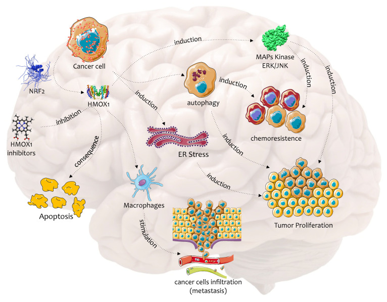
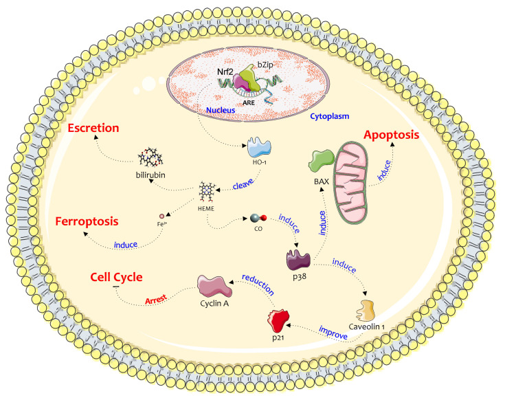
Central nervous system tumors are the most common pediatric solid tumors and account for 20%-25% of all childhood malignancies. Several lines of evidence suggest that brain tumors show altered redox homeostasis that triggers the activation of various survival pathways, leading to disease progression and chemoresistance. Among these pathways, heme oxygenase-1 (HO-1) plays an important role. HO-1 catalyzes the enzymatic degradation of heme with the simultaneous release of carbon monoxide (CO), ferrous iron (Fe2+), and biliverdin. The biological effects of HO-1 in tumor cells have been shown to be cell-specific since, in some tumors, its upregulation promotes cell cycle arrest and cellular death, whereas, in other neoplasms, it is associated with tumor survival and progression. This review focuses on the role of HO-1 in central nervous system malignancies and the possibility of exploiting such a target to improve the outcome of well-established therapeutic regimens. Finally, several studies show that HO-1 overexpression is involved in the development and resistance of brain tumors to chemotherapy and radiotherapy, suggesting the use of HO-1 as an innovative therapeutic target to overcome drug resistance. The following keywords were used to search the literature related to this topic: nuclear factor erythroid 2 p45-related factor 2, heme oxygenase, neuroblastoma, medulloblastoma, meningioma, astrocytoma, oligodendroglioma, glioblastoma multiforme, and gliomas.

Keywords: NRF2; ROS; brain cancer; nuclear factor erythroid 2 p45-related factor 2; oxidative stress.


Conflict of interest statement

The authors declare no conflict of interest.

Figures

Figure 1
Figure 1
Biochemical pathways of NRF2 activation in cancer cells. Elevated metabolic rate is responsible for increased ROS production that induces Nrf2 release from Keap1 and its nuclear translocation in cancer cells. NRF2 in the nucleus binds to antioxidant response elements (ARE) and induces the expression of several antioxidant and detoxifying enzymes, such as γ-glutamate-cysteine ligase (γ-GCL), glutathione peroxidase (GPx), glutathione reductase (GR), NAD(P)H quinone dehydrogenase 1 (NQO1) and heme oxygenase-1 (HO-1). This figure was drawn using the software CorelDraw and the vector image bank of Servier Medical Art (http://smart.servier.com/). Servier Medical Art by Servier is licensed under a Creative Commons Attribution 3.0 Unported License (https://creativecommons.org/licenses/by/3.0/).
Figure 2
Figure 2
Biochemical pathway of HO-1 in the chemoresistance and progression of brain cancer. This figure was drawn using the software CorelDraw and the vector image bank of Servier Medical Art (http://smart.servier.com/). Servier Medical Art by Servier is licensed under a Creative Commons Attribution 3.0 Unported License (https://creativecommons.org/licenses/by/3.0/).
Figure 3
Figure 3
A possible anticancer pathway of HO-1 in neuroblastoma cells. The figure shows different pathways induced by NRF2 action on HO-1. All pathways converge through the stress of other factors, such as the splitting of the heme group and the release of Fe + ions, the arrest of the cell cycle with induction of p21 or through BAX and classic apoptosis. This figure was drawn using the software CorelDraw and the vector image bank of Servier Medical Art (http://smart.servier.com/). Servier Medical Art by Servier is licensed under a Creative Commons Attribution 3.0 Unported License (https://creativecommons.org/licenses/by/3.0/).

References

    1. Ostrom Q.T., Gittleman H., Truitt G., Boscia A., Kruchko C., Barnholtz-Sloan J.S. CBTRUS Statistical Report: Primary Brain and Other Central Nervous System Tumors Diagnosed in the United States in 2011–2015. Neuro Oncol. 2018;20:iv1–iv86. doi: 10.1093/neuonc/noy131. - DOI - PMC - PubMed
    1. Jensen O.M., Estève J., Møller H., Renard H. Cancer in the European Community and its member states. Eur. J. Cancer. 1990;26:1167–1256. doi: 10.1016/0277-5379(90)90278-2. - DOI - PubMed
    1. DeBerardinis R.J., Chandel N.S. Fundamentals of cancer metabolism. Sci. Adv. 2016;2:e1600200. doi: 10.1126/sciadv.1600200. - DOI - PMC - PubMed
    1. Reuter S., Gupta S.C., Chaturvedi M.M., Aggarwal B.B. Oxidative stress, inflammation, and cancer: How are they linked? Free Radic. Biol. Med. 2010;49:1603–1616. doi: 10.1016/j.freeradbiomed.2010.09.006. - DOI - PMC - PubMed
    1. Kensler T.W., Wakabayashi N., Biswal S. Cell survival responses to environmental stresses via the Keap1-Nrf2-ARE pathway. Annu. Rev. Pharmacol. Toxicol. 2007;47:89–116. doi: 10.1146/annurev.pharmtox.46.120604.141046. - DOI - PubMed

LinkOut - more resources