. 2020 May 22;9(5):1290.
doi: 10.3390/cells9051290.

Central Modulation of Selective Sphingosine-1-Phosphate Receptor 1 Ameliorates Experimental Multiple Sclerosis

Affiliations

Central Modulation of Selective Sphingosine-1-Phosphate Receptor 1 Ameliorates Experimental Multiple Sclerosis

Alessandra Musella et al. Cells. .

Abstract

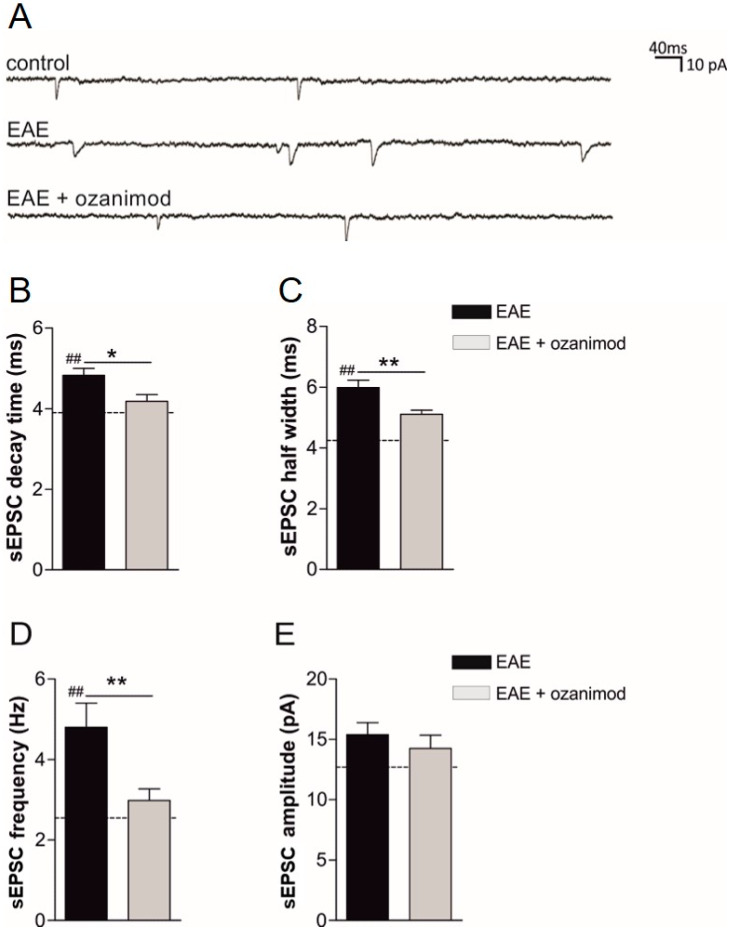
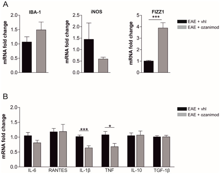
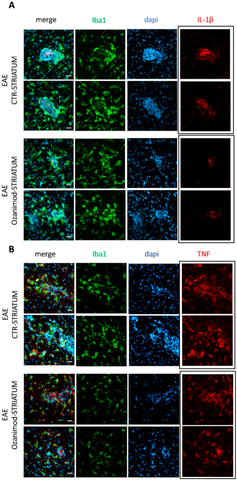
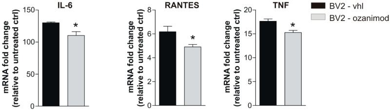
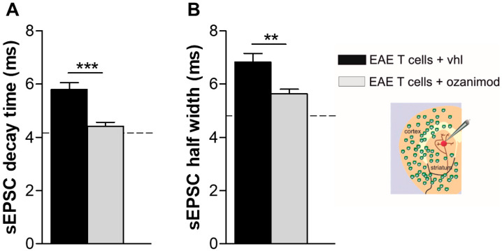
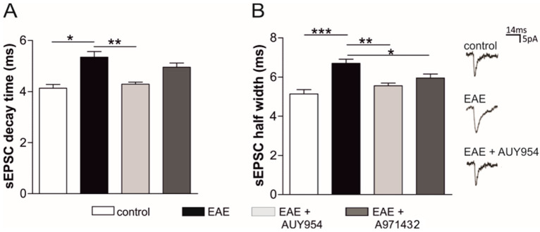
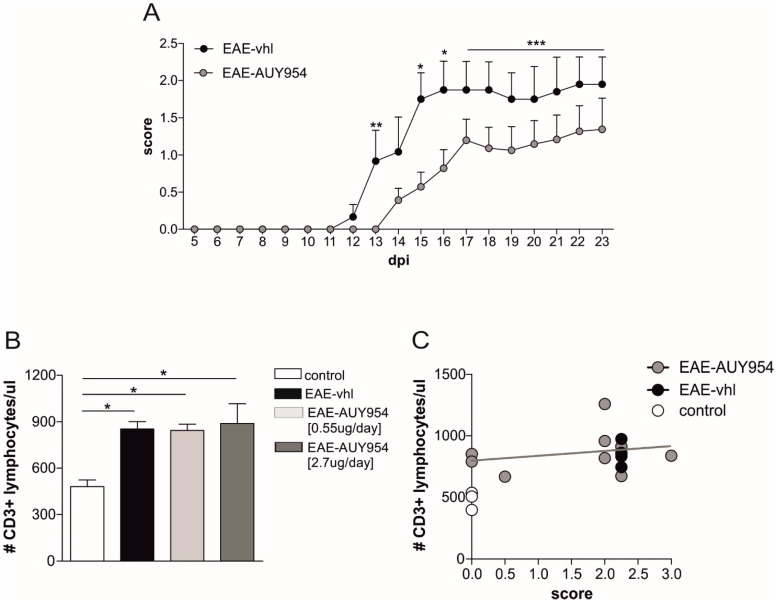
Future treatments of multiple sclerosis (MS), a chronic autoimmune neurodegenerative disease of the central nervous system (CNS), aim for simultaneous early targeting of peripheral immune function and neuroinflammation. Sphingosine-1-phosphate (S1P) receptor modulators are among the most promising drugs with both "immunological" and "non-immunological" actions. Selective S1P receptor modulators have been recently approved for MS and shown clinical efficacy in its mouse model, the experimental autoimmune encephalomyelitis (EAE). Here, we investigated the anti-inflammatory/neuroprotective effects of ozanimod (RPC1063), a S1P1/5 modulator recently approved in the United States for the treatment of MS, by performing ex vivo studies in EAE brain. Electrophysiological experiments, supported by molecular and immunofluorescence analysis, revealed that ozanimod was able to dampen the EAE glutamatergic synaptic alterations, through attenuation of local inflammatory response driven by activated microglia and infiltrating T cells, the main CNS-cellular players of EAE synaptopathy. Electrophysiological studies with selective S1P1 (AUY954) and S1P5 (A971432) agonists suggested that S1P1 modulation is the main driver of the anti-excitotoxic activity mediated by ozanimod. Accordingly, in vivo intra-cerebroventricular treatment of EAE mice with AUY954 ameliorated clinical disability. Altogether these results strengthened the relevance of S1P1 agonists as immunomodulatory and neuroprotective drugs for MS therapy.

Keywords: A971432; AUY954; S1P1; S1P5; T lymphocytes; experimental autoimmune encephalomyelitis (EAE); glutamate synaptic dysfunction; microglia; neuroinflammation; ozanimod; pro-inflammatory cytokines; sphingosine-1-phosphate receptors.


Conflict of interest statement

DC is the recipient of an Institutional grant from Celgene. No personal compensation was received. The founding sponsors had no role in the design of the study; in the collection, analyses, or interpretation of data; in the writing of the manuscript, and in the decision to publish the results. The other authors declare that they have no competing interests.

Figures

Figure 1
Figure 1
Ex vivo treatment of corticostriatal slices with ozanimod recovers synaptic alterations induced by experimental autoimmune encephalomyelitis (EAE). (A) Examples of spontaneous glutamate-mediated excitatory postsynaptic currents (sEPSC) traces recorded from medium spiny neurons (MSNs) in corticostriatal slices in the different experimental conditions (healthy control, EAE, EAE + ozanimod). Bath incubation corticostriatal slices with ozanimod (1000 nM, 2 h) recovers EAE-induced alterations of glutamatergic transmission, in terms of decay time (B), half width (C), and frequency (D). sEPSC amplitude is not affected by ozanimod treatment (E). Dotted lines refer to healthy mouse values. Data are expressed as mean ± SEM. Unpaired t-test, * p < 0.05; ** p < 0.01 EAE vs. EAE + ozanimod; ## p < 0.01 EAE vs. healthy mice.
Figure 2
Figure 2
Ex vivo treatment of EAE corticostriatal slices with ozanimod modulates inflammatory markers related to microglial activation and lowers IL-1β and TNF mRNA levels. (A) qPCR quantification of microglial markers from EAE striatal slices incubated with ozanimod (1000 nM,2 h) shows a significant upregulation of the M2 marker FIZZ1 (resistin like alpha, Retnla) without changing the expression of iNOS (inducible nitric oxide synthetase) and IBA1 (binding adaptor molecule 1). (B) qPCR quantification of cytokines from EAE striatal slices incubated with ozanimod (1000 nM, 2 h) shows a significant downregulation of IL-1β and TNF mRNAs. All data are expressed as mean ± SEM and as fold change of EAE vehicle samples. Unpaired t-test, * p < 0.05; *** p < 0.001.
Figure 3
Figure 3
Ozanimod attenuates IL-1β immunolabelling with negligible effect on TNF in EAE striatal slices. Confocal images of EAE striatal slices incubated with vehicle or ozanimod (1000 nM, 2 h) stained for IBA1 (green), cell nuclei (cyan), IL-1 β (red in A), and TNF (red in B) show that ozanimod treatment leads to a significantly milder expression of IL-1 β within lesioned area, highlighted by IBA1 and dapi staining, with minor effect on TNF. Scale bars: 20 µm.
Figure 4
Figure 4
In vitro treatment of BV2 cell line with ozanimod modulates mRNA levels of pro-inflammatory cytokines. qPCR experiments performed on BV2 microglial cells activated by Th1 Mix for 2 h and incubated with ozanimod (1000 nM, 1 h pretreatment) or vehicle (DMSO) show a downregulation of IL-6, TNF and RANTES mRNAs. All data are expressed as mean ± SEM and as fold change of untreated controls. Unpaired t-test, * p < 0.05.
Figure 5
Figure 5
In vitro treatment of EAE T cells with ozanimod abolishes T cell synaptotoxicity. (A,B) The enhancement of sEPSC decay time (A) and sEPSC half width (B),typically induced by EAE lymphocytes, was significantly reduced by in vitro treatment of EAE T cells with ozanimod (1000 nM). Dotted lines refer to control condition (control T cells). Data are presented as ± SEM. Unpaired t-test, ** p < 0.01; *** p < 0.001.
Figure 6
Figure 6
Ex vivo treatment with S1P1 and S1P5 agonists ameliorates glutamatergic dysfunction in EAE striatal slices. Whole-cell patch clamp recordings from MSNs show that the kinetics of the glutamatergic currents, decay time (A) and half width (B), were increased in EAE striatum and were completely rescued after 2 h incubation with AUY954 (300 nM, S1P1 specific). A971432 (200 nM, S1P5 specific) only partially rescued the EAE sEPSC kinetics. On the right, representative peaks of electrophysiological recordings in the different experimental conditions are shown. Data are presented as mean ± SEM. ANOVA, * p < 0.05; ** p < 0.01; *** p < 0.001.
Figure 7
Figure 7
AUY954 icv treatment ameliorates EAE clinical disability without affecting T cell absolute count. (A) The graph shows representative clinical course of EAE mice treated for four weeks with two different AUY954 dosages (2.7 µg/day and 0.55 µg/day) or vehicle, preventively delivered by icv infusion. AUY954 icv treatment significantly ameliorated EAE disease progression. Mann-Whitney test on day 13, 15, 16, and from 17 to 23 days post immunization (dpi) by cumulating the data, * p < 0.05; ** p < 0.01; *** p < 0.001). (B) CD3+ lymphocytes were counted in the peripheral blood of EAE mice (21 dpi) receiving vehicle (vhl) or different AUY954 dosages, 2.7 µg/day and 0.55 µg/day. No significant reduction was observed at any dosage in comparison to EAE-vhl mice. Conversely, T cell count was significantly less in healthy mice in comparison to other EAE groups. Data are presented as mean ± SEM; ANOVA Tukey’s p < 0.05. (C) Statistical analysis between clinical score and T cell count shows the lack of correlation between these two parameters in AUY954-EAE mice (r2 = 0.06).

Similar articles

Cited by

References

    1. Frischer J.M., Bramow S., Dal-Bianco A., Lucchinetti C.F., Rauschka H., Schmidbauer M., Laursen H., Sorensen P.S., Lassmann H. The relation between inflammation and neurodegeneration in multiple sclerosis brains. Brain. 2009;132:1175–1189. doi: 10.1093/brain/awp070. - DOI - PMC - PubMed
    1. Centonze D., Muzio L., Rossi S., Furlan R., Bernardi G., Martino G. The link between inflammation, synaptic transmission and neurodegeneration in multiple sclerosis. Cell Death Differ. 2010;17:1083–1091. doi: 10.1038/cdd.2009.179. - DOI - PubMed
    1. Rossi S., Furlan R., De Chiara V., Motta C., Studer V., Mori F., Musella A., Bergami A., Muzio L., Bernardi G., et al. Interleukin-1β causes synaptic hyperexcitability in multiple sclerosis. Ann. Neurol. 2012;71:76–83. doi: 10.1002/ana.22512. - DOI - PubMed
    1. Reich D.S., Lucchinetti C.F., Calabresi P.A. Multiple Sclerosis. N. Engl. J. Med. 2018;378:169–180. doi: 10.1056/NEJMra1401483. - DOI - PMC - PubMed
    1. Mandolesi G., Gentile A., Musella A., Fresegna D., De Vito F., Bullitta S., Sepman H., Marfia G.A., Centonze D. Synaptopathy connects inflammation and neurodegeneration in multiple sclerosis. Nat. Rev. Neurol. 2015;11:711. doi: 10.1038/nrneurol.2015.222. - DOI - PubMed

Publication types

MeSH terms