Review
. 2020 May 22;8(5):130.
doi: 10.3390/biomedicines8050130.

Spinal Cord Involvement in MS and Other Demyelinating Diseases

Affiliations
Review

Spinal Cord Involvement in MS and Other Demyelinating Diseases

Mariano Marrodan et al. Biomedicines. .

Abstract

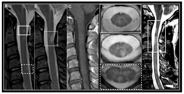
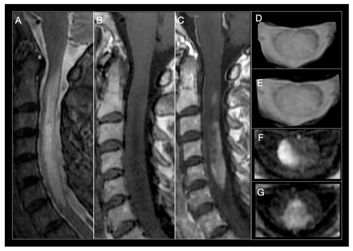
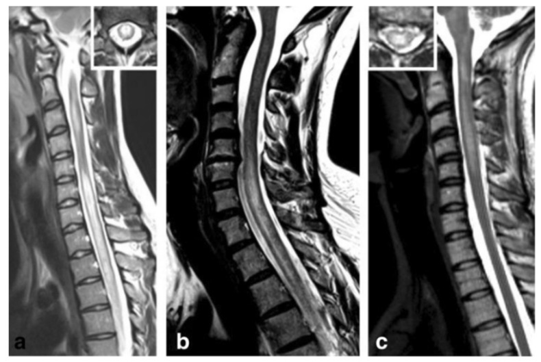
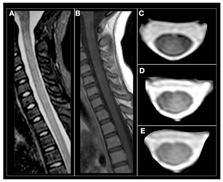
Diagnostic accuracy is poor in demyelinating myelopathies, and therefore a challenge for neurologists in daily practice, mainly because of the multiple underlying pathophysiologic mechanisms involved in each subtype. A systematic diagnostic approach combining data from the clinical setting and presentation with magnetic resonance imaging (MRI) lesion patterns, cerebrospinal fluid (CSF) findings, and autoantibody markers can help to better distinguish between subtypes. In this review, we describe spinal cord involvement, and summarize clinical findings, MRI and diagnostic characteristics, as well as treatment options and prognostic implications in different demyelinating disorders including: multiple sclerosis (MS), neuromyelitis optica spectrum disorder, acute disseminated encephalomyelitis, anti-myelin oligodendrocyte glycoprotein antibody-associated disease, and glial fibrillary acidic protein IgG-associated disease. Thorough understanding of individual case etiology is crucial, not only to provide valuable prognostic information on whether the disorder is likely to relapse, but also to make therapeutic decision-making easier and reduce treatment failures which may lead to new relapses and long-term disability. Identifying patients with monophasic disease who may only require acute management, symptomatic treatment, and subsequent rehabilitation, rather than immunosuppression, is also important.

Keywords: acute disseminated encephalomyelitis; glial fibrillary acidic protein; multiple sclerosis; myelin oligodendrocyte glycoprotein; myelitis; neuromyelitis optica; spinal cord.


Conflict of interest statement

M.M. has nothing to disclose. M.I.G. has received reimbursement for developing educational presentations, from Merck Argentina, Biogen Argentina, Sanofi-Genzyme Argentina, Bayer Inc Argentina and Novartis Argentina, and has received travel/accommodations stipends from Merck Argentina, Biogen Argentina, Roche Argentina, Novartis Argentina, and TEVA Argentina. J.C. is a board member of Merck-Serono Argentina, Biogen-Idec LATAM, Merck-Serono. LATAM, Novartis and Genzyme global. Correale has received reimbursement for developing educational presentations for Merck-Serono Argentina, Merck-Serono LATAM, Biogen-Idec Argentina, Genzyme Argentina, and Novartis Argentina and Roche Argentina as well as professional travel/accommodations stipends. The funders had no role in the design of the study; in the collection, analysis, or interpretation of data; in the writing of the manuscript, or in the decision to publish the results.

Figures

Figure 1
Figure 1
Multiple Sclerosis myelitis. (AF) 32-year-old woman diagnosed with relapsing remitting course (RRMS) 2 years earlier, EDSS 0. (A,B) Sagittal short-tau inversion recovery (STIR) showing small, focal, chronic, peripheral lesions. (C) Sagittal post-contrast T1 weighted, absence of enhancement, T2 lesions are isointense. (DF) axial T2 multiple-echo recombined gradient echo (MERGE). (D) right paramedian posterior lesion corresponds to lesion framed by a box in (A). (E) left paramedian posterior lesion corresponds to lesion framed by a dotted box in (A). (F) posterior lesion corresponds to lesion framed by a dotted line in (A). (G) 46-year-old man diagnosed with primary progressive multiple sclerosis (PPMS) in 2011, EDSS 6. Sagittal T2-weighted, framed area shows multiple sclerosis (MS) lesions and spinal cord atrophy.
Figure 2
Figure 2
Neuromyelitis optica (NMO) myelitis. Images from a 58-year-old woman with acute longitudinally extensive myelitis (C1–C7). (A) Sagittal STIR showing an extensive lesion, involving more than 3 segments, that widens the cervical spinal cord. (B) Sagittal T1-weighted sequences show an extensive T1-hypointense lesion. (C) T1-weighted images after contrast administration, extensive enhancement of cervical lesion. (D,E) Axial T2-MERGE hyperintense area that involves more than half the diameter of the spinal cord. (E,F) Axial T1-weighted, intense contrast enhancement of lateral (E) and central-posterior (F,G) areas.
Figure 3
Figure 3
Anti-myelin oligodendrocyte glycoprotein (MOG) antibody myelitis. (a) Sagittal T2-weighted spinal MRI performed at disease onset revealed a large longitudinal centrally-located lesion extending over the entire spinal cord, as well as swelling of the cord. (b) Longitudinally extensive central spinal cord T2 lesion in another patient. (c) T2-hyperintense lesions extending from the pontomedullary junction throughout the cervical cord to C5, in a third patient. Insets in (a) and C show axial sections of the thoracic cord at lesion level [172]. Figure is extracted from Jarius, S. et al., J Neuroinflammation 2016, 13, 280 (http://creativecommons.org/licenses/by/4.0/).
Figure 4
Figure 4
Anti-MOG antibody myelitis. A 12-year-old girl with relapse in the cervical spine. (A) sagittal STIR, subtle and diffuse hyperintensity of the cervical spinal cord. (B) Sagittal T1-weighted, spinal cord is isointense without contrast enhancement. (CE) axial T2-weighted images showing subtle and diffuse spinal cord hyperintensity (Courtesy Dr. Angeles Schteinschnaider).

Similar articles

Cited by

References

    1. Barreras P., Fitzgerald K.C., Mealy M.A., Jimenez J.A., Becker D., Newsome S.D., Levy M., Gailloud P., Pardo C.A. Clinical biomarkers differentiate myelitis from vascular and other causes of myelopathy. Neurology. 2018;90:E12–E21. doi: 10.1212/WNL.0000000000004765. - DOI - PMC - PubMed
    1. Transverse Myelitis Consortium Working Group Proposed diagnostic criteria and nosology of acute transverse myelitis. Neurology. 2002;59:499–505. doi: 10.1212/WNL.59.4.499. - DOI - PubMed
    1. de Seze J., Lanctin C., Lebrun C., Malikova I., Papeix C., Wiertlewski S., Pelletier J., Gout O., Clerc C., Moreau C., et al. Idiopathic acute transverse myelitis: Application of the recent diagnostic criteria. Neurology. 2005;65:1950–1953. doi: 10.1212/01.wnl.0000188896.48308.26. - DOI - PubMed
    1. Jacob A., Weinshenker B.G. An approach to the diagnosis of acute transverse myelitis. Semin. Neurol. 2008;28:105–120. doi: 10.1055/s-2007-1019132. - DOI - PubMed
    1. GBD 2015 Neurological Disorders Collaborator Group Global, regional, and national burden of neurological disorders during 1990–2015: A systematic analysis for the Global Burden of Disease Study 2015. Lancet Neurol. 2017;16:877–897. doi: 10.1016/S1474-4422(17)30299-5. - DOI - PMC - PubMed

LinkOut - more resources