Review
. 2020 May 22;12(5):570.
doi: 10.3390/v12050570.

Molecular, Evolutionary, and Structural Analysis of the Terminal Protein Domain of Hepatitis B Virus Polymerase, a Potential Drug Target

Affiliations
Review

Molecular, Evolutionary, and Structural Analysis of the Terminal Protein Domain of Hepatitis B Virus Polymerase, a Potential Drug Target

Timothy S Buhlig et al. Viruses. .

Abstract

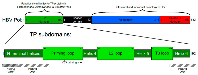
Approximately 250 million people are living with chronic hepatitis B virus (HBV) infections, which claim nearly a million lives annually. The target of all current HBV drug therapies (except interferon) is the viral polymerase; specifically, the reverse transcriptase domain. Although no high-resolution structure exists for the HBV polymerase, several recent advances have helped to map its functions to specific domains. The terminal protein (TP) domain, unique to hepadnaviruses such as HBV, has been implicated in the binding and packaging of the viral RNA, as well as the initial priming of and downstream synthesis of viral DNA-all of which make the TP domain an attractive novel drug target. This review encompasses three types of analysis: sequence conservation analysis, secondary structure prediction, and the results from mutational studies. It is concluded that the TP domain of HBV polymerase is comprised of seven subdomains (three unstructured loops and four helical regions) and that all three loop subdomains and Helix 5 are the major determinants of HBV function within the TP domain. Further studies, such as modeling inhibitors of these critical TP subdomains, will advance the TP domain of HBV polymerase as a therapeutic drug target in the progression towards a cure.

Keywords: hepatitis B virus; protein priming; terminal protein.


Conflict of interest statement

The authors declare no conflict of interest.

Figures

Figure 1
Figure 1
Schematic representation of hepatitis B virus polymerase domains and terminal protein (TP) subdomains. The polymerase protein is responsible for DNA synthesis, which is carried out by the catalytic reverse transcriptase (RT) domain. While the RNA template is copied into viral DNA, the RNase H domain degrades the RNA template. The TP domain acts as a primer for the initial DNA synthesis steps, and the DNA remains attached to the TP domain throughout synthesis. The spacer domain is thought to allow for flexibility while the TP is attached at one end of the nascent viral DNA and the RT domain synthesizes the other end. Regions of similarity with other proteins are highlighted. Within the TP domain (green), the function of the helical subdomains is likely to provide structure, while the loop subdomains are involved in activities critical to the viral replication cycle, such as protein priming that initiates from the tyrosine at position 63 (Y63). The regions of the TP domain that overlap with other open reading frames (ORF) are shown as striped boxes. These regions are under additional selection pressure when mutations occur. HBV: hepatitis B virus; HIV: human immunodeficiency virus; Pol: polymerase.
Figure 2
Figure 2
Polymerase-dependent functions of the replication cycle of hepatitis B virus (HBV) and four functional assays of HBV polymerase (HBV Pol) activity. (a) Starting with covalently closed circular DNA (cccDNA), which is located inside the nucleus of infected liver cells, the pre-genomic RNA (pgRNA) is transcribed by the host cell. Next, pgRNA is sent to the cytoplasm and translated into viral proteins. The translated HBV Pol binds to pgRNA at the 5′ epsilon stem-loop structure. The interaction between the HBV Pol and the pgRNA facilitates the encapsidation (packaging) of the HBV Pol and pgRNA. Priming most likely occurs after RNA packaging, within the nucleocapsid. Using a free hydroxyl group of the tyrosine at position 63 (Y63), HBV Pol performs the dual functions of primer and polymerase. The DNA strand remains attached to HBV Pol while synthesizing the initial GAA nucleotides. HBV Pol and these bases then change templates to the 3′ end of pgRNA and carry on minus-strand DNA synthesis. As the template pgRNA is copied, it is degraded by the RNase H activity of HBV Pol. This ssDNA-containing nucleocapsid will mature as the second strand of DNA is copied by HBV Pol, forming relaxed circular DNA (rcDNA). This rcDNA-containing nucleocapsid is the infective particle. (b) In RNA binding assays, the HBV Pol can be purified with in vitro synthesized RNA or epsilon RNA from cell culture (referred to as in vivo), which is co-purified with HBV Pol. Together, they can be evaluated for RNA binding activity by measuring RNA levels. Priming is measured from polymerase-epsilon constructs that synthesize DNA in the in vitro priming assay. Due to protein priming, HBV Pol becomes covalently labeled with the radionucleotides and can be detected by autoradiography. RNA packaging assays use HBV Pol and a polymerase-minus construct. The RNA in the purified nucleocapsid is compared to capsid protein levels, a ratio that describes RNA packaging levels. DNA synthesis can also be measured from purified nucleocapsids using HBV-specific Southern blotting. ε: epsilon RNA secondary structure motif.
Figure 3
Figure 3
Evolutionary relationships and conservation among hepatitis B virus (HBV) isolates. (a) Amino acid sequences from 66 piscine (blue), avian (red), and mammalian (gray) HBV isolates were aligned and compared phylogenetically. Branch lengths represent the evolutionary distances between isolates of HBV that infect different host species. (b) Genotypes of human HBV isolates are grouped phylogenetically. Amino acid sequences from 584 human HBV isolates were compared, using reference sequences for genotypes A through G for determining genotype branching (colorized branches). Genetically identical isolates were consolidated into single lines. For both (a) and (b), the evolutionary history was inferred using the unweighted pair group method with arithmetic means. Trees are drawn to scale, with branch lengths in the same units as those of the evolutionary distances used to infer the phylogenetic tree. Evolutionary analyses were conducted in MEGA [55]. (c) Alignment of secondary structure predictions from representative HBV species of mammalian, avian, and piscine isolates. Alpha helices and beta sheets are shown for each sequence, and similar secondary-structure patterns were aligned based on three unstructured loops (regions between helices). The amino acid positions that are 100% conserved among mammalian, avian, and piscine HBV isolates are numbered and shown highlighted at the top, along the human HBV sequence for genotype D. A basic map of the subdomains of the human HBV TP domain is shown for reference, at the bottom in green.
Figure 4
Figure 4
Two-dimensional and three-dimensional interactions of the terminal protein (TP) domain of the hepatitis B virus (HBV) polymerase protein. (a) Amino acid sequence for the TP domain, numbered according to genotype D. Overlap with other open reading frames (ORF) is indicated by striped bars. The TP domain is grouped into seven subdomains according to secondary structure predictions, with the helical regions in gray boxes. All known HBV (uppercase) and duck HBV (lowercase) mutants that have been tested are marked to represent the four tested functions. If a letter is absent, the mutant was not tested in that assay. Defective phenotypes are highlighted by blue circles representing either an extensive loss of function (<20% activity) or partial loss of function (<70% activity), whereas mutants exhibiting a wild-type phenotype are not circled. The T3 motif has been extensively tested in mutational studies. Yellow stars show TP domain residues with >99% homology to duck HBV (DHBV). Homology among human samples is graphed above the sequence (black line), where peaks indicate regions of high homology. Homology is shown as percent conservation, a moving average of amino acid homology among 584 isolates of human HBV in 11 bp windows (five amino acids upstream and downstream from each position). (b) The three-dimensional structure of TP as predicted using QUARK. The seven predicted subdomains of TP are indicated and color-coded. The functional loop subdomains extend outward.

References

    1. Smith S., Harmanci H., Hutin Y., Hess S., Bulterys M., Peck R., Rewari B., Mozalevskis A., Shibeshi M., Mumba M. Global progress on the elimination of viral hepatitis as a major public health threat: An analysis of WHO member state responses 2017. JHEP Rep. 2019;1:81–89. doi: 10.1016/j.jhepr.2019.04.002. - DOI - PMC - PubMed
    1. Alonso S., Guerra A.-R., Carreira L., Ferrer J.-Á., Gutiérrez M.-L., Fernandez-Rodriguez C.M. Upcoming pharmacological developments in chronic hepatitis B: Can we glimpse a cure on the horizon? BMC Gastroenterol. 2017;17:168. doi: 10.1186/s12876-017-0726-2. - DOI - PMC - PubMed
    1. Asselah T., Loureiro D., Boyer N., Mansouri A. Targets and future direct-acting antiviral approaches to achieve hepatitis B virus cure. Lancet. Gastroenterol. Hepatol. 2019;4:883–892. doi: 10.1016/S2468-1253(19)30190-6. - DOI - PubMed
    1. Martinez M.G., Villeret F., Testoni B., Zoulim F. Can we cure hepatitis B virus with novel direct-acting antivirals? Liver Int. 2020;40:27–34. doi: 10.1111/liv.14364. - DOI - PubMed
    1. Hepatitis B Foundation: Drug Watch. [(accessed on 21 May 2020)]; Available online: http://www.hepb.org/treatment-and-management/drug-watch/

LinkOut - more resources