. 2020 May 22;12(5):1330.
doi: 10.3390/cancers12051330.

From CENTRAL to SENTRAL (SErum aNgiogenesis cenTRAL): Circulating Predictive Biomarkers to Anti-VEGFR Therapy

Affiliations

From CENTRAL to SENTRAL (SErum aNgiogenesis cenTRAL): Circulating Predictive Biomarkers to Anti-VEGFR Therapy

Riccardo Giampieri et al. Cancers (Basel). .

Abstract

Background: In the last decade, a series of analyses failed to identify predictive biomarkers of resistance/susceptibility for anti-angiogenic drugs in metastatic colorectal cancer (mCRC). We conducted an exploratory preplanned analysis of serum pro-angiogenic factors (SErum aNgiogenesis-cenTRAL) in 72 mCRC patients enrolled in the phase II CENTRAL (ColorEctalavastiNTRiAlLdh) trial, with the aim to identify potential predictive factors for sensitivity/resistance to first line folinic acid-fluorouracil-irinotecan regimen (FOLFIRI) plus bevacizumab.

Methods: First-line FOLFIRI/bevacizumab patients were prospectively assessed for the following circulating pro-angiogenic factors, evaluated with ELISA (enzyme-linked immunosorbent assay)-based technique at baseline and at every cycle: Vascular endothelial growth factor A (VEGF-A), hepatocyte growth factor (HGF), stromal derived factor-1 (SDF-1), placental derived growth factor (PlGF), fibroblast growth factor-2 (FGF-2), monocyte chemotactic protein-3 (MCP-3), interleukin-8 (IL-8).

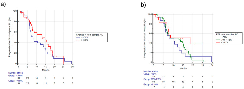
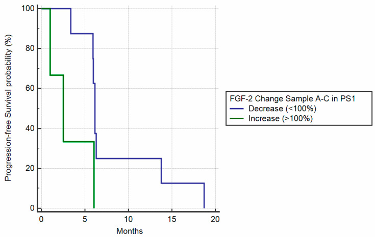
Results: Changes in circulating FGF-2 levels among different blood samples seemed to correlate with clinical outcome. Patients who experienced an increase in FGF-2 levels at the second cycle of chemotherapy compared to baseline, had a median Progression Free Survival (mPFS) of 12.85 vs. 7.57 months (Hazard Ratio-HR: 0.73, 95% Confidence Interval-CI: 0.43-1.27, p = 0.23). Similar results were seen when comparing FGF-2 concentrations between baseline and eight-week time point (mPFS 12.98 vs. 8.00 months, HR: 0.78, 95% CI: 0.46-1.33, p = 0.35).

Conclusions: Our pre-planned, prospective analysis suggests that circulating FGF-2 levels' early increase could be used as a marker to identify patients who are more likely to gain benefit from FOLFIRI/bevacizumab first-line therapy.

Keywords: FGF2; PlGF; VEGF; angiogenesis; bevacizumab; circulating biomarkers; colon cancer.


Conflict of interest statement

The authors declare no conflict of financial or nonfinancial interest.

Figures

Figure 1
Figure 1
Dot plot of FGF-2 values for blood samples A, B, and C. High inter-patient variability for FGF-2 plasma levels at baseline (sample A), after two weeks (sample B), and at eight weeks (sample C). Blood sample A median FGF-2 serum level was 44.963 pg/mL (range: 13.203–73.618) and mean FGF-2 serum level was 46.355 pg/mL (standard deviation, SD: 11.060). Blood sample B median FGF-2 serum level was 44.919 (range 12.537–72) and mean FGF-2 serum level was 47.103 (SD: 11.763). Similarly, in blood sample C, median FGF-2 serum level was 43.509 (range 0.544–78.735) and mean FGF-2 serum level was 44.582 (SD: 14.141).
Figure 2
Figure 2
Progression Free Survival (PFS) based on baseline FGF-2 levels. (a) PFS stratified by median FGF-2, median Progression Free Survival (mPFS), respectively, for higher or lower than median: 8.52 vs. 8.60 months, Hazard Ratio (HR): 1.16, 95% Confidence Interval (CI): 0.70–1.92, p = 0.53). (b) PFS stratified in quartiles (first quartile: <40.945 pg/mL, second quartile: 40.945–44.964 pg/mL, third quartile: 44.945–50.819 pg/mL, fourth quartile >50.819 pg/mL), respectively, 10.3 vs. 7.54 vs. 8.52 vs. 8.49 months, p = 0.90.
Figure 3
Figure 3
PFS based on ratio of FGF-2 levels between sample A/B. (a) PFS stratified by reduction (<100% concentration of FGF-2 between sample A/B) vs. increase (>100% concentration of FGF-2 between sample A/B). The mPFS for increase vs. reduction was, respectively, 12.85 vs. 7.57 months, HR: 0.73, 95% CI: 0.43–1.27, p = 0.23. (b) PFS stratified by different FGF-2 concentration ratios between sample A/B. The 10th percentile was 80%, the 25th percentile was 90%, the 75th percentile was 114%, and the 90th percentile was 123%. Using the 25th and 75th percentiles, we identified three groups (<90%, 90%–114%, >114% ratio between A/B), mPFS, respectively, was 6.95 vs. 8.49 vs. 14.66 months, p = 0.32.
Figure 4
Figure 4
PFS based on ratio of FGF-2 levels between sample A/C. (a) PFS stratified by reduction (<100% concentration of FGF-2 between sample A/C) vs. increase (>100% concentration of FGF-2 between sample A/C). The mPFS for increase vs. reduction was, respectively, 12.98 vs. 8 months, HR: 0.78, 95% CI: 0.46–1.33, p = 0.35. (b) PFS stratified by different FGF-2 concentration ratio between sample A/C. The 10th percentile was 59%, the 25th percentile was 79%, the 75th percentile was 118%, and the 90th percentile was 137%. Using as cutoff the 25th and 75th percentiles, we identified three groups (<79%, 79%–118%, >118% ratio between A/C), mPFS was, respectively, 8.00 vs. 12.85 vs. 18.91 months, p = 0.35.
Figure 5
Figure 5
Circulating FGF-2 levels’ changes throughout various time points. FGF-2 levels’ concentration (pg/mL) in blood samples at baseline (sample A), two weeks (sample B), eight weeks (sample C), 10 weeks, 24 weeks, and, for patients who did not progress prior, sample at PD time (sample LAST).
Figure 6
Figure 6
Circulating FGF-2 levels’ changes throughout various time points. FGF-2 levels’ percentage change (ratio) between baseline levels (sample A) and two weeks (sample B), eight weeks (sample C), 10 weeks, 24 weeks and, for patients who did not progress prior, sample at PD time (sample LAST).
Figure 7
Figure 7
PFS in symptomatic (ECOG PS-1) patients stratified by increase of FGF-2 between baseline and eight weeks sample. Patients with symptomatic disease (ECOG PS-1) with FGF-2 levels’ increase between baseline and eight weeks’ treatment had a statistically significantly improved mPFS (6.13 vs. 2.52 months, HR: 3.67, p = 0.0294).

References

    1. Oza A.M., Cook A.D., Pfisterer J., Embleton A., Ledermann J.A., Pujade-Lauraine E., Kristensen G., Carey M.S., Beale P., Cervantes A., et al. Standard chemotherapy with or without bevacizumab for women with newly diagnosed ovarian cancer (ICON7): Overall survival results of a phase 3 randomised trial. Lancet Oncol. 2015;16:928–936. doi: 10.1016/S1470-2045(15)00086-8. - DOI - PMC - PubMed
    1. Miller K., Wang M., Gralow J., Dickler M., Cobleigh M., Perez E.A., Shenkier T., Cella D., Davidson N.E. Paclitaxel plus bevacizumab versus paclitaxel alone for metastatic breast cancer. N. Engl. J. Med. 2007;357:2666–2676. doi: 10.1056/NEJMoa072113. - DOI - PubMed
    1. Sandler A., Gray R., Perry M.C., Brahmer J., Schiller J.H., Dowlati A., Lilenbaum R., Johnson D.H. Paclitaxel carboplatin alone or with bevacizumab for non-small-cell lung cancer. N. Engl. J. Med. 2006;355:2542–2550. doi: 10.1056/NEJMoa061884. - DOI - PubMed
    1. Yang J.C., Haworth L., Sherry R.M., Hwu P., Schwartzentruber D.J., Topalian S.L., Steinberg S.M., Chen H.X., Rosenberg S.A. A randomized trial of bevacizumab, an anti-vascular endothelial growth. factor antibody, for metastatic renal cancer. N. Engl. J. Med. 2003;349:427–434. doi: 10.1056/NEJMoa021491. - DOI - PMC - PubMed
    1. Hurwitz H., Fehrenbacher L., Novotny W., Cartwright T., Hainsworth J., Heim W., Berlin J., Baron A., Griffing S., Holmgren E., et al. Bevacizumab plus irinotecan, fluorouracil, and leucovorin for metastatic colorectal cancer. N. Engl. J. Med. 2004;350:2335–2342. doi: 10.1056/NEJMoa032691. - DOI - PubMed

LinkOut - more resources