. 2020 May 24;21(10):3695.
doi: 10.3390/ijms21103695.

Down-Regulation of miR-23a-3p Mediates Irradiation-Induced Neuronal Apoptosis

Affiliations

Down-Regulation of miR-23a-3p Mediates Irradiation-Induced Neuronal Apoptosis

Boris Sabirzhanov et al. Int J Mol Sci. .

Abstract

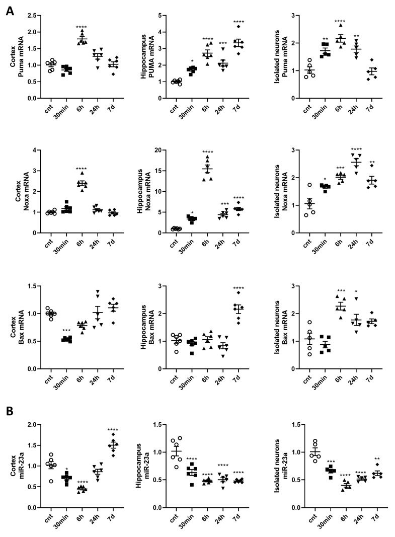
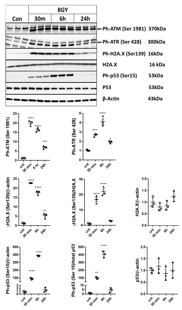
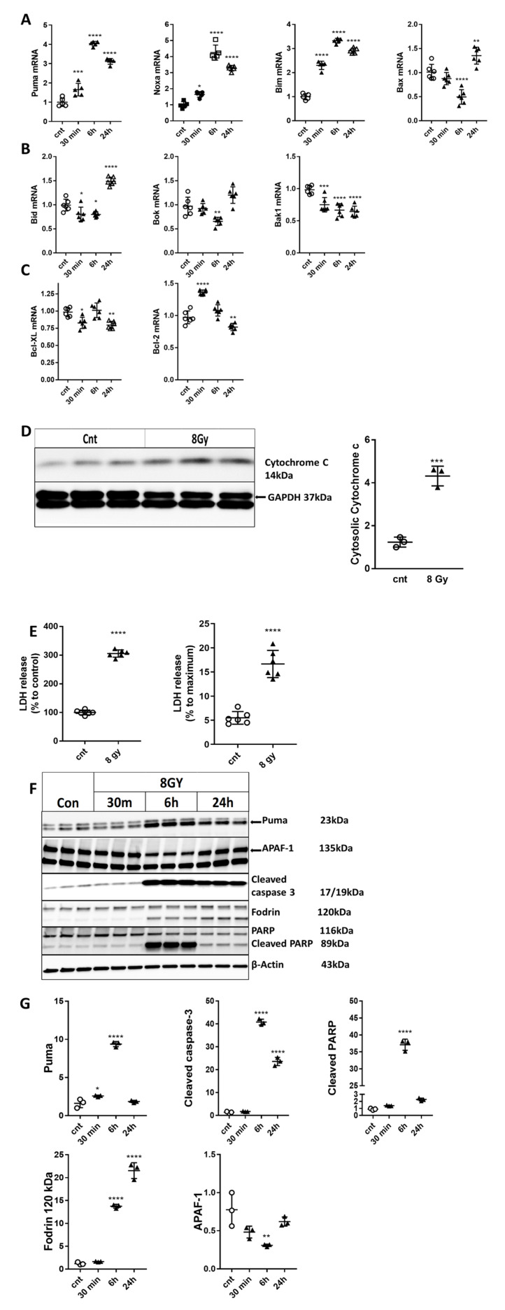
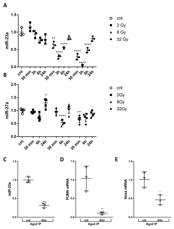
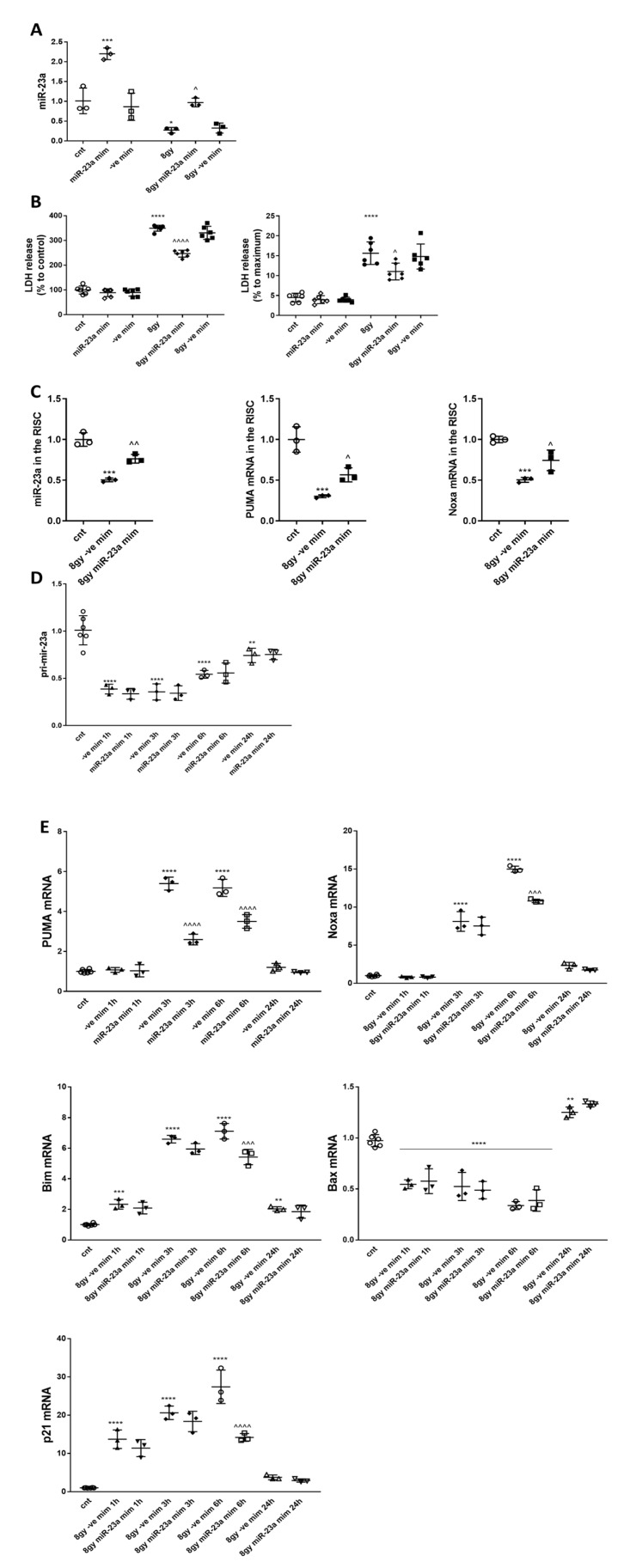
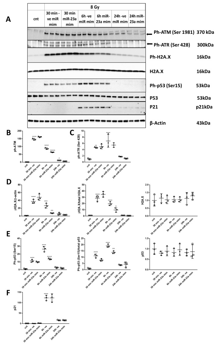
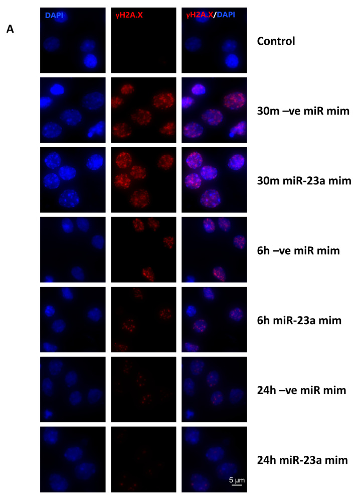
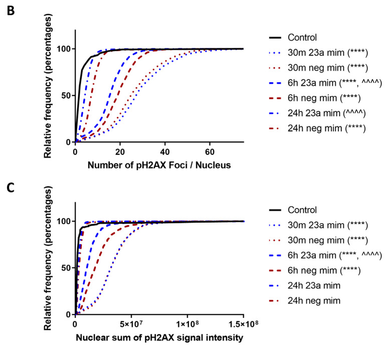
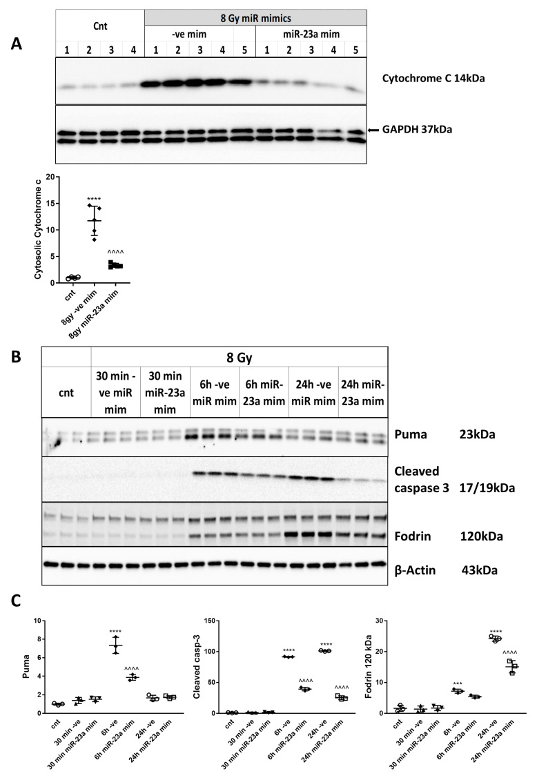
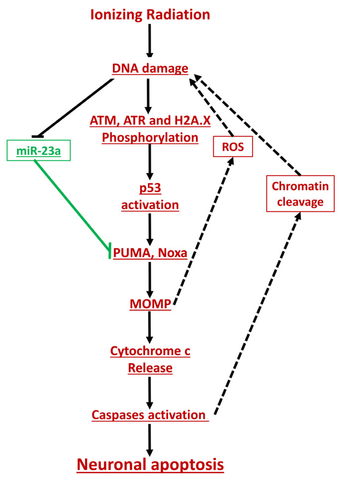
Radiation-induced central nervous system toxicity is a significant risk factor for patients receiving cancer radiotherapy. Surprisingly, the mechanisms responsible for the DNA damage-triggered neuronal cell death following irradiation have yet to be deciphered. Using primary cortical neuronal cultures in vitro, we demonstrated that X-ray exposure induces the mitochondrial pathway of intrinsic apoptosis and that miR-23a-3p plays a significant role in the regulation of this process. Primary cortical neurons exposed to irradiation show the activation of DNA-damage response pathways, including the sequential phosphorylation of ATM kinase, histone H2AX, and p53. This is followed by the p53-dependent up-regulation of the pro-apoptotic Bcl2 family molecules, including the BH3-only molecules PUMA, Noxa, and Bim, leading to mitochondrial outer membrane permeabilization (MOMP) and the release of cytochrome c, which activates caspase-dependent apoptosis. miR-23a-3p, a negative regulator of specific pro-apoptotic Bcl-2 family molecules, is rapidly decreased after neuronal irradiation. By increasing the degradation of PUMA and Noxa mRNAs in the RNA-induced silencing complex (RISC), the administration of the miR-23a-3p mimic inhibits the irradiation-induced up-regulation of Noxa and Puma. These changes result in an attenuation of apoptotic processes such as MOMP, the release of cytochrome c and caspases activation, and a reduction in neuronal cell death. The neuroprotective effects of miR-23a-3p administration may not only involve the direct inhibition of pro-apoptotic Bcl-2 molecules downstream of p53 but also include the attenuation of secondary DNA damage upstream of p53. Importantly, we demonstrated that brain irradiation in vivo results in the down-regulation of miR-23a-3p and the elevation of pro-apoptotic Bcl2-family molecules PUMA, Noxa, and Bax, not only broadly in the cortex and hippocampus, except for Bax, which was up-regulated only in the hippocampus but also selectively in isolated neuronal populations from the irradiated brain. Overall, our data suggest that miR-23a-3p down-regulation contributes to irradiation-induced intrinsic pathways of neuronal apoptosis. These regulated pathways of neurodegeneration may be the target of effective neuroprotective strategies using miR-23a-3p mimics to block their development and increase neuronal survival after irradiation.

Keywords: Bim; MOMP; Noxa; microRNA (miR), Puma; neuronal apoptosis; radiation.


Conflict of interest statement

The authors declare no conflict of interest.

Figures

Figure 1
Figure 1
The expression of pro-apoptotic members of the Bcl-2 family is upregulated, and miR-23a-3p is downregulated in the cortex and hippocampus of irradiated mice. Tissues and neurons were collected at 30 min, 6 h, 24 h, and 7 days after 10 Gy whole-brain irradiation. Total RNA was used for qPCR analysis. qPCR quantification of Puma, Noxa, Bax mRNA levels (A); and miR-23a-3p (B) in cortex, hippocampus, and ex vivo neurons, n = 6/group for brain tissues, n = 5/group for ex vivo neurons, with two technical replicates. Data represent the mean ± SD of one-way ANOVA and Tukey post-hoc analysis, * p < 0.05, ** p < 0.01, *** p < 0.001, **** p < 0.0001 vs. control animals.
Figure 2
Figure 2
Irradiation induces the activation of DNA damage and p53 pathways in primary cortical neurons. Neurons were collected 30 min, 6 h, and 24 h after 8 Gy irradiation. Image of a representative experiment. Whole-cell lysates were separated by SDS-polyacrylamide gel and immunoblotted with antibodies against Ph-ATM (Ser1981), Ph-ATR (Ser428) (approximately 300kDa), γ-H2A.X (Ser139), and Ph-p53 (Ser15) (representative images). Protein levels were quantified by densitometry; then, they were normalized to β-actin and to the parent protein. Data are presented as fold change compared with untreated control levels. The experiment was repeated 3 times with similar results, n = 3/group in each experiment. Data represent the mean ± SD of one-way ANOVA and Tukey post-hoc analysis, * p < 0.05, ** p < 0.01, *** p < 0.001, **** p < 0.0001 vs. control RCN.
Figure 3
Figure 3
IR up-regulated select pro-apoptotic members of the Bcl-2 family, mitochondrial membrane permeabilization, and release of pro-apoptotic molecules. Neurons were collected at 30 min, 6 h, and 24 h after 8 Gy irradiation. Total RNA was used for qPCR analysis. qPCR quantification of Puma, Noxa, Bim, and Bax (A); Bid, Bok, and Bak1 (B), and Bcl-xL and Bcl-2 (C) mRNA levels in a representative experiment. The experiment was repeated three times. N = 5/group in each experiment, with two technical replicates. Data represent the mean ± SD of one-way ANOVA and Tukey post-hoc analysis, * p < 0.05, ** p < 0.01, *** p < 0.001, **** p < 0.0001 vs. control RCN. RCNs were collected at 24 h after 8 Gy irradiation. Cytosolic fractions were separated by SDS-polyacrylamide gel and immunoblotted with antibodies against cytochrome c and GAPDH. Image of the representative experiment (D). Protein levels were quantified by densitometry, normalized to approximately a 37 kDa band of GAPDH, and presented as a fold change compared to control levels. The experiment was repeated 3 times with similar results, n = 3/group in each experiment. Data represent the mean ± SD. Statistical significance assigned by one-tailed t-test, ***p < 0.001 versus control. Irradiation induces neuronal cell death. Neurons were irradiated with 8 Gy. Twenty-four hours later, LDH release was measured. Data are expressed as a percentage of control untreated neurons as well as completely permeabilized cells (100% cell death) (E). The experiment was repeated six times with similar results, n = 6/group. Data represent the mean ± SD. Statistical significance was assigned by the one-tailed t-test, p < 0.0001 vs. control. Rat cortical neurons (RCNs) were collected at 30 min, 6 h, and 24 h after ionizing radiation (IR). Whole-cell lysates were separated by SDS-polyacrylamide gel and immunoblotted with antibodies against Puma, cleaved caspase-3, poly (ADP-ribose) polymerase family, member 1 (PARP), cleaved α-fodrin, and apoptotic peptidase activating factor 1 (Apaf-1). Image of a representative experiment (F). Protein levels were quantified by densitometry, normalized to β-actin, and presented as a fold change compared with untreated control levels (G). The experiment was repeated three times. N = 3/group in each experiment. Data represent mean ± SD of one-way ANOVA and Tukey post-hoc analysis, * p < 0.05, ** p < 0.01, *** p < 0.001, **** p < 0.0001 vs. control RCN.
Figure 4
Figure 4
Irradiation decreased levels of cellular miR-23a-3p, and miR-23a-3p, PUMA, and Noxa mRNAs within the RNA-induced silencing complex. Neurons collected at 30 min, 6 h, and 24 h after 2, 8, and 32 Gy irradiation. Total RNA was used for qPCR analysis. qPCR quantification of miR-23a-3p (A) and miR-27a-3p (B) levels in a representative experiment. The experiment was repeated three times. N = 3/group in each experiment for miR-23a-3p, n = 5/group in each experiment for miR-27a-3p, with 2 technical replicates. Data represent the mean ± SD of one-way ANOVA and Tukey post-hoc analysis * p < 0.05, ** p < 0.01, *** p < 0.001, **** p < 0.0001 vs. control RCN. Neurons were collected 3 h after 8 Gy treatment, subjected to RIP with Ago2 antibodies, and samples used for qPCR analysis. qPCR quantification of miR-23a-3p (C), Puma (D) and Noxa (E) levels in precipitates after RIP in a representative experiment. The experiment was repeated two times, n = 3/group in each experiment with 2 technical replicates. Data represent the mean ± SD. Statistical significance assigned by one-tailed t-test, *** p < 0.001 versus control, n = 3/group.
Figure 5
Figure 5
miR-23a-3p mimic reverses the decrease of endogenous miR-23a-3p in irradiated primary cortical neurons and attenuates neuronal cell death. RCNs were transfected with miR-23a-3p mimics and negative control mimics before irradiation. At 3 h after exposure to 8 Gy, neurons were harvested for RNA isolation. qPCR quantification of miR-23a-3p in a representative experiment (A). The experiment was repeated two times. N = 3/group in each experiment with 2 technical replicates. Data represent mean ± SD. Statistical significance assigned by one-way ANOVA and Tukey post hoc analysis; * p < 0.05, *** p < 0.001 vs. control RCN; ^ p < 0.05 vs. irradiated negative control mimics. LDH release was measured at 24 h after irradiation as a percentage of control untreated neurons as well as completely permeabilized cells (100% cell death). Analysis of a representative experiment (B). The experiment was repeated four times with similar results, n = 6/group in each experiment. Data represent mean ± SD. Statistical significance assigned by one-way ANOVA and Tukey post hoc analysis, * p < 0.05, *** p < 0.001, vs. control RCN. ^ p < 0.05, ^^^^ p < 0.0001 vs. negative control mimic transfected cells (-ve mim) RCN. RCNs were treated as described above and harvested 3 h after 8 Gy irradiation. Lysates from each sample were subjected to RNA-binding protein immunoprecipitation (RIP) using Ago2 antibodies, followed by qPCR analysis for levels of miR-23a-3p, Puma, and Noxa in the RISC. Analysis of a representative experiment (C) n = 3/group in each experiment with 2 technical replicates. Data represent mean ± SD. Significance assigned by one-way ANOVA and Tukey post hoc analysis, miR-23a-3p *** p = 0.0001 vs. control RCNs; ^^ p < 0.0039 vs. 8 Gy + miR-ve mimic; Puma *** p = 0.0004 vs. control RCNs; ^ p < 0.0471 vs. 8 Gy + miR-ve mimic; Noxa *** p = 0.0006 vs. control RCNs; ^ p < 0.0228 vs. 8 Gy + miR-ve mimic. IR causes the down-regulation of miR-23a-3p at the transcription stage. qPCR quantification of pri-miR-23a-3p. Analysis of a representative experiment (D). Data represent the mean ± SD of one-way ANOVA and Tukey post-hoc analysis, n = 6 for the control group, n = 3 for all other groups with 2 technical replicates in each experiment; * p < 0.05, ****p < 0.0001 vs. control RCN. miR-23a-3p mimic attenuates the irradiation-induced elevation of Puma, Noxa, and Bim in primary cortical neurons. RCNs were transfected with miR-23a-3p mimics and negative control mimics before irradiation. Neurons were harvested for RNA isolation at 1 h, 3 h, 6 h, and 24 h after exposure to 8 Gy. qPCR quantification of Puma, Noxa, Bim, and Bax and p21. Analysis of a representative experiment (E). The experiment was repeated three times, n = 6 for control, n = 3 for all other groups in each experiment with 2 technical replicates. Data represent mean ± SD. Statistical significance assigned by one-way ANOVA and Tukey post hoc analysis, ** p < 0.01, *** p < 0.001, **** p < 0.0001 vs. control RCN, ^^^ p < 0.001, ^^^^ p < 0.0001 vs. negative control mimic transfected cells (-ve mim) RCN.
Figure 6
Figure 6
miR-23a-3p attenuates DNA damage response and p53 activation in primary cortical neurons following irradiation. RCNs were transfected with miR-23a-3p mimics, and negative control mimics before irradiation. Neurons were collected at 30 min, 6 h, and 24 h after 8 Gy irradiation. Whole-cell lysates were separated by SDS-polyacrylamide gel and immunoblotted with antibodies against Ph-ATM (Ser1981), Ph-ATR (Ser428) (approximately 300 kDa), γ-H2A.X (Ser139), H2A.X, Ph-p53 (Ser15), p53, p21 and β-actin (representative image). Image of a representative experiment (A). Protein levels were quantified by densitometry, normalized to β-actin and the parent proteins for γ-H2A.X and Ph-p53, and presented as fold change compared with untreated control levels (BF). The experiment was repeated 3 times with similar results, n = 3/group in each experiment. Data represent mean ± SD. Statistical significance assigned by one-way ANOVA and Tukey post-hoc analysis, * p < 0.05, ** p < 0.01, *** p < 0.001, **** p < 0.0001 vs. control RCN, ^^ p < 0.01, ^^^^ p < 0.0001 vs. negative control mimic transfected cells (-ve mim) RCN.
Figure 7
Figure 7
miR-23a-3p attenuates DNA damage response in primary cortical neurons following irradiation. Representative microscopy images from 30 min, 6 h, and 24 h of RCNs stained for γ-H2A.X (red), and 4′,6-diamidino-2-phenylindole (DAPI) (blue) (A). Data were calculated and plotted for all fields together as a cumulative frequency distribution without binning for γ-H2A.X foci count (B) and nuclear staining intensity (C). Eight fields were acquired per treatment as detailed above, and an average of 117 nuclei were identified per field.
Figure 7
Figure 7
miR-23a-3p attenuates DNA damage response in primary cortical neurons following irradiation. Representative microscopy images from 30 min, 6 h, and 24 h of RCNs stained for γ-H2A.X (red), and 4′,6-diamidino-2-phenylindole (DAPI) (blue) (A). Data were calculated and plotted for all fields together as a cumulative frequency distribution without binning for γ-H2A.X foci count (B) and nuclear staining intensity (C). Eight fields were acquired per treatment as detailed above, and an average of 117 nuclei were identified per field.
Figure 8
Figure 8
miR-23a-3p attenuates caspase-dependent neuronal apoptosis after irradiation. Neurons were transfected with miR-23a-3p mimics and miR-ve mimics and collected at 6 h after 8 Gy irradiation. Cytosolic fractions were separated by SDS-polyacrylamide gel and immunoblotted with antibodies against cytochrome c and GAPDH. Image of a representative experiment (A). Protein levels were quantified by densitometry, normalized to an approximately 37 kDa band of GAPDH, and presented as a fold change compared to control levels, n = 4 for control, n = 5 for other groups in each experiment. The experiment was repeated three times. Data represent mean ± SD. Significance assigned by one-way ANOVA and Tukey post-hoc analysis, **** p < 0.0001 vs. control RCNs; ^^^^ p < 0.0001 vs. 8 Gy + miR-ve mimic. RCNs were transfected with miR-23a-3p mimics, and negative control mimics before irradiation. Neurons were collected at 30 min, 6 h, and 24 h after 8 Gy irradiation. Whole-cell lysates were separated by SDS-polyacrylamide gel and immunoblotted with antibodies against Puma, α-fodrin, and cleaved caspase-3. Image of a representative experiment. (B). Protein levels were quantified by densitometry, normalized to β-actin, and presented as fold change compared with untreated control levels (C). The experiment was repeated 3 times with similar results, n = 3/group in each experiment. Data represent mean ± SD of one-way ANOVA and Tukey post hoc analysis, *** p < 0.001, **** p < 0.0001 vs. control RCN, ^^^^ p < 0.0001 vs. negative control mimic transfected cells (-ve mim) RCN.
Figure 9
Figure 9
The schematic illustration of the role of miR-23a on the IR-induced neuronal outcome. Pro-apoptotic events are shown in red, anti-apoptotic events are shown in green. Confirmed changes are underlined and shown in solid arrows.

References

    1. Li L., Wang W., Welford S., Zhang T., Wang X., Zhu X. Ionizing radiation causes increased tau phosphorylation in primary neurons. J. Neurochem. 2014;131:86–93. doi: 10.1111/jnc.12769. - DOI - PubMed
    1. Imperato J.P., Paleologos N.A., Vick N.A. Effects of treatment on long-term survivors with malignant astrocytomas. Ann. Neurol. 1990;28:818–822. doi: 10.1002/ana.410280614. - DOI - PubMed
    1. DeAngelis L.M., Delattre J.Y., Posner J.B. Radiation-induced dementia in patients cured of brain metastases. Neurology. 1989;39:789–796. doi: 10.1212/WNL.39.6.789. - DOI - PubMed
    1. Gale R.P. Medical and policy considerations for nuclear and radiation accidents, incidents and terrorism. Curr. Opin. Hematol. 2017;24:496–501. doi: 10.1097/MOH.0000000000000380. - DOI - PubMed
    1. Cekanaviciute E., Rosi S., Costes S.V. Central Nervous System Responses to Simulated Galactic Cosmic Rays. Int. J. Mol. Sci. 2018;19:3669. doi: 10.3390/ijms19113669. - DOI - PMC - PubMed

MeSH terms