. 2020 May 24;11(5):534.
doi: 10.3390/mi11050534.

High-Speed Manipulation of Microobjects Using an Automated Two-Fingered Microhand for 3D Microassembly

Affiliations

High-Speed Manipulation of Microobjects Using an Automated Two-Fingered Microhand for 3D Microassembly

Eunhye Kim et al. Micromachines (Basel). .

Abstract

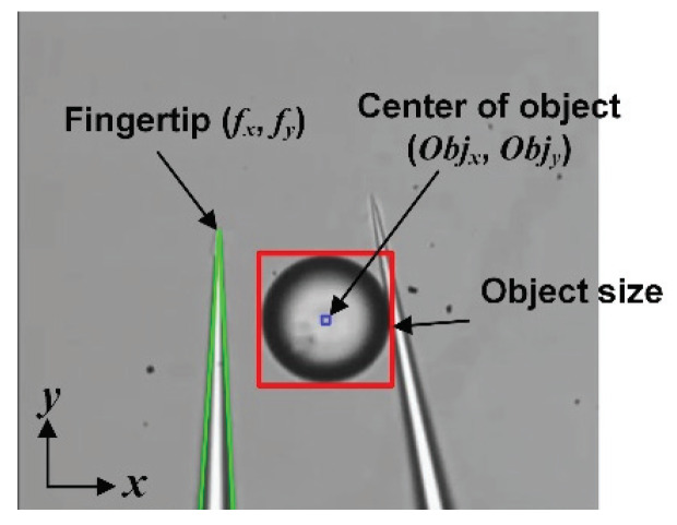
To assemble microobjects including biological cells quickly and precisely, a fully automated pick-and-place operation is applied. In micromanipulation in liquid, the challenges include strong adhesion forces and high dynamic viscosity. To solve these problems, a reliable manipulation system and special releasing techniques are indispensable. A microhand having dexterous motion is utilized to grasp an object stably, and an automated stage transports the object quickly. To detach the object adhered to one of the end effectors, two releasing methods-local stream and a dynamic releasing-are utilized. A system using vision-based techniques for the recognition of two fingertips and an object, as well automated releasing methods, can increase the manipulation speed to faster than 800 ms/sphere with a 100% success rate (N = 100). To extend this manipulation technique, 2D and 3D assembly that manipulates several objects is attained by compensating the positional error. Finally, we succeed in assembling 80-120 µm of microbeads and spheroids integrated by NIH3T3 cells.

Keywords: 3D assembly; automatic releasing; high-speed motion; local stream; micromanipulation; tissue engineering.


Conflict of interest statement

The authors declare no conflict of interest.

Figures

Figure 1
Figure 1
System of microhand. (a) Overall system of microhand; (b) System architecture.
Figure 2
Figure 2
Manipulation process of an object.
Figure 3
Figure 3
Position detection of the left fingertip and the center of an object.
Figure 4
Figure 4
Transportation system. (a) Moving two end effectors; (b) Moving substrate.
Figure 5
Figure 5
Detecting the grasped objects. (a) Before removing two fingers; (b) After removing two fingers.
Figure 6
Figure 6
Automatic releasing (a) When the object is adhered to left; (b) When the object is adhered to right.
Figure 7
Figure 7
Releasing process for improving success rate. (a) Flowchart; (b) Image sequence of releasing process.
Figure 8
Figure 8
Manipulation of several objects outside of the visible space. (a) Moving the stage to find the second object; (b) Searching and grasping the second object; (c) Transporting the second object to the desired position; (d) Releasing the second object.
Figure 9
Figure 9
Result of the automated manipulation of an object; (a) Placing position of 100 µm microbeads; (b) Placing position of 55 µm microbeads.
Figure 10
Figure 10
Time-lapse images of the automated manipulation of an object.
Figure 11
Figure 11
Error recovery to compensate the position error based on a prearranged object. (a) Manipulating the second object by adjusting X-axis; (b) Manipulating the fourth object by adjusting X and Y-axis.
Figure 12
Figure 12
Results of the manipulation. (a) Several objects for making special characters (“O” and “U”); (b) Nine objects for making 2D structure; (c) Five objects for making 3D structure.
Figure 13
Figure 13
Results of the manipulation of three objects. (a) 85−120 µm microbeads, (b) 100−120 µm spheroids.

References

    1. Norotte C., Marga F.S., Niklason L.E., Forgacs G. Scaffold-free vascular tissue engineering using bioprinting. Biomaterials. 2009;30:5910–5917. doi: 10.1016/j.biomaterials.2009.06.034. - DOI - PMC - PubMed
    1. Richards D., Jia J., Yost M., Markwald R., Mei Y. 3D bioprinting for vascularized tissue fabrication. Ann. Biomed. Eng. 2017;45:132–147. doi: 10.1007/s10439-016-1653-z. - DOI - PMC - PubMed
    1. Kolesky D.B., Homan K.A., Skylar-Scott M.A., Lewis J.A. Three-dimensional bioprinting of thick vascularized tissues. Proc. Nat. Acad. Sci. USA. 2016;113:3179–3184. doi: 10.1073/pnas.1521342113. - DOI - PMC - PubMed
    1. Haraguchi Y., Shimizu T., Sasagawa T., Sekine H., Sakaguchi K., Kikuchi T., Sekine W., Sekiya S., Yamato M., Umezu M. Fabrication of functional three-dimensional tissues by stacking cell sheets in vitro. Nat. Protoc. 2012;7:850. doi: 10.1038/nprot.2012.027. - DOI - PubMed
    1. Owaki T., Shimizu T., Yamato M., Okano T. Cell sheet engineering for regenerative medicine: Current challenges and strategies. Biotechnol. J. 2014;9:904–914. doi: 10.1002/biot.201300432. - DOI - PubMed

LinkOut - more resources