. 2020 Jun 9;117(23):12847-12855.
doi: 10.1073/pnas.2003191117. Epub 2020 May 26.

Collective effects of XMAP215, EB1, CLASP2, and MCAK lead to robust microtubule treadmilling

Affiliations

Collective effects of XMAP215, EB1, CLASP2, and MCAK lead to robust microtubule treadmilling

Göker Arpağ et al. Proc Natl Acad Sci U S A. .

Abstract

Microtubule network remodeling is essential for fundamental cellular processes including cell division, differentiation, and motility. Microtubules are active biological polymers whose ends stochastically and independently switch between phases of growth and shrinkage. Microtubule treadmilling, in which the microtubule plus end grows while the minus end shrinks, is observed in cells; however, the underlying mechanisms are not known. Here, we use a combination of computational and in vitro reconstitution approaches to determine the conditions leading to robust microtubule treadmilling. We find that microtubules polymerized from tubulin alone can treadmill, albeit with opposite directionality and order-of-magnitude slower rates than observed in cells. We then employ computational simulations to predict that the combinatory effects of four microtubule-associated proteins (MAPs), namely EB1, XMAP215, CLASP2, and MCAK, can promote fast and sustained plus-end-leading treadmilling. Finally, we experimentally confirm the predictions of our computational model using a multi-MAP, in vitro microtubule dynamics assay to reconstitute robust plus-end-leading treadmilling, consistent with observations in cells. Our results demonstrate how microtubule dynamics can be modulated to achieve a dynamic balance between assembly and disassembly at opposite polymer ends, resulting in treadmilling over long periods of time. Overall, we show how the collective effects of multiple components give rise to complex microtubule behavior that may be used for global network remodeling in cells.

Keywords: dynamic instability; in vitro reconstitution; microtubule; microtubule-associated proteins; treadmilling.


Conflict of interest statement

The authors declare no competing interest.

Figures

Fig. 1.
Fig. 1.
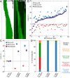
Population-level measurements predict microtubule treadmilling with leading minus ends. (A) Schematic of the assay used to study the dynamics of GTP-tubulin extensions grown from GMPCPP-stabilized microtubule seeds using TIRF microscopy. (B) Representative kymograph showing dynamic microtubule extensions (green) growing from a stable microtubule “seed” (magenta). Quantification of (C) growth rate, (D) shrinkage rate, (E) catastrophe frequency, and (F) rescue frequency of both microtubule ends over a range of tubulin concentrations. (G) Analytically calculated flux (net assembly/disassembly rate) on microtubule ends as a function of tubulin concentration. Blue and red lines are weighted linear fits in C and G. The area highlighted in gray indicates a range of tubulin concentrations consistent with minus-end-leading treadmilling behavior (Materials and Methods). Error bars are SEM.
Fig. 2.
Fig. 2.
Individual microtubules grown with tubulin alone can treadmill in vitro. (A) Schematic of a “seedless” assay that permits individual polymer treadmilling. (B) Representative kymographs showing examples of distinct dynamic modes used for classification. Yellow lines show the net flux at each microtubule end. (C) Color bar shows the number of microtubules observed in each dynamic mode (total n = 183). (D) Measured flux values at individual microtubule plus and minus ends. Fluxes at given ends were determined empirically by dividing the net length gain/loss by observation time. Net length gain is associated with positive flux and net length loss with negative flux. The total flux on a given microtubule is the sum of the fluxes at its plus and minus ends. (Right) Mean (±SD) of the individual fluxes (n = 183). Observation times ranged from 10 to 35 min, with a median of 33.8 min (29.4 ± 7.3 min, mean ± SD). Initial polymer lengths at the beginning of analysis ranged between 0.4 µm and 26.0 µm, with median of 4.6 µm (5.4 ± 3.4 µm, mean ± SD). Data were obtained from three independent experiments.
Fig. 3.
Fig. 3.
Simulations predict robust plus-end-leading treadmilling in the presence of MAPs. (A) Simulated conditions with different combinations of MAPs. The top two rows show fields of view at 0 min and at 5 min of simulation time. Green microtubules are chosen for example kymographs (bottom row), while the rest of the polymers are shown in red. (B) Average net flux rates showing the effects of MAPs in the seedless assay. Error bars are SEM (n = 93, 96, 81, and 95 for conditions 1 through 4, respectively). (C) Classification of simulated microtubules into four different dynamic modes. n = 100 microtubules were simulated for each condition at the start of simulations. The longest duration that both ends of a given microtubule remained within the field of view was used to determine the empirical flux rates and classify dynamic modes. Microtubules observed for less than 30 s were discarded. See SI Appendix, Table S2 for further details.
Fig. 4.
Fig. 4.
Reconstitution of robust plus-end-leading treadmilling in vitro. (A) Representative kymographs of a simulated (Left) and in vitro (Right) microtubule in the presence of XMAP215, EB1, CLASP2, and MCAK. (B) Empirically measured fluxes of individual microtubules in vitro. Observation times ranged from 2 to 30 min, with median of 12.4 min (14.6 ± 9.3 min, mean ± SD). (C) Average net flux rates comparing tubulin alone in vitro, multicomponent in silico, and multicomponent in vitro experiments in the presence of MAPs. Error bars are SEM (n = 183, 95, and 48, respectively). In vitro data in the presence of MAPs were obtained from four independent experiments. (D) Percentage of microtubules classified into distinct dynamic modes, comparing the three conditions as in C.

References

    1. Bugyi B., Carlier M. F., Control of actin filament treadmilling in cell motility. Annu. Rev. Biophys. 39, 449–470 (2010). - PubMed
    1. Bisson-Filho A. W. et al. ., Treadmilling by FtsZ filaments drives peptidoglycan synthesis and bacterial cell division. Science 355, 739–743 (2017). - PMC - PubMed
    1. Deng X. et al. ., Four-stranded mini microtubules formed by Prosthecobacter BtubAB show dynamic instability. Proc. Natl. Acad. Sci. U.S.A. 114, E5950–E5958 (2017). - PMC - PubMed
    1. Chaikeeratisak V. et al. ., Viral capsid trafficking along treadmilling tubulin filaments in bacteria. Cell 177, 1771–1780 e12 (2019). - PMC - PubMed
    1. Erickson H. P., Microtubule assembly from single flared protofilaments-forget the cozy corner? Biophys. J. 116, 2240–2245 (2019). - PMC - PubMed

Publication types

Substances