Case Reports
. 2020 May 26;7(5):e789.
doi: 10.1212/NXI.0000000000000789. Print 2020 Sep 3.

COVID-19-related acute necrotizing encephalopathy with brain stem involvement in a patient with aplastic anemia

Affiliations
Case Reports

COVID-19-related acute necrotizing encephalopathy with brain stem involvement in a patient with aplastic anemia

Luke Dixon et al. Neurol Neuroimmunol Neuroinflamm. .

Abstract

Objective: To describe a novel case of coronavirus disease 2019 (COVID-19)-associated acute necrotizing encephalopathy (ANE) in a patient with aplastic anemia where there was early brain stem-predominant involvement.

Methods: Evaluation of cause, clinical symptoms, and treatment response.

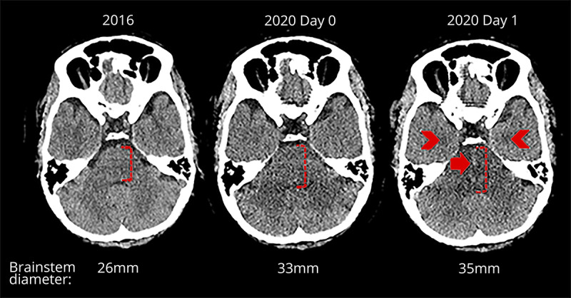
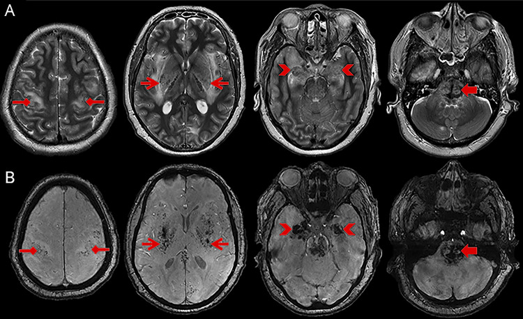
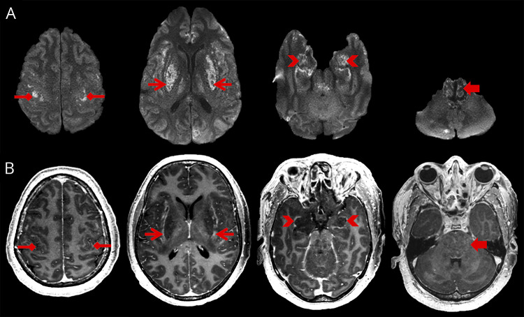
Results: A 59-year-old woman with a background of transfusion-dependent aplastic anemia presented with seizures and reduced level of consciousness 10 days after the onset of subjective fever, cough, and headache. Nasopharyngeal swab testing for severe acute respiratory syndrome coronavirus (SARS-CoV-2) was positive, and CT during admission demonstrated diffuse swelling of the brain stem. She required intubation and mechanical ventilation for airway protection, given her reduced level of consciousness. The patient's condition deteriorated, and MRI on day 6 demonstrated worsening brain stem swelling with symmetrical hemorrhagic lesions in the brain stem, amygdalae, putamina, and thalamic nuclei. Appearances were consistent with hemorrhagic ANE with early brain stem involvement. The patient showed no response to steroid therapy and died on the eighth day of admission.

Conclusions: COVID-19 may be associated with an acute severe encephalopathy and, in this case, was considered most likely to represent an immune-mediated phenomenon. As the pandemic continues, we anticipate that the spectrum of neurologic presentation will broaden. It will be important to delineate the full clinical range of emergent COVID-19-related neurologic disease.


Figures

Figure 1
Figure 1. CT of the head findings over time
Axial CT head images on different dates. From left to right, premorbid previous CT performed in 2016, 2020 day 0 admission CT, and day 1 follow-up CT. Early admission CT demonstrates subtle new swelling of the brain stem, and the follow-up CT 1 day later shows progression of the swelling with new central hemorrhagic foci (closed arrow) and symmetrical hypodensities in both amygdalae (chevrons). On day 1 of the follow-up CT, there was also hypodensity in both thalami and dorsolateral putamina (not shown).
Figure 2
Figure 2. T2-weighted and susceptibility-weighted MRI head at day 6
Serial T2-weighted (A) and susceptibility-weighted (B) axial MRI images of the brain demonstrating abnormal swelling and T2-weighted signal (A) with intrinsic hemorrhage (B) in the subcortical perirolandic regions (diamond arrow heads), dorsolateral putamina, ventrolateral thalamic nuclei and subinsular regions (open arrow heads), amygdalae (chevrons) and, pons (closed arrow heads). Abnormal signal is also shown in the splenium of the corpus callosum and cingulate gyri (not labeled).
Figure 3
Figure 3. Diffusion weighted and contrast-enhanced T1-weighted MRI head at day 6
Serial diffusion-weighted imaging (A) and contrast-enhanced T1-weighted (B) axial MRI images of the brain demonstrating abnormal restricted diffusion (A) and peripheral enhancement (B) in the same areas as the abnormal T2-weighted signal. Namely, the subcortical perirolandic regions (diamond arrow heads), dorsolateral putamina, ventrolateral thalamic nuclei and subinsular regions (open arrow heads), amygdalae (chevrons), and pons (closed arrow heads). Restricted diffusion is also shown in the splenium of the corpus callosum and cingulate gyri (not labeled).

References

    1. Mao L, Jin H, Wang M, et al. . Neurologic manifestations of hospitalized patients with coronavirus disease 2019 in Wuhan, China. JAMA Neurol Epub 2020 April 10. doi: 10.1001/jamaneurol.2020.1127. - DOI - PMC - PubMed
    1. Poyiadji N, Shahin G, Noujaim D, Stone M, Patel S, Griffith B. COVID-19–associated acute hemorrhagic necrotizing encephalopathy: CT and MRI features. Radiology Epub 2020 Mar 31. doi: 10.1148/radiol.2020201187. - DOI - PMC - PubMed
    1. Mashima K, Yano S, Yokoyama H, et al. . Epstein-barr virus-associated lymphoproliferative disorder with encephalitis following anti-thymocyte globulin for aplastic anemia resolved with rituximab therapy: a case report and literature review. Intern Med 2017;56:701–706. - PMC - PubMed
    1. Tam DYS, Cheng FWT, Chan PKS, et al. . Intact survival of refractory CMV limbic encephalitis in a patient with severe aplastic anemia after unrelated bone marrow transplantation. J Pediatr Hematol Oncol 2012;34:472–474. - PubMed
    1. Wong AM, Simon EM, Zimmerman RA, Wang HS, Toh CH, Ng SH. Acute necrotizing encephalopathy of childhood: correlation of MR findings and clinical outcome. Am J Neuroradiol 2006;27:1919–1923. - PMC - PubMed

Publication types

MeSH terms