. 2020 May 26;10(1):8692.
doi: 10.1038/s41598-020-65277-6.

Novel cultivated endophytic Verrucomicrobia reveal deep-rooting traits of bacteria to associate with plants

Affiliations

Novel cultivated endophytic Verrucomicrobia reveal deep-rooting traits of bacteria to associate with plants

Wiebke Bünger et al. Sci Rep. .

Abstract

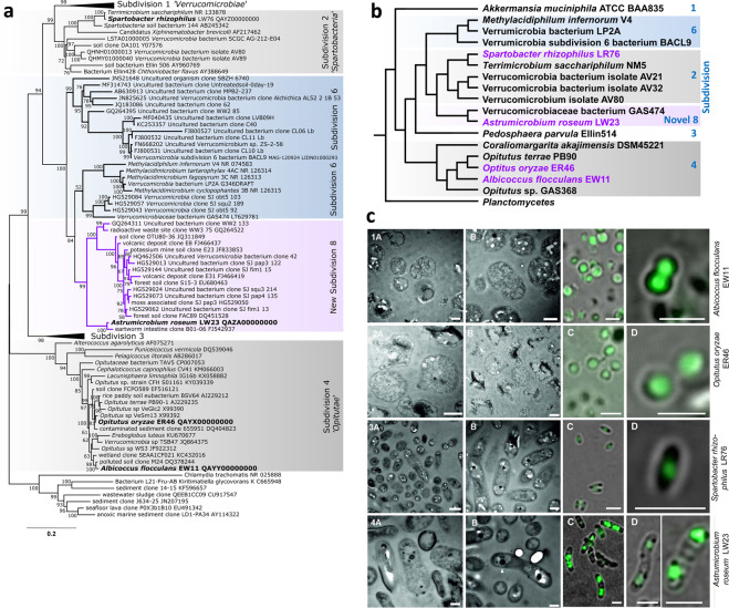
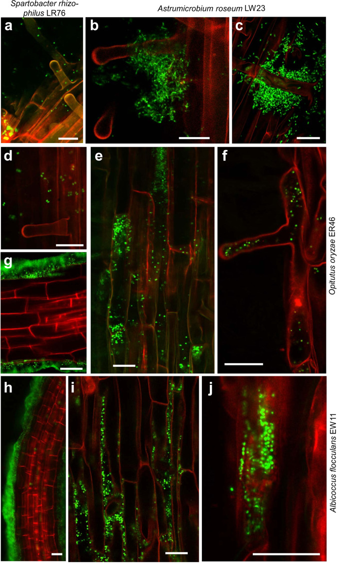
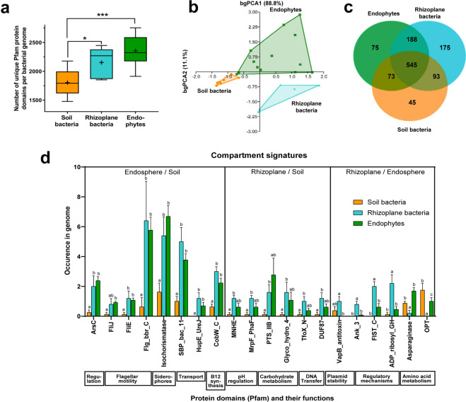
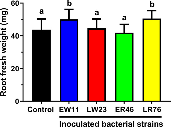
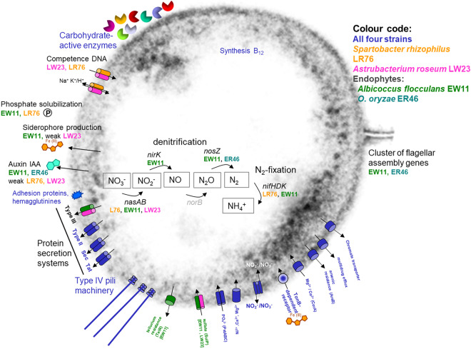
Despite the relevance of complex root microbial communities for plant health, growth and productivity, the molecular basis of these plant-microbe interactions is not well understood. Verrucomicrobia are cosmopolitans in the rhizosphere, nevertheless their adaptations and functions are enigmatic since the proportion of cultured members is low. Here we report four cultivated Verrucomicrobia isolated from rice, putatively representing four novel species, and a novel subdivision. The aerobic strains were isolated from roots or rhizomes of Oryza sativa and O. longistaminata. Two of them are the first cultivated endophytes of Verrucomicrobia, as validated by confocal laser scanning microscopy inside rice roots after re-infection under sterile conditions. This extended known verrucomicrobial niche spaces. Two strains were promoting root growth of rice. Discovery of root compartment-specific Verrucomicrobia permitted an across-phylum comparison of the genomic conformance to life in soil, rhizoplane or inside roots. Genome-wide protein domain comparison with niche-specific reference bacteria from distant phyla revealed signature protein domains which differentiated lifestyles in these microhabitats. Our study enabled us to shed light into the dark microbial matter of root Verrucomicrobia, to define genetic drivers for niche adaptation of bacteria to plant roots, and provides cultured strains for revealing causal relationships in plant-microbe interactions by reductionist approaches.


Conflict of interest statement

The authors declare no competing interests.

Figures

Figure 1
Figure 1
Molecular phylogenetic (a), phylogenomic (b) and cell morphology (c) analysis of the novel Verrucomicrobia isolates. (a) Molecular phylogeny based on based 16S rRNA gene sequences. Novel isolates of Verrucomicrobia in bold, putative novel subdivision 8 in purple. Almost complete 16S rRNA gene sequences were aligned with reference sequences from representatives of every subdivision of the phylum Verrucomicrobia. The evolutionary history was inferred by using the Maximum Likelihood method implemented in IQ-TREE. The percentage of trees in which the associated taxa clustered together is shown next to the branches. The tree is drawn to scale, with branch lengths measured in the number of substitutions per site. There were a total of 1228 positions in the final dataset, and Chlamydia trachomatis (NR 025888) was used as outgroup. (b) Phylogenomic relationships of Verrucomicrobia inferred by the whole-genome-based CVTree approach. Genomes or metagenomes covering all known subdivisions were selected, and novel isolates (purple) included. Analysis by CVTree3 with K = 6. (c) Cell morphology of strains EW11(1), ER46(2), LR76(3), and LW23(4). (A, B) Transmission electron micrographs of thin sections. (C, D) Images of cells stained with SYBR green to reveal condensed nucleic acids inside cells by Confocal Laser Scanning Microscopy. White arrowhead, electron-dense material. Scale bars indicate 500 nm (1–2 A + B, 3–4 B), 1000 nm (3–4 A) or 2 µm (C,D). Endospores were not observed. Specimen preparation through conventional fixation and dehydration lead to an irregular, scattered shape of some cells (2B) as described for other Verrucomicrobia.
Figure 2
Figure 2
Confocal Laser Scanning Microscopy of rice roots at 8 days post inoculation in hydroponic gnotobiotic culture. Rice variety O. sativa ssp. japonica cv. Nipponbare, inoculated under gnotobiotic conditions and incubated for 7d. Roots were stained with SYBR Green to visualize bacteria. (a) Colonization of strain LR76, of strain LW23 (b,c), of strain EW11 (e,f), and of strain ER46 (d,g–j). Bars indicate 100μm (a), 50 μm (b–e,g,h) or 20 μm (f), respectively. Streaking of plant medium on RSA agar did not reveal any contamination. The non-inoculated control pants showed microscopically no bacterial colonization (Supplementary Fig. 1), and generally in this seed batch, intrinsic endophytes were not detected in any experiment.
Figure 3
Figure 3
Protein domains differentiating bacterial lifestyles in endophere, rhizoplane and soil across bacterial phyla. Protein domains in (meta)genomes of well-established model organisms typically colonizing endosphere, rhizoplane or soil were compared. (a) Abundance of unique protein domains in (meta)genomes of bacteria from different compartments. Box-and-whisker plots for median (center lines), 25th and 75th percentiles (box edges), extreme data points (whiskers), and mean value (plus). Horizontal lines: statistical significance according to analysis of variance (ANOVA) followed by Holm-Sidak’s multiple comparison test, P ≤ 0.05 (*) or P ≤ 0.001 (***). (b) Between-group principal component analysis (bgPCA) of all Pfam protein domains in (meta)genomes of bacteria from the three compartments (P < 0.005 for all 3 pairwise comparisons according to PERMANOVA). (c) Venn diagram indicating differences and commonalities in Pfam domains of structural proteins encoded in the (meta)genomes from different compartments. The inner circle indicates the number of core protein families shared between all strains. The number of domains shared by all strains of one compartment are given in the outer circles (based on data from Supplementary Table S5). (d) Compartment signatures of protein domains. Examples of Pfam domains are given that are significantly enriched in (meta)genomes from endosphere or rhizoplane bacteria; for a given Pfam domain, bars are labelled by different letters if they are significantly different from each other at P < 0.05 using the Holm-Sidak method without assuming equal variance. Functions related to the respective domains are given below. Compartments depicted in the same colour in A-D. Model organisms were for soil: Verrucomicrobia bacterium isolates AV21, AV32, AV80, Acidobacteria bacterium isolate gp1 AA112, Gemmatimonadetes bacterium isolate AG11 [soil metagenomes obtained by whole genome shotgun sequencing (WGS)], Opitutus terrae PB90-1, Opitutus sp. GAS368, Azoarcus aromaticum EbN1 (complete genomes); for rhizoplane: Verrucomicrobia Astrumicrobium roseum LW23 and Spartobacter rhizophilus LR76, Azospirillum halopraeferans Au4, Azoarcus communis SWuB3, Azospirillum brasilense Sp7 (all WGS), and endophytes: Opitutaceae Albicoccus flocculans EW11 and Opitutus terrae ER46 (WGS), Azoarcus olearius BH72, Herbaspirillum seropedicae Z67, Gluconacetobacter diazotrophicus PAl 5, Serratia proteamaculans 568, Azospirillum sp. B510, Pseudomonas putida W619, Pseudomonas stutzeri A1501, Klebsiella variicola 342 (previously: K. pneumoniae 342), Enterobacter sp. 638, Methylobacterium populi BJ001, and Stenotrophomonas maltophilia R551-3 (complete genome). Based on data from Supplementary Table 7 where details are given. Graphs done with GraphPad Prism.
Figure 4
Figure 4
Root growth promotion by verrucomicrobial isolates. Sterile seedlings of O. sativa cv. Nipponbare were grown in hydroponic culture without (control) or with inoculation of single verrucomicrobial strains, and root fresh weight was evaluated 7 d post inoculation. Error bars above columns indicate standard deviations from three independent experiments (6–12 plants each). Columns headed by different letters are significantly different from each other at P < 0.05 according to analysis of variance (ANOVA) followed by uncorrected Fisher’s LSD.
Figure 5
Figure 5
Overview of selected features of plant-colonizing Verrucomicrobia strains based on genomic data or experimental tests. Features common to all four isolates are shown in blue, colour codes for occurrence in Spartobacter rhizophilus LR76, Astrumicrobium roseum LW23, or endophytes Albicoccus flocculans EW11 or Opitutus terrae ER46 are given in the figure. In addition to carbohydrate metabolism, selected transporters and nitrogen cycling pathways, features likely related to plant-microbe interactions are shown.

References

    1. Reinhold-Hurek B, Bünger W, Burbano CS, Sabale M, Hurek T. Roots shaping their microbiome: Global hot spots for microbial activity. Annu. Rev. Phytopathol. 2015;53:403–424. - PubMed
    1. Krings M, et al. Endophytic cyanobacteria in a 400-million-yr-old land plant: A scenario for the origin of a symbiosis? Rev. Paleaobot. Palynol. 2009;153:62–69.
    1. Kenrick P, Strullu-Derrien C. The origin and early evolution of roots. Plant Physiol. Bioch. 2014;166:570–580. - PMC - PubMed
    1. Kroll S, Agler MT, Kemen E. Genomic dissection of host-microbe and microbe-microbe interactions for advanced plant breeding. Curr. Opin. Plant Biol. 2017;36:71–78. - PubMed
    1. Foll M, Gaggiotti OE, Daub JT, Vatsiou A, Excoffier L. Widespread signals of convergent adaptation to high altitude in Asia and America. Am. J. Hum. Gen. 2014;95:394–407. - PMC - PubMed

Publication types

Substances