. 2020 Sep;14(9):2275-2287.
doi: 10.1038/s41396-020-0670-y. Epub 2020 May 26.

Global distribution of a chlorophyll f cyanobacterial marker

Affiliations

Global distribution of a chlorophyll f cyanobacterial marker

Laura A Antonaru et al. ISME J. 2020 Sep.

Erratum in

Abstract

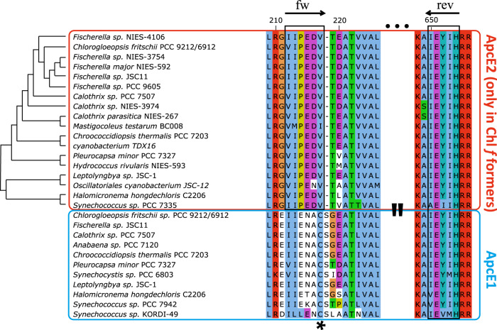
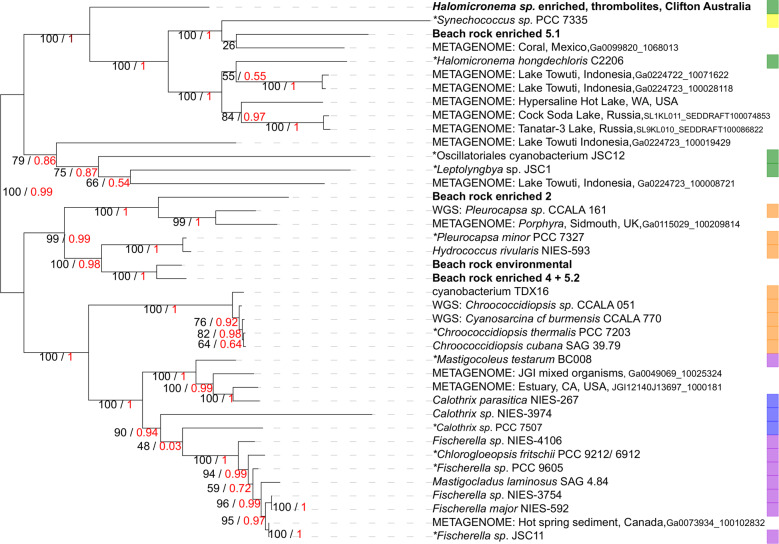
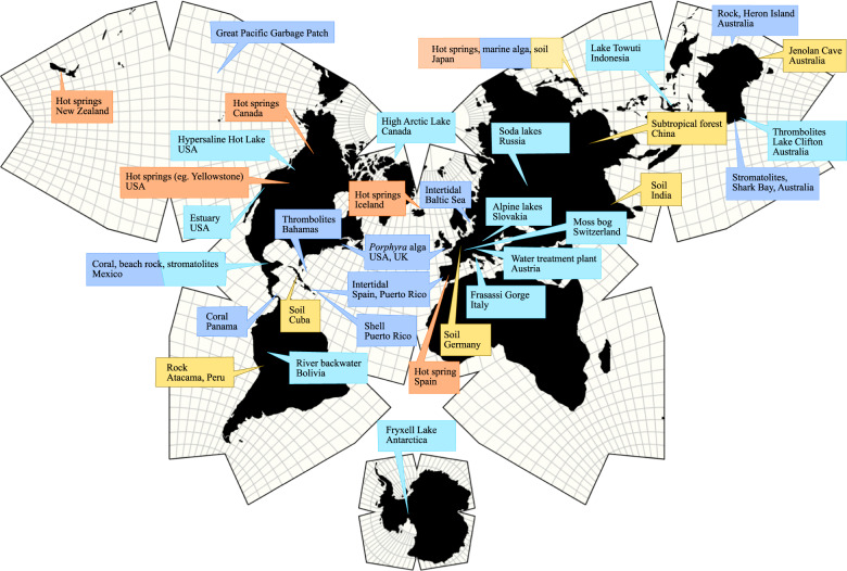
Some cyanobacteria use light outside the visible spectrum for oxygenic photosynthesis. The far-red light (FRL) region is made accessible through a complex acclimation process that involves the formation of new phycobilisomes and photosystems containing chlorophyll f. Diverse cyanobacteria ranging from unicellular to branched-filamentous forms show this response. These organisms have been isolated from shaded environments such as microbial mats, soil, rock, and stromatolites. However, the full spread of chlorophyll f-containing species in nature is still unknown. Currently, discovering new chlorophyll f cyanobacteria involves lengthy incubation times under selective far-red light. We have used a marker gene to detect chlorophyll f organisms in environmental samples and metagenomic data. This marker, apcE2, encodes a phycobilisome linker associated with FRL-photosynthesis. By focusing on a far-red motif within the sequence, degenerate PCR and BLAST searches can effectively discriminate against the normal chlorophyll a-associated apcE. Even short recovered sequences carry enough information for phylogenetic placement. Markers of chlorophyll f photosynthesis were found in metagenomic datasets from diverse environments around the globe, including cyanobacterial symbionts, hypersaline lakes, corals, and the Arctic/Antarctic regions. This additional information enabled higher phylogenetic resolution supporting the hypothesis that vertical descent, as opposed to horizontal gene transfer, is largely responsible for this phenotype's distribution.


Conflict of interest statement

The authors declare that they have no conflict of interest.

Figures

Fig. 1
Fig. 1. Conserved far-red specific motifs are present in ApcE2 fragments.
Outlined in red are ApcE2 sequences associated with the far-red cluster, including close homologues found in this study. A phylogeny of the full-length DNA sequences is shown on the left-hand side and in Fig. 5 in more detail. The blue box encompasses conventional (white-light) ApcE sequences. Conventional ApcE is found in chlorophyll f-containing organisms, along far-red ApcE2. The phytochrome-binding cysteine (residue 217, asterisk) is missing in far-red sequences, and instead they show a conserved VIPEDV-like motif (residues 204–209 in C. thermalis). Black boxes and arrows highlight areas covered by the primers designed in this study, both in a far-red-specific area (fw) and a conserved ApcE-specific area (rev).
Fig. 2
Fig. 2. apcE2 is a marker of chlorophyll f. apcE2 primers are specific for chlorophyll f-forming cyanobacteria, and the amplicons they form can be seen as ~1.2 kb bands on agarose gels.
They can be used to efficiently distinguish between (a) strains that have the far-red acclimation cluster and (b) strains that lack it. Primers of moderate degeneracy were used unless otherwise mentioned. Even at higher degeneracy, no false positives appeared in the negative controls. c Newly-sequenced strains from the Japanese NIES culture collection tested positive for apcE2. The gene was originally found via BLAST searches. d Particularly significant, the method is useful for discovering new strains. The apcE2 gene is present in enriched samples from Heron Island beach rock (“beach rock 2,4,5”) and thrombolites from Lake Clifton (Halomicronema, Clifton). It was also recovered directly from a beach rock environmental sample (rightmost, separate PCR). The environmental sample band is the result of three consecutive PCR runs; however, a faint band was visible after the first run. This strongly suggests that chlorophyll f cyanobacteria are present in these environments. Molecular weight marker: 2-Log Ladder.
Fig. 3
Fig. 3. Phylogenetic tree reconstruction of apcE2 variants illustrates previously unknown diversity.
The tree was built with PhyML (red, aLRT values) and RaxML (black, 1000 bootstrap). With the given root point, this gene tree resembles a cyanobacterial species tree. The analysis includes sequences recovered from metagenomics databases (labeled as “metagenome”) or the whole-genome-sequencing NCBI database (WGS). This gene is present in a diverse array of far-red photosynthesizing cyanobacteria, including unicellular (Section I, labeled yellow), aggregates (Section II, orange), filamentous (Section III, green), heterocyst-forming (Section IV, blue) as well as branched and heterocyst-producing forms (Section V, purple). Sequences from the beach rock strains (2, 4, 5.1, and 5.2) and the Halomicronema Clifton isolate were recovered experimentally. Asterisks mark strains associated with FR acclimation before this study.
Fig. 4
Fig. 4. Confocal imaging of cyanobacteria from beach rock enrichment samples.
Sample “beach rock 5” (a) contains unicellular and filamentous strains. Genetic data suggests the presence of a filamentous Halomicronema, aggregate-forming Pleurocapsa and unicellular Acaryochloris. Sample “beach rock 2” (b) represents a relatively pure culture of aggregate-forming cyanobacteria similar to Pleurocapsa. The strains in the image contain chlorophyll a and phycobilisomes (magenta, fluorescence emission range 650–680 nm), but also chlorophyll d/f (yellow, 720–750 nm). See right-hand column for overlay. Scale bar 15 μm (b) and 25 µm (a). Spectral scans of individual cells shown in (a) are displayed in (c). Cells were divided in filamentous (fil, black) and unicellular forms (uni, red). The spectral scans of cells shown in (b) are given in (d). The error bars indicate the difference in fluorescence between individual cells relative to the maximum intensity.
Fig. 5
Fig. 5. Chlorophyll f-containing cyanobacteria are distibuted in a variety of environments across the globe.
The illustration is based on previous pigment and genomic data as well as recent metagenomic and environmental data. Locations are color-coded based on environment. Dark blue: marine. Red: hot springs. Yellow: terrestrial. Light blue: terrestrial aquatic (including a wide range from oligotrophic to hypersaline to nutrient-rich).

References

    1. Ting CS, Rocap G, King J, Chisholm SW. Cyanobacterial photosynthesis in the oceans: the origins and significance of divergent light-harvesting strategies. Trends Microbiol. 2002;10:134–42. - PubMed
    1. Chen M, Schliep M, Willows RD, Cai ZL, Neilan BA, Scheer H. A red-shifted chlorophyll. Science. 2010;329:1318–9. - PubMed
    1. Gan F, Zhang S, Rockwell NC, Martin SS, Lagarias JC, Bryant DA. Extensive remodeling of a cyanobacterial photosynthetic apparatus in far-red light. Science. 2014;345:1312–7. - PubMed
    1. Nürnberg DJ, Morton J, Santabarbara S, Telfer A, Joliot P, Antonaru LA, et al. Photochemistry beyond the red limit in chlorophyll f–containing photosystems. Science. 2018;360:1210–3. - PubMed
    1. Gan F, Bryant DA. Adaptive and acclimative responses of cyanobacteria to far‐red light. Environ Microbiol. 2015;17:3450–65. - PubMed

Publication types