Review
. 2020 Oct;21(10):585-606.
doi: 10.1038/s41580-020-0251-y. Epub 2020 May 26.

RNA delivery by extracellular vesicles in mammalian cells and its applications

Affiliations
Review

RNA delivery by extracellular vesicles in mammalian cells and its applications

Killian O'Brien et al. Nat Rev Mol Cell Biol. 2020 Oct.

Abstract

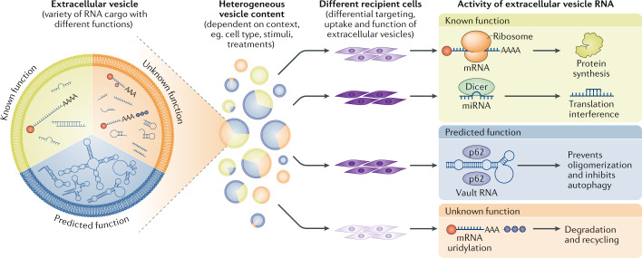
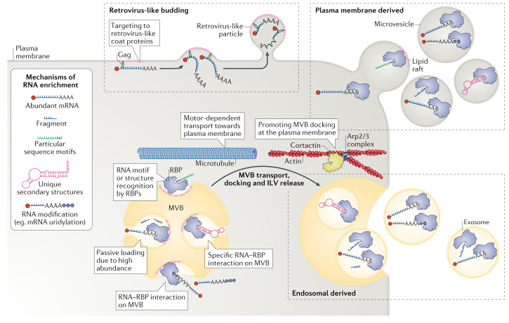
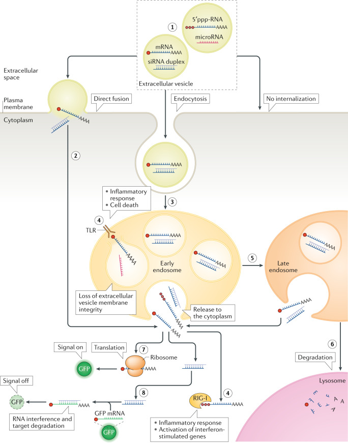
The term 'extracellular vesicles' refers to a heterogeneous population of vesicular bodies of cellular origin that derive either from the endosomal compartment (exosomes) or as a result of shedding from the plasma membrane (microvesicles, oncosomes and apoptotic bodies). Extracellular vesicles carry a variety of cargo, including RNAs, proteins, lipids and DNA, which can be taken up by other cells, both in the direct vicinity of the source cell and at distant sites in the body via biofluids, and elicit a variety of phenotypic responses. Owing to their unique biology and roles in cell-cell communication, extracellular vesicles have attracted strong interest, which is further enhanced by their potential clinical utility. Because extracellular vesicles derive their cargo from the contents of the cells that produce them, they are attractive sources of biomarkers for a variety of diseases. Furthermore, studies demonstrating phenotypic effects of specific extracellular vesicle-associated cargo on target cells have stoked interest in extracellular vesicles as therapeutic vehicles. There is particularly strong evidence that the RNA cargo of extracellular vesicles can alter recipient cell gene expression and function. During the past decade, extracellular vesicles and their RNA cargo have become better defined, but many aspects of extracellular vesicle biology remain to be elucidated. These include selective cargo loading resulting in substantial differences between the composition of extracellular vesicles and source cells; heterogeneity in extracellular vesicle size and composition; and undefined mechanisms for the uptake of extracellular vesicles into recipient cells and the fates of their cargo. Further progress in unravelling the basic mechanisms of extracellular vesicle biogenesis, transport, and cargo delivery and function is needed for successful clinical implementation. This Review focuses on the current state of knowledge pertaining to packaging, transport and function of RNAs in extracellular vesicles and outlines the progress made thus far towards their clinical applications.


Conflict of interest statement

The authors declare no competing interests.

Figures

Fig. 1
Fig. 1. Principles of functional cell communication by extracellular vesicle RNA.
Extracellular vesicles are generated as highly heterogeneous populations with different types of RNA cargo within them and in different amounts and proportions. Functionally, these RNAs can be divided into those with known functions, for example some mRNA, microRNA (miRNA) and small interfering RNA (green zone), those with predicted functions, for example, some transfer RNA, small nucleolar RNA, small nuclear RNA, Y RNA and vault RNA (blue zone) and those with unknown functions, for example, fragmented and degraded (methylated and uridylidated) RNA species (orange zone). This heterogeneity is further enhanced by the fact that extracellular vesicle cargo content strongly depends on the context (for example, cell type, stimuli and treatments). The effect that different kinds of RNA in vesicles can have on recipient cells is dictated in part by the nature of these cells, which will show differential capability for recognizing specific vesicles, their uptake and ultimately their functional effect.
Fig. 2
Fig. 2. RNA packaging into extracellular vesicles and their release into the extracellular space.
A variety of different RNA species can be packaged into extracellular vesicles. A number of modalities have been proposed for incorporation of (specific) RNAs into extracellular vesicles. First, RNAs can be targeted to the plasma membrane and released as microvesicles. They can also be targeted to the endosomal compartment and incorporated into intraluminal vesicles (ILVs) of the multivesicular body (MVB), which then can be targeted to the plasma membrane, where it fuses to release ILVs as as exosomes. Both these modes of biogenesis share many factors, and hence the vesicle type and vesicle origin are typically difficult to ascertain and control. Membrane microdomains (lipid rafts) have been strongly associated with the release of extracellular vesicles. Also, cytoskeletal components are implicated in extracellular vesicle biogenesis, in particular for exosomes, which are transported via microtubules, and their docking at the plasma membrane is supported by Arp2/3-generated branched-actin filaments stabilized by the actin-bundling activity of cortactin. Cortical actin remodelling is also an important event in membrane shaping during microvesicle release (not shown). Loading of RNA into extracellular vesicles can occur via multiple routes: passively due to an abundance of the RNA in the cytosol; by recognition via a number of RNA-binding proteins (RBPs), such as Argonaute, annexin A2, major vault protein (MVP), heterogeneous nuclear ribonucleoproteins A2/B1 (HNRNPA2B1),YBX1, SYNCRIP and lupus La protein, that bind particular sequence motifs in the RNA or that recognize unique secondary RNA structures; and through specific modifications, such as uridylation. Packaging of RNA into extracellular vesicles can also be promoted by its recognition by retroviral coat proteins such as Gag (and their silent copies present in animal genomes), which efficiently target RNAs they recognize to the plasma membrane (or the membrane of the MVB; not shown), resulting in virus-like particle release.
Fig. 3
Fig. 3. Extracellular vesicle RNA cargo interaction with recipient cells and its functional delivery.
After encountering the recipient cell, the extracellular vesicle is typically bound to its surface via cell-surface receptors (although extracellular vesicles can also be engulfed from the environment in a process known as macropinocytosis; not shown). After establishing an interaction with the cell surface, the vesicle can remain bound on the surface or can be internalized (1). One possible means of internalization is direct fusion with the plasma membrane (2), but the most common mechanism of internalization involves endocytosis, whereby extracellular vesicles are taken up to endosomes (3). In the endosome, RNA content might be released into the luminal space (if the integrity of the extracellular vesicle membrane is perturbed) or it might be released into the cytoplasm (of note, the frequency of these events is low, and endosomal escape of extracellular vesicle cargo is currently an important bottleneck in functional RNA cargo delivery by extracellular vesicles). In both cases RNAs can be recognized by pattern recognition receptors, such as Toll-like receptors (TLRs) and RIG-I or NOD-like receptors that reside in the endosome and in the cytoplasm, respectively, raising innate immune response signalling (4). Early endosomes will gradually transform into late endosomes with progressive internal acidification and possible release of RNA (stimulated by the decreasing pH) (5). Further down the endocytic pathway, endosomes will mature into lysosomes, in which the cargo that has not been released to the cytoplasm will be degraded (6). RNA cargo that reaches the cytoplasm can elicit its functional effect. For example, mRNA can be translated into a functional protein, such as green fluorescent protein (GFP), and the resulting fluorescence can act as a reporter of functional delivery of extracellular vesicle cargo (7). When small interfering RNA (siRNA) cargo is released into the cytoplasm, it can inhibit translation of specific transcripts, such as those encoding fluorescent proteins. In this case, disappearance of fluorescence will report on functional delivery of extracellular vesicle cargo (8). Extracellular vesicles can be tracked along this route with use of different reagents or labelling strategies (Table 3). 5′ppp-RNA, 5′- triphosphorylated RNA.
Fig. 4
Fig. 4. Different strategies for using extracellular vesicles for therapeutic applications.
Examples of therapeutic applications of extracellular vesicles that have resulted in either preclinical assessment or clinical trials (references to primary research or clinical trials are given). Generally, there are five strategies that can be used: implantation of donor cells, such as mesenchymal stem cells (MSCs), which release extracellular vesicles on-site (1); injection of purified, unmodified extracellular vesicles from cultured donor cells (2); injection of purified extracellular vesicles that have been modified for targeting or loading of cargo in vitro (3); injection of extracellular vesicles enriched in the cargo of interest and/or with enhanced targeting/uptake properties from genetically modified cells (4); and implantation of genetically modified cells that release modified extracellular vesicles in vivo (5). AAV, adeno-associated virus; siRNA, small interfering RNA.

References

    1. Danielson KM, et al. Plasma circulating extracellular RNAs in left ventricular remodeling post-myocardial infarction. EBioMedicine. 2018 doi: 10.1016/j.ebiom.2018.05.013. - DOI - PMC - PubMed
    1. Varcianna A, et al. Micro-RNAs secreted through astrocyte-derived extracellular vesicles cause neuronal network degeneration in C9orf72 ALS. EBioMedicine. 2019 doi: 10.1016/j.ebiom.2018.11.067. - DOI - PMC - PubMed
    1. Abels ER, et al. Glioblastoma-associated microglia reprogramming is mediated by functional transfer of extracellular miR-21. Cell Rep. 2019 doi: 10.1016/j.celrep.2019.08.036. - DOI - PMC - PubMed
    1. Hoshino A, et al. Tumour exosome integrins determine organotropic metastasis. Nature. 2015 doi: 10.1038/nature15756. - DOI - PMC - PubMed
    1. Mathieu M, Martin-Jaular L, Lavieu G, Théry C. Specificities of secretion and uptake of exosomes and other extracellular vesicles for cell-to-cell communication. Nat. Cell Biol. 2019 doi: 10.1038/s41556-018-0250-9. - DOI - PubMed

Publication types