Review
. 2020 Aug;21(8):421-438.
doi: 10.1038/s41580-020-0250-z. Epub 2020 May 26.

Mechanisms, regulation and functions of the unfolded protein response

Affiliations
Review

Mechanisms, regulation and functions of the unfolded protein response

Claudio Hetz et al. Nat Rev Mol Cell Biol. 2020 Aug.

Abstract

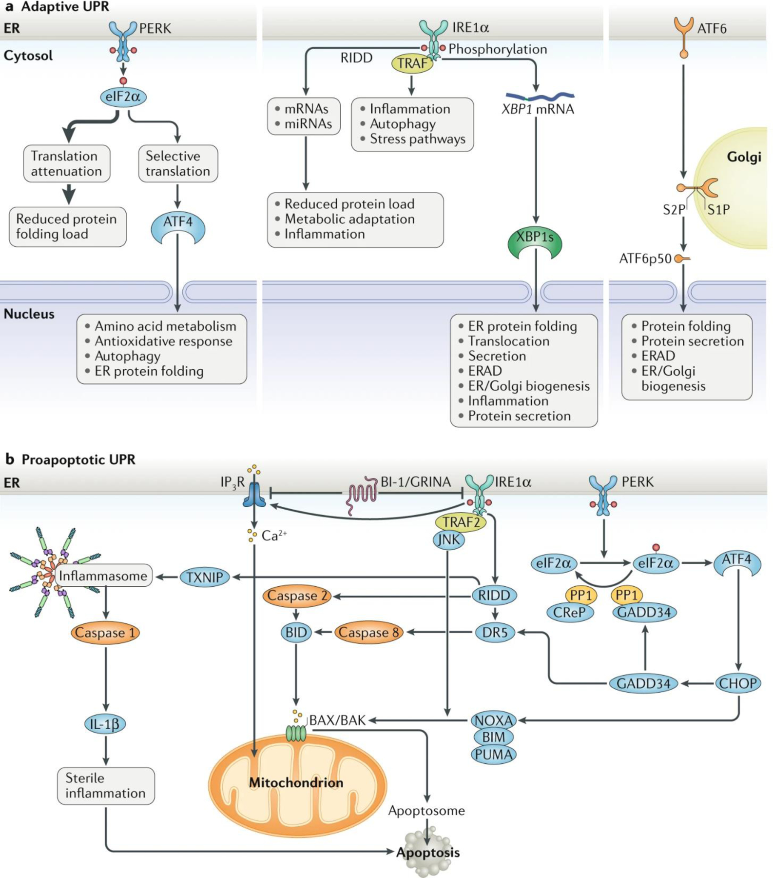
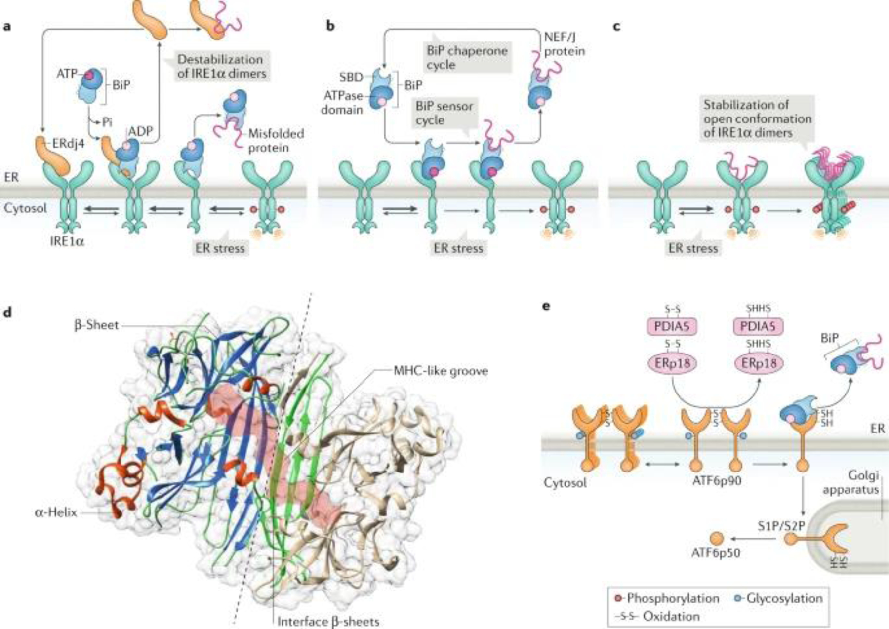
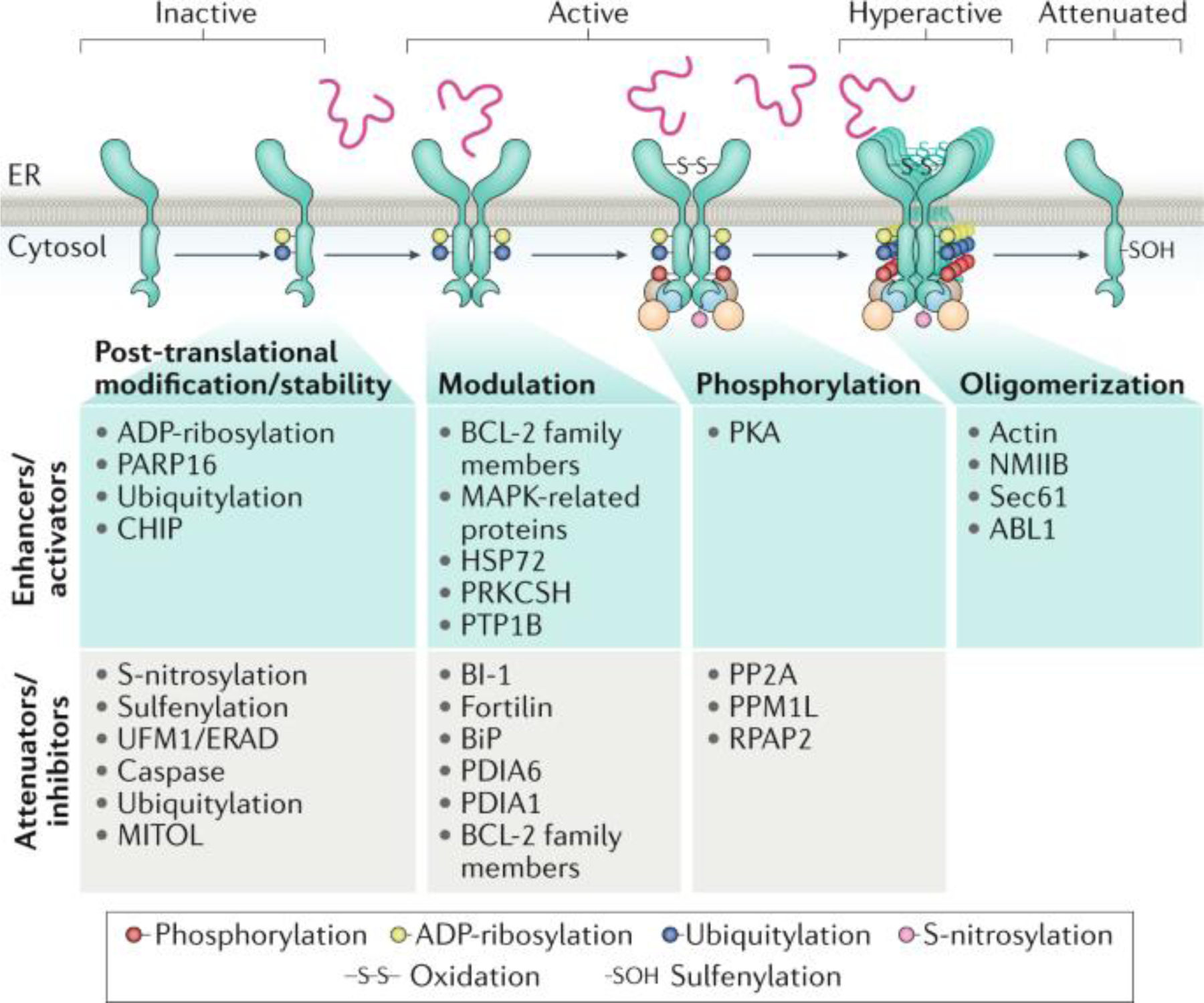
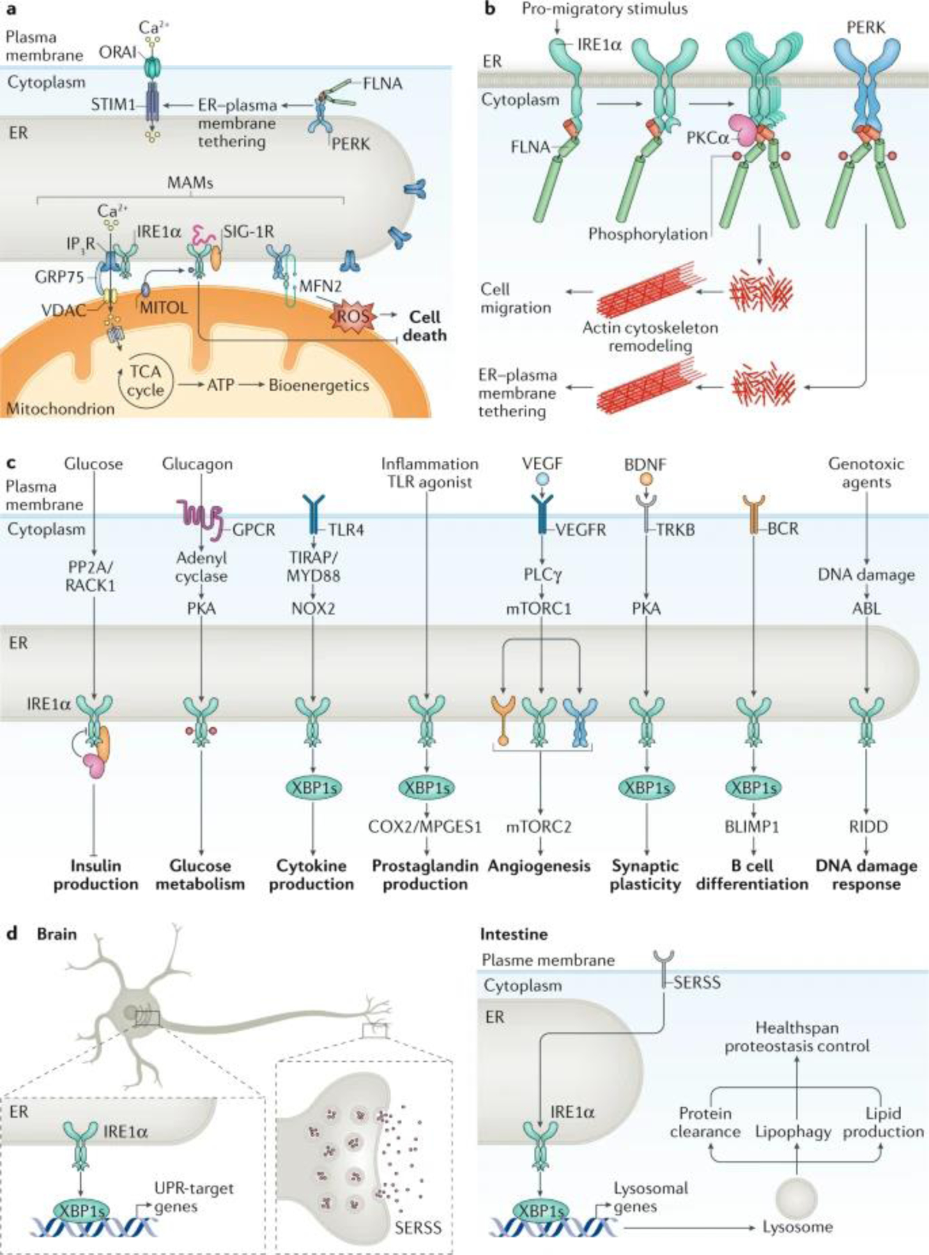
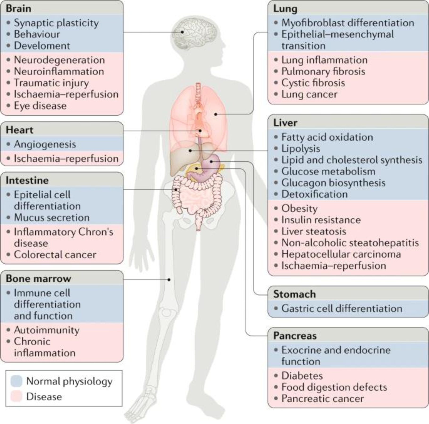
Cellular stress induced by the abnormal accumulation of unfolded or misfolded proteins at the endoplasmic reticulum (ER) is emerging as a possible driver of human diseases, including cancer, diabetes, obesity and neurodegeneration. ER proteostasis surveillance is mediated by the unfolded protein response (UPR), a signal transduction pathway that senses the fidelity of protein folding in the ER lumen. The UPR transmits information about protein folding status to the nucleus and cytosol to adjust the protein folding capacity of the cell or, in the event of chronic damage, induce apoptotic cell death. Recent advances in the understanding of the regulation of UPR signalling and its implications in the pathophysiology of disease might open new therapeutic avenues.


Conflict of interest statement

Competing interests: The authors declare no competing interests.

Figures

Fig. 1:
Fig. 1:. The major UPR pathways initiated from the ER.
a | Adaptive unfolded protein response (UPR). Under endoplasmic reticulum (ER) stress, three major UPR branches are activated: (1) PERK phosphorylates eukaryotic translation initiation factor 2 subunit-α (eIF2α), reducing the overall frequency of mRNA translation initiation. However, selective mRNAs, such as ATF4 mRNA, are preferentially translated in the presence of phosphorylated eIF2α. ATF4 activates the transcription of UPR target genes encoding factors involved in amino acid biosynthesis, the antioxidative response, autophagy and apoptosis. (2) IRE1α RNase splices XBP1 mRNA, which encodes a potent transcription factor that activates expression of UPR target genes involved in ER proteostasis and cell pathophysiology. IRE1α RNase can also cleave ER-associated mRNAs or non-coding functional RNAs, leading to their degradation through regulated IRE1-dependent decay (RIDD), which modulates the protein folding load, cell metabolism, inflammation and inflammasome signalling pathways. The IRE1α cytosolic domain may also serve as a scaffold to recruit adaptor proteins, for example tumour necrosis factor receptor-associated factor (TRAF) family members, thereby activating inflammatory responses under non-canonical ER stress conditions. (3) ATF6 transits from the ER to the Golgi apparatus, where it is cleaved by site-1 protease (S1P) and site-2 protease (S2P), yielding an active cytosolic ATF6 fragment (ATF6p50). This fragment migrates to the nucleus, activating transcription of the UPR target genes involved in ER protein folding homeostasis and cell physiology. Additionally, unfolded or misfolded proteins accumulated in the ER lumen may be degraded through the proteasome-based ER-associated protein degradation (ERAD) machinery that is regulated by the ATF6-mediated and/or IRE1α–X-box-binding protein 1 (XBP1)-mediated UPR branches. b | Proapoptotic UPR. Under ER stress, the PERK–eIF2α UPR branch induces translation of ATF4, which can activate expression of the proapoptotic factor CCAAT/enhancer-binding protein homologous protein (CHOP) and GADD34. GADD34 targets protein phosphatase 1 (PP1) to dephosphorylate eIF2α and thereby restore mRNA translation. Constitutive repressor of eIF2α phosphorylation (CReP) also serves as a cofactor to provide PP1 specificity for phosphorylated eIF2α under ER stress. CHOP promotes ER stress-induced apoptosis by modulating GADD34, death receptor 5 (DR5) and the members of the BCL-2 or BH3-only family, including NOXA, BIM and PUMA, to stimulate protein synthesis and exacerbating protein folding defect. Furthermore, the IRE1α UPR branch is involved in caspase 2-dependent, caspase 8-dependent or BAX/BAK-dependent apoptosis through RIDD or activation of TRAF2–JUN N-terminal kinase (JNK) signalling. The IRE1α-mediated RIDD also regulates thioredoxin-interacting protein (TXNIP) to activate inflammasome-dependent and caspase 1–IL-1β-dependent sterile inflammation, leading to apoptosis. In addition, Ca2+ release from the ER via inositol 1,4,5-trisphosphate receptor (IP3R), which interacts with the ER-located antiapoptotic proteins BAX inhibitor 1 (BI-1) and GRINA, contributes to mitochondrial reactive oxygen species release and the activation of the BAX/BAK-dependent apoptosome. miRNA, microRNA.
Fig. 2:
Fig. 2:. Regulation of IRE1α and PERK signalling.
a | Indirect endoplasmic reticulum (ER) stress sensing model. In resting cells, the ER stress sensor IRE1α is maintained in an inactive state through its association with the ER chaperone BiP (also known as GRP78). On accumulation of unfolded proteins, BiP preferentially binds to unfolded protein peptides, thereby releasing the ER stress sensor to allow its spontaneous dimerization and activation. In this model, BiP has the capacity to destabilize IRE1α dimers and maintain the unfolded protein response transducer in an inactive state. In addition, the BiP co-chaperone ERdj4 is required for BiP binding to IRE1α and repression of IRE1α activation. b | Alternatively, BiP might bind misfolded proteins through the substrate-binding domain (SBD), which transduces a signal to the ATPase domain to release the repressive interaction over IRE1α and PERK. c | A direct recognition model proposes that unfolded proteins bind directly to the luminal domains of IRE1α, facilitating the assembly of highly ordered IRE1α clusters. This may orient the cytosolic region of the dimer to create a ribonuclease site and generate an mRNA docking region. d | The 3D structure of the ER luminal domain of yeast Ire1p is shown, depicting the dimeric interphase (dashed line) and the major histocompatibility complex (MHC) class-I like groove (pink surface), where misfolded peptides might bind. Protein Data Bank accession number 2BE1. e | ATF6 is regulated by its glycosylation and redox state, in addition to the binding of various disulfide isomerases, including PDIA5 and ERp18. ATF6p90, full-length AFT6; J protein, J-domain protien; NEF, nucleotide exchange factor; S1P, site-1 protease; S2P, site-2 protease.
Fig. 3:
Fig. 3:. Regulation of IRE1α signalling through protein–protein interactions and post-translational modifications.
Several proteins can form a complex with IRE1α. A schematic representation is presented for negative and positive regulators of IRE1α signalling (attenuators and enhancers of unfolded protein response signalling). The effects of these regulators at different stages of the IRE1α signalling process are indicated, including IRE1α dimerization, oligomerization and phosphorylation. In addition, the occurrence of several post-translational modifications that could modify the stability or the activity of IRE1α is indicated. BI-1, BAX inhibitor 1; ER, endoplasmic reticulum; ERAD endoplasmic reticulum-associated protein degradation; NMIIB, non-muscle myosin heavy chain IIB; PKA, protein kinase A; PP2A, protein phosphatase 2A.
Fig. 4:
Fig. 4:. ER stress-independent functions of the UPR.
a | IRE1α and PERK localize to endoplasmic reticulum (ER)–mitochondrion contact sites that form structures known as mitochondria-associated membranes (MAMs). PERK regulates reactive oxygen species (ROS) propagation under ER stress at MAMs, in addition to affecting ER-to-mitochondrion tethering through the interaction with mitofusin 2 (MFN2). PERK also associates with filamin A (FLNA) to regulate ER–plasma membrane contact sites and calcium entry into the cell through ORAI–stromal interaction molecule (STIM) channels. The activity and stability of IRE1α is differentially regulated at MAMs through interaction with σ1 receptor (SIG-1R). IRE1α also docks the inositol 1,4,5-trisphosphate receptor (IP3R) at MAMs to control the transfer of calcium into the mitochondria and the activation of the tricarboxylic acid (TCA) cycle to produce ATP. b | Cell migration is regulated by IRE1α and PERK through the direct binding of filamin A, a regulator of actin cytoskeleton dynamics. IRE1α recruits protein kinase Cα (PKCα) as a scaffold to trigger filamin A phosphorylation, leading to its activation as a crosslinker of actin filaments. c | Plasma membrane receptor signalling pathways undergo crosstalk with unfolded protein response (UPR) signalling by leading to the activation of UPR sensors in an ER stress-independent manner. In addition, genotoxic stress might trigger a non-canonical activation of IRE1α to trigger regulated IRE1-dependent decay (RIDD) and modulate the DNA damage response. d | Cell-non-autonomous UPR activation. Expression of spliced X-box-binding protein 1 (XBP1s) in neurons signals for distal tissues to activate IRE1α−XBP1 and drive proteostatic changes that control healthspan and lifespan in simple model organisms such as Caenorhabditis elegans. XBP1s regulates different cellular processes to extend healthspan, including lipophagy, lysosomal function, proteostasis and lipid production. Neurotransmitter release mediates non-autonomous signalling downstream of XBP1s, suggesting that a secreted ER stress signal (SERSS) promotes ER stress resistance and longevity. BCR, B cell receptor; BDNF, brain-derived neurotrophic factor; GPCR, G protein-coupled receptor; mTORC1, mTOR complex 1; PKA, protein kinase A; PLCγ, phospholipase Cγ; PP2A, protein phosphatase 2A; TLR, Toll-like receptor; UFM1, ubiquitin-fold modifier 1, VDAC, voltage-dependent anion-selective channel; VEGFR, vascular endothelial growth factor receptor.
Fig. 5:
Fig. 5:. Role of the UPR in physiology and diseases.
Genetic and pharmacological manipulation of major unfolded protein response (UPR) components has revealed that the UPR pathways play a part in the functions of diverse organs and cell types. Preclinical models have also shown that dysregulation of UPR signalling, mediated by specific UPR components, contributes to a variety of diseases. The figure illustrates the roles of the UPR in organ physiology (blue) or pathological conditions (pink) affecting the same tissues.

References

    1. Hebert DN & Molinari M In and out of the ER: protein folding, quality control, degradation, and related human diseases. Physiol. Rev 87, 1377–1408 (2007). - PubMed
    1. Balch WE, Morimoto RI, Dillin A & Kelly JW Adapting proteostasis for disease intervention. Science 319, 916–919 (2008). - PubMed
    1. Kaufman RJ Orchestrating the unfolded protein response in health and disease. J. Clin. Invest 110, 1389–1398 (2002). - PMC - PubMed
    1. Ron D & Walter P Signal integration in the endoplasmic reticulum unfolded protein response. Nat. Rev. Mol. Cell Biol 8, 519–529 (2007). - PubMed
    1. Dever TE et al. Phosphorylation of initiation factor 2 alpha by protein kinase GCN2 mediates gene-specific translational control of GCN4 in yeast. Cell 68, 585–596 (1992). - PubMed

Publication types