

Age-Related Changes in Pain Perception Are Associated With Altered Functional Connectivity During Resting State
- PMID: 32457594
- PMCID: PMC7221150
- DOI: 10.3389/fnagi.2020.00116
Age-Related Changes in Pain Perception Are Associated With Altered Functional Connectivity During Resting State
Abstract
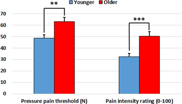
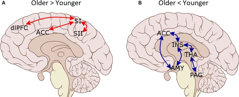
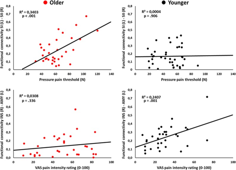
Aging affects pain experience and brain functioning. However, how aging leads to changes in pain perception and brain functional connectivity has not yet been completely understood. To investigate resting-state and pain perception changes in old and young participants, this study employed region of interest (ROI) to ROI resting-state functional connectivity (rsFC) analysis of imaging data by using regions implicated in sensory and affective dimensions of pain, descending pain modulation, and the default-mode networks (DMNs). Thirty-seven older (66.86 ± 4.04 years; 16 males) and 38 younger healthy participants (20.74 ± 4.15 years; 19 males) underwent 10 min' eyes-closed resting-state scanning. We examined the relationship between rsFC parameters with pressure pain thresholds. Older participants showed higher pain thresholds than younger. Regarding rsFC, older adults displayed increased connectivity of pain-related sensory brain regions in comparison to younger participants: increased rsFC between bilateral primary somatosensory area (SI) and anterior cingulate cortex (ACC), and between SI(L) and secondary somatosensory area (SII)-(R) and dorsolateral prefrontal cortex (PFC). Moreover, decreased connectivity in the older compared to the younger group was found among descending pain modulatory regions: between the amygdala(R) and bilateral insula(R), thalamus(R), ACC, and amygdala(L); between the amygdala(L) and insula(R) and bilateral thalamus; between ACC and bilateral insula, and between periaqueductal gray (PAG) and bilateral thalamus. Regarding the DMN, the posterior parietal cortex and lateral parietal (LP; R) were more strongly connected in the older group than in the younger group. Correlational analyses also showed that SI(L)-SII(R) rsFC was positively associated with pressure pain thresholds in older participants. In conclusion, these findings suggest a compensatory mechanism for the sensory changes that typically accompanies aging. Furthermore, older participants showed reduced functional connectivity between key nodes of the descending pain inhibitory pathway.
Keywords: aging; functional connectivity; pain perception; pain-related network; resting-state.
Copyright © 2020 González-Roldán, Terrasa, Sitges, van der Meulen, Anton and Montoya.
Figures
References
LinkOut - more resources
Full Text Sources
Miscellaneous