. 2020 May 8:8:385.
doi: 10.3389/fbioe.2020.00385. eCollection 2020.

Additively Manufactured Patient-Specific Anthropomorphic Thorax Phantom With Realistic Radiation Attenuation Properties

Affiliations

Additively Manufactured Patient-Specific Anthropomorphic Thorax Phantom With Realistic Radiation Attenuation Properties

Sepideh Hatamikia et al. Front Bioeng Biotechnol. .

Abstract

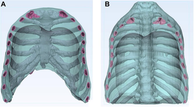
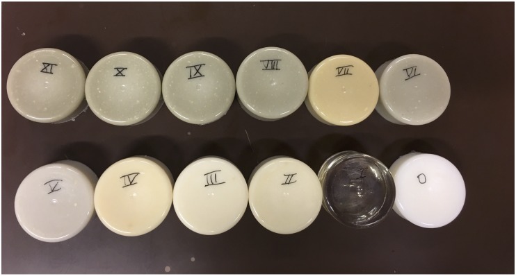
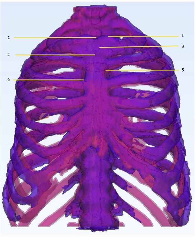
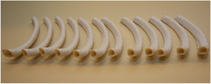
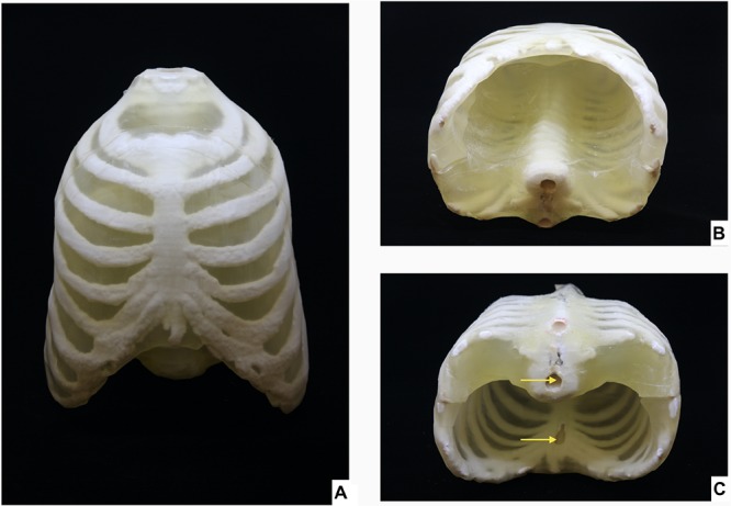
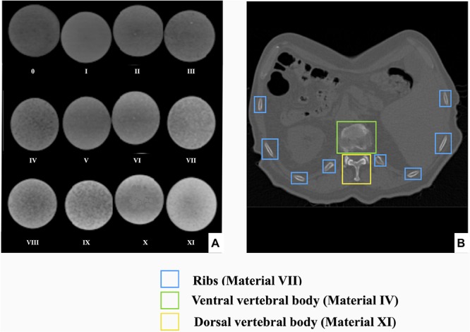
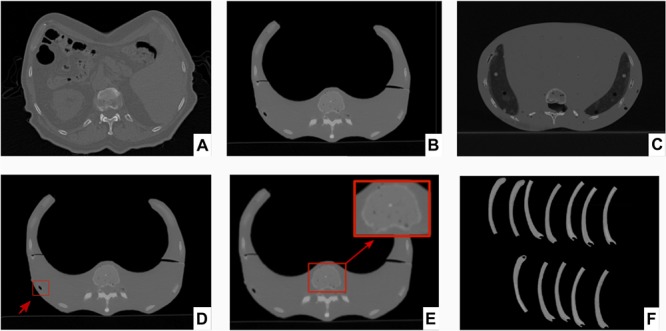
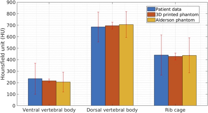
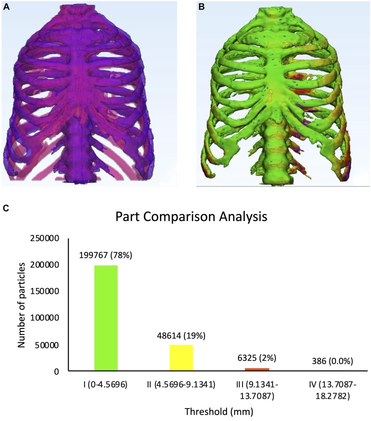
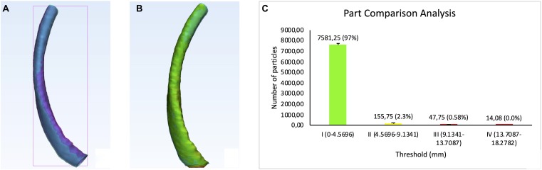
Conventional medical imaging phantoms are limited by simplified geometry and radiographic skeletal homogeneity, which confines their usability for image quality assessment and radiation dosimetry. These challenges can be addressed by additive manufacturing technology, colloquially called 3D printing, which provides accurate anatomical replication and flexibility in material manipulation. In this study, we used Computed Tomography (CT)-based modified PolyJetTM 3D printing technology to print a hollow thorax phantom simulating skeletal morphology of the patient. To achieve realistic heterogenous skeletal radiation attenuation, we developed a novel radiopaque amalgamate constituting of epoxy, polypropylene and bone meal powder in twelve different ratios. We performed CT analysis for quantification of material radiodensity (in Hounsfield Units, HU) and for identification of specific compositions corresponding to the various skeletal structures in the thorax. We filled the skeletal structures with their respective radiopaque amalgamates. The phantom and isolated 3D printed rib specimens were rescanned by CT for reproducibility tests regarding verification of radiodensity and geometry. Our results showed that structural densities in the range of 42-705HU could be achieved. The radiodensity of the reconstructed phantom was comparable to the three skeletal structures investigated in a real patient thorax CT: ribs, ventral vertebral body and dorsal vertebral body. Reproducibility tests based on physical dimensional comparison between the patient and phantom CT-based segmentation displayed 97% of overlap in the range of 0.00-4.57 mm embracing the anatomical accuracy. Thus, the additively manufactured anthropomorphic thorax phantom opens new vistas for imaging- and radiation-based patient care in precision medicine.

Keywords: 3D printed thorax; additive manufacturing; computed tomography (CT) imaging; patient-specific phantom; precision medicine; radiation attenuation.


Figures

FIGURE 1
FIGURE 1
Two-dimensional image of the hollow thorax phantom. The images display the hollow skeletal integument (pink) consisting of ribs, sternum and vertebrae, embedded in a transparent body (blue). (A) Ventral segment, (B) Dorsal segment.
FIGURE 2
FIGURE 2
Radiopaque amalgamate samples used for analyzing radiodensities. The image displays cured radiopaque amalgamate samples in 12 different compositions labeled from 0 to XI.
FIGURE 3
FIGURE 3
Physical dimensional comparison of 3D printed thorax phantom. Registration of thorax phantom STL (purple) on patient STL (pink) in 3-Matic 13.0 software using the following anatomic landmarks: (1) Clavicular notch, (2) Jugular notch, (3) Manubrium, (4) Sternal angle, (5) Superior articular angle, (6) Inferior articular angle.
FIGURE 4
FIGURE 4
Twelve 3D Printed rib specimens. Twelve 3D Printed replicates of the dorsal segment from the body of rib 7.
FIGURE 5
FIGURE 5
3D Printed thorax phantom. Thorax phantom consisting of the skeletal integument embedded in the transparent 3D printed body (A) Ventral view, (B) Caudal view of thorax phantom in supine position, (C) Caudal view of thorax phantom is prone position showing inlets (indicated by yellow arrows) for filling the radiopaque amalgamate in the sternum and ventral vertebral body.
FIGURE 6
FIGURE 6
Radiodensity analysis of radiopaque amalgamate samples and their corresponding anatomic structures in human thorax. (A) CT scan of 12 different compositions of the cured radiopaque amalgamates for radiodensity analysis, (B) Axial section of CT from a patient thorax displaying that the radiodensities of the ribs, ventral and dorsal vertebral bodies were replicated by radiopaque amalgamates VII, IV, and XI, respectively.
FIGURE 7
FIGURE 7
Corresponding axial sections from CT scans of patient, 3D printed thorax phantom and Alderson phantom and twelve printed ribs. Axial sections from CT scans of (A) Patient, (B) 3D printed thorax phantom, (C) Alderson Rando anthropomorphic thorax phantom. Axial sections from CT scans of 3D printed thorax phantom showing (D) Incorporation of air bubble (indicated by red arrow and box), (E) Replication of inhomogeneity in the ventral vertebral cancellous bone (magnified view in the red box), (F) CT scan of the rib specimens for reproducibility tests of the radiopaque amalgamate and model geometry.
FIGURE 8
FIGURE 8
Results from comparison of radiodensities of patient, 3D printed thorax and Alderson Rando phantom. Graph showing average and standard deviation in radiodensities (represented by Hounsfield Units, HU) of the three anatomic structures, analyzed by CT scan of patient, 3D printed thorax and Alderson Rando phantom.
FIGURE 9
FIGURE 9
Physical dimensional comparison of 3D printed thorax phantom with the patient (A) Registration of a CT-derived STL of the 3D printed thorax phantom (purple) on the patient thorax CT-derived STL(pink) using anatomic landmarks in 3-Matic 13.0 software, (B) Color-coded result of Part comparison analysis, (C) Graph explaining the color-coding based quantification of element overlap; 78% elements on the patient thorax overlap with the 3D printed thorax phantom.
FIGURE 10
FIGURE 10
Physical dimensional comparison of 3D printed rib specimens. (A) Registration of one of the twelve CT-derived STLs of the 3D printed rib specimens (purple) on the patient thorax CT-derived STL (pink) using anatomic landmarks in 3-Matic 13.0 software, (B) Color-coded result of Part comparison analysis, (C) Graph explaining the color-coding based quantification of element overlap; 97% elements on the patient rib overlap with the 3D printed ribs.

References

    1. Brouwers L., Teutelink A., van Tilborg F. A. J. B., de Jongh M. A. C., Lansink K. W. W., Bemelman M. (2018). Validation study of 3D-printed anatomical models using 2 PLA printers for preoperative planning in trauma surgery, a human cadaver study. Eur. J. Trauma Emerg. Surg. 45 1013–1020. 10.1007/s00068-018-0970-3 - DOI - PMC - PubMed
    1. Ehler Barney B. M., Higgins P. D., Dusenbery K. E. (2014). Patient specific 3D printed phantom for IMRT quality assurance. Phys. Med. Biol. 59 5763–5773. 10.1088/0031-9155/59/19/5763 - DOI - PubMed
    1. Filippou V., Tsoumpas C. (2018). Recent advances on the development of phantoms using 3D printing for imaging with CT, MRI, PET, SPECT, and ultrasound. Med. Phys. 45 e740–e760. 10.1002/mp.13058 - DOI - PMC - PubMed
    1. Hatamikia S., Biguri A., Kronreif G., Furtado H., Kettenbach J., Figl M., et al. (2019a). “Source-detector trajectory optimization for C-arm CBCT,” in Proceedings of the Supplement of the International Journal of CARS (IJCARS), June 18–21, Rennes.
    1. Hatamikia S., Biguri A., Kronreif G., Kettenbach J., Birkfellner W. (2019b). “CBCT reconstruction based on arbitrary trajectories using TIGRE software tool,” in Proceedings of the 19th Joint Conference on New Technologies for Computer/Robot Assisted Surgery (CRAS), 21-22 March, Genova.

LinkOut - more resources