. 2020 May 27:9:e56312.
doi: 10.7554/eLife.56312.

Transgenesis and web resources in quail

Affiliations

Transgenesis and web resources in quail

Olivier Serralbo et al. Elife. .

Abstract

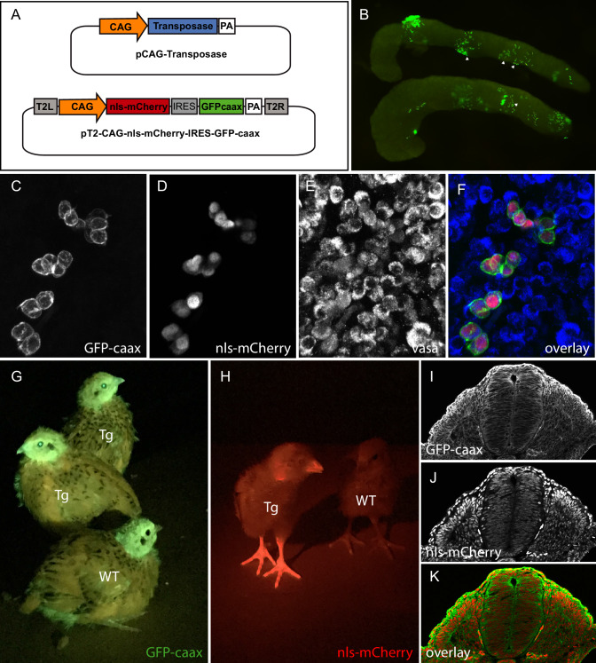
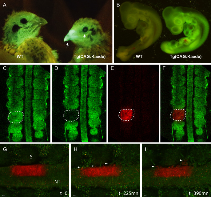
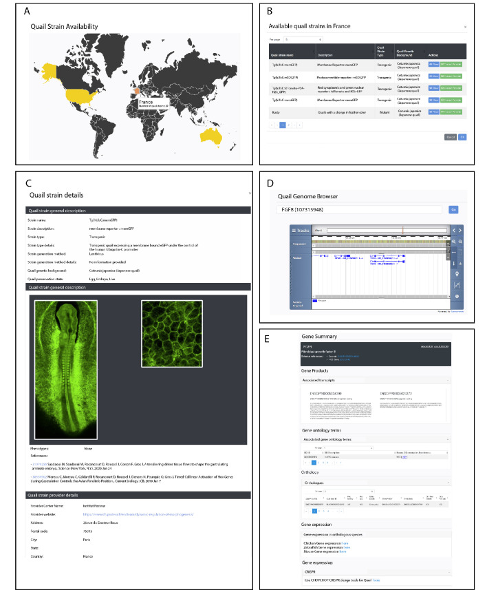
Due to its amenability to manipulations, to live observation and its striking similarities to mammals, the chicken embryo has been one of the major animal models in biomedical research. Although it is technically possible to genome-edit the chicken, its long generation time (6 months to sexual maturity) makes it an impractical lab model and has prevented it widespread use in research. The Japanese quail (Coturnix coturnix japonica) is an attractive alternative, very similar to the chicken, but with the decisive asset of a much shorter generation time (1.5 months). In recent years, transgenic quail lines have been described. Most of them were generated using replication-deficient lentiviruses, a technique that presents diverse limitations. Here, we introduce a novel technology to perform transgenesis in quail, based on the in vivo transfection of plasmids in circulating Primordial Germ Cells (PGCs). This technique is simple, efficient and allows using the infinite variety of genome engineering approaches developed in other models. Furthermore, we present a website centralizing quail genomic and technological information to facilitate the design of genome-editing strategies, showcase the past and future transgenic quail lines and foster collaborative work within the avian community.

Keywords: genetics; genomics; primordial germ cells; quail; transfection; transgenesis.


Conflict of interest statement

OS, DS, NV, CC, MD, TD, JG, CM No competing interests declared

Figures

Figure 1.
Figure 1.. PGC transfection in vivo and generation of a quail line ubiquitously expressing membranal GFP and nuclear mCherry.
(A) Vectors used in the injection mix. (B) Gonads from E7 embryo dissected 5 days after in vivo PGC transfection, showing GFP-positive transfected PGCs (arrowheads). (C–F) Confocal views of transfected (GFP- and mCherry-positive) PGCs among non-transfected PGCs, in the gonads of E7 injected embryo. PGCs are recognized by their expression of the Vasa marker (E,F). (G–H) Transgenic (Tg) and wild-type (WT) chicks showing ubiquitous expression of membranal GFP and nuclear mCherry when observed with UV goggles. (I–J) cross-section of an E3 transgenic quail embryo, showing strong and ubiquitous expression of the transgenes in all cells of the embryo.
Figure 2.
Figure 2.. Design and use of the CrystallGFP mini gene.
(A–D) Cross-section of the head of an E4 embryo, electroporated one day earlier in the optic cup with a CrystallGFP minigene. (A) DAPI, (B) electroporation marker CAG-RFP plasmid, (C) GFP, (D) overlay. (E–G) Cross-section of the head of a 3-day-old embryo of the Tg(MLC:GFP-IRES-NLS-mCherry,CRYBB1:GFP) transgenic line showing the specific expression of GFP throughout the lens. (H) Electroporation constructs used to express the CrystallGFP minigene in a muscle-specific transgenic line (see Figure 4). (I) Transgenic and WT adults of the muscle-specific transgenic line showing GFP expression in lens.
Figure 3.
Figure 3.. Generation of the photoconvertible Kaede transgenic quail line TgT2(CAG:Kaede).
(A) Two-week-old WT and transgenic quails showing the ubiquitous expression of the green fluorescent Kaede in the beak and eye (arrows). (B) WT and transgenic 3-day-old embryos showing strong ubiquitous expression of the protein. (C–F) A newly formed somite before (C) and after (D–F) photoconversion. (G–I) Snapshots from a time-lapse video (see Video 3) showing the morphogenic movements of photoconverted neural tube cells. Arrowheads in H and I show neural crest cells initiating their lateral migration. NT: Neural Tube, S: Somite.
Figure 4.
Figure 4.. Description of muscle specific transgenic quail TgT2(Mmu.MLC1F/3F:GFP-CAAX-IRES-NLS-mCherry,Gga.CRYBB1:GFP).
(A–D) Cross-section of E3 transgenic embryo stained for the indicated markers, showing the expression of the transgene throughout the primary myotome. (A) GFP-CAAX, (B) NLS-mCherry, (C) Pax7, (D) Merge. Insets in (A–D) Magnifications of the regions indicated in (A–D) showing the cellular localisation of the markers. (E–H) E5 Transgenic embryo showing GFP-CAAX (E) and NLS-mCherry (F) in the transition zone (TZ, arrow) where progenitors from the dermomyotome translocate to elongate and differentiate. (I–K) E5 embryos showing strong and specific expression of the muscle-specific reporter in somites (arrowheads). In this quail line, transgenic embryos can be selected at hatching by tηε GFP expression in lens due to the CrystallGFP minigene (arrows). (H–J) E7 transgenic embryo showing muscle-specific expression of the transgene in the head, limbs and trunk.
Figure 5.
Figure 5.. Description of QuailNet features.
(A) Interactive world map displaying the number of quail strains available by country. (B) List of quail strains together with a general description by country. (C) Detailed description of a specific quail strain (e.g. Tg(hUbC:memGFP)). (D) Quail genome browser displaying genomic information and location of a queried gene (e.g. FGF8). (E) Information associated with a queried gene (e.g. FGF8).

Similar articles

Cited by

References

    1. Ahn J, Shin S, Suh Y, Park JY, Hwang S, Lee K. Identification of the avian RBP7 gene as a new adipose-specific gene and RBP7 promoter-driven GFP expression in adipose tissue of transgenic quail. PLOS ONE. 2015;10:e0124768. doi: 10.1371/journal.pone.0124768. - DOI - PMC - PubMed
    1. Ando R, Hama H, Yamamoto-Hino M, Mizuno H, Miyawaki A. An optical marker based on the UV-induced green-to-red photoconversion of a fluorescent protein. PNAS. 2002;99:12651–12656. doi: 10.1073/pnas.202320599. - DOI - PMC - PubMed
    1. Antin PB, Yatskievych TA, Davey S, Darnell DK. GEISHA: an evolving gene expression resource for the chicken embryo. Nucleic Acids Research. 2014;42:D933–D937. doi: 10.1093/nar/gkt962. - DOI - PMC - PubMed
    1. Barnes HJ. Diseases of quail. Veterinary Clinics of North America: Small Animal Practice. 1987;17:1109–1144. doi: 10.1016/S0195-5616(87)50107-3. - DOI - PMC - PubMed
    1. Belle M, Godefroy D, Couly G, Malone SA, Collier F, Giacobini P, Chédotal A. Tridimensional visualization and analysis of early human development. Cell. 2017;169:161–173. doi: 10.1016/j.cell.2017.03.008. - DOI - PubMed

Publication types