

Mitochondrial light switches: optogenetic approaches to control metabolism
- PMID: 32459870
- PMCID: PMC7672652
- DOI: 10.1111/febs.15424
Mitochondrial light switches: optogenetic approaches to control metabolism
Abstract
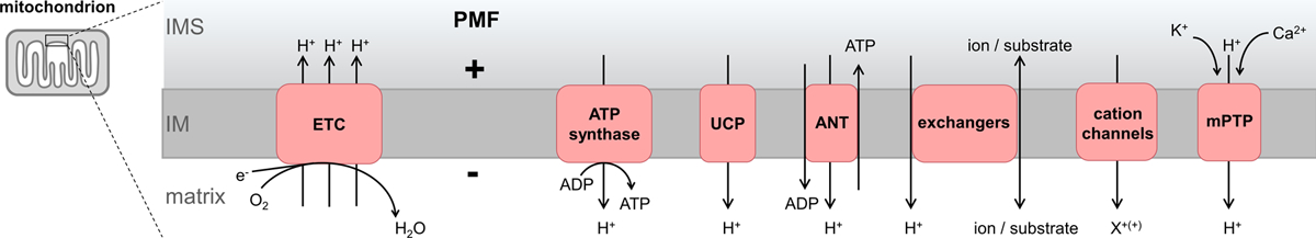
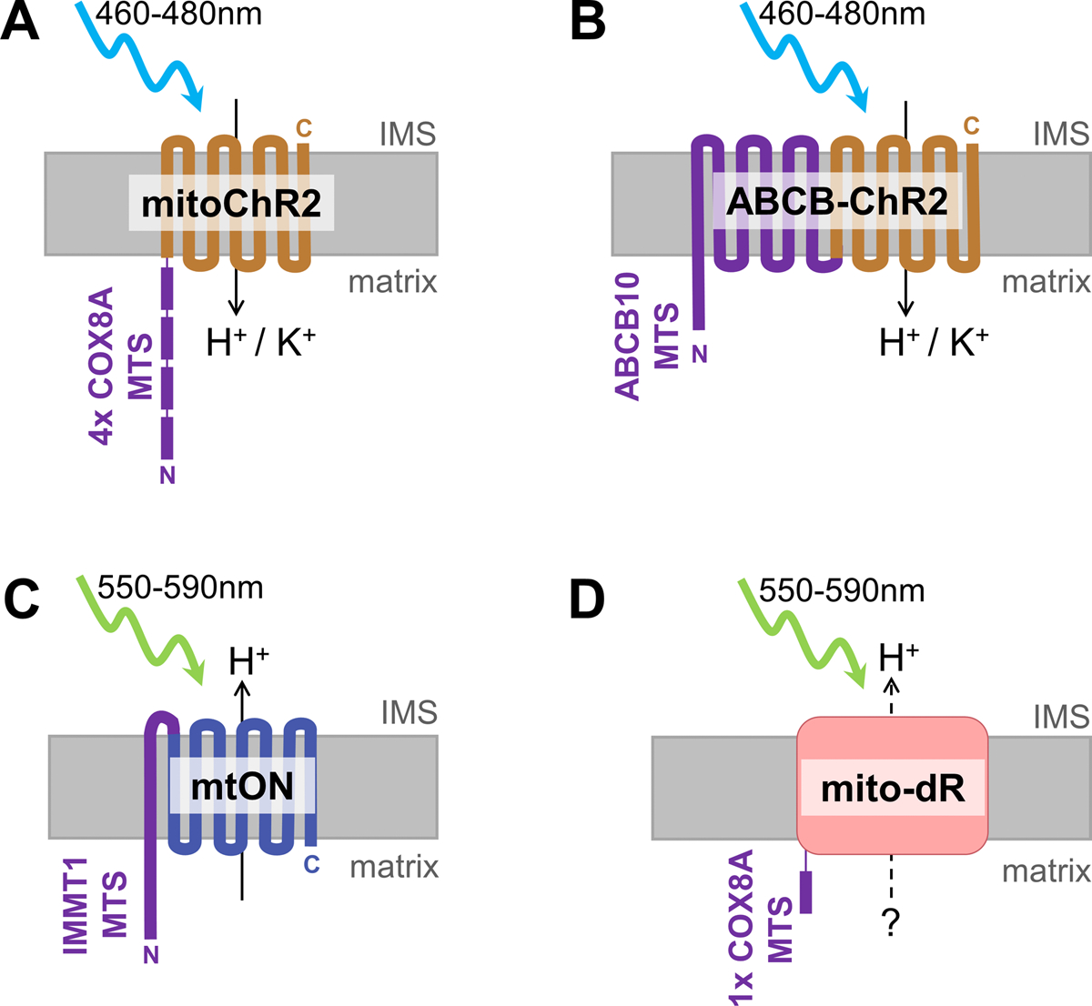
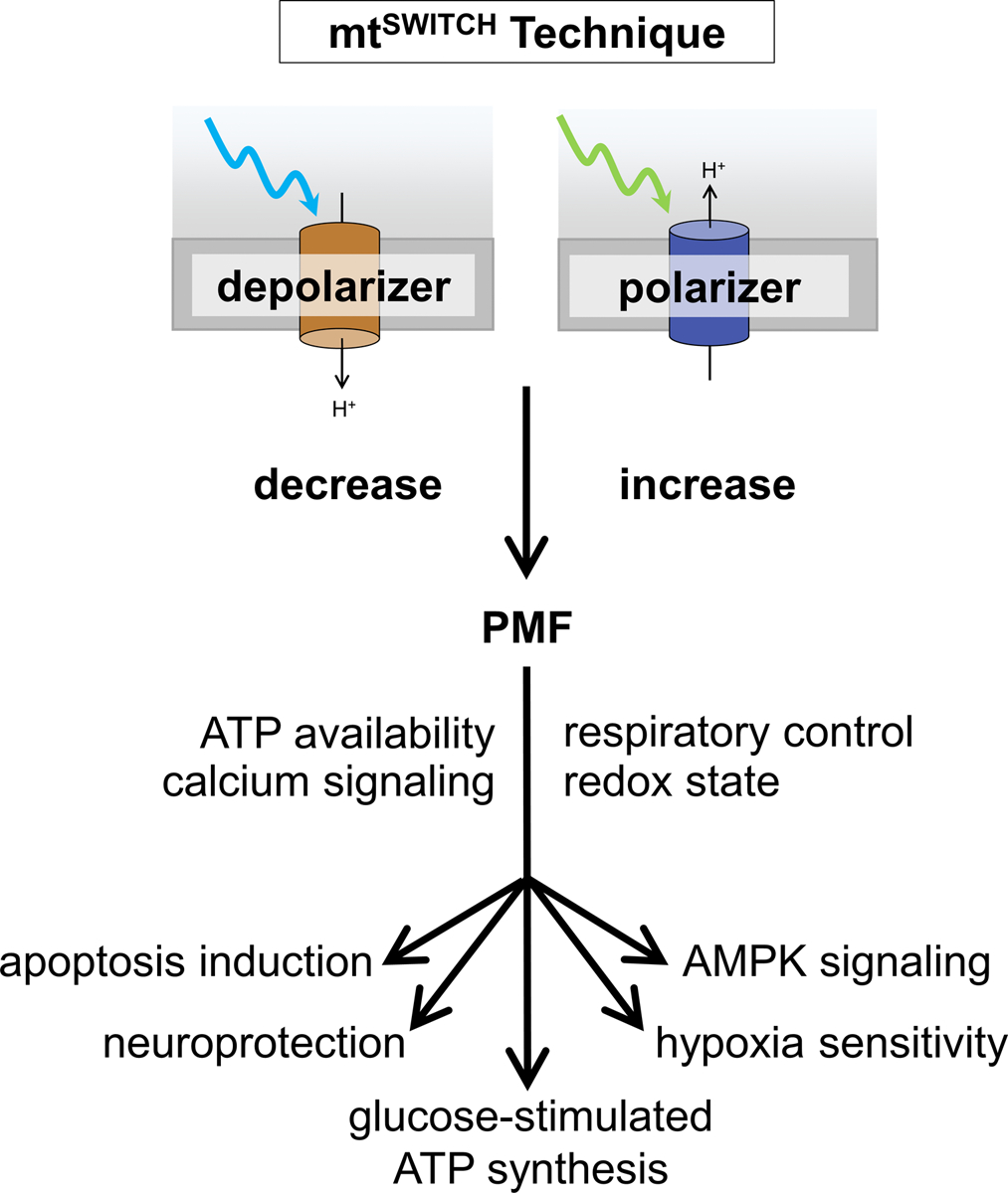
Developing new technologies to study metabolism is increasingly important as metabolic disease prevalence increases. Mitochondria control cellular metabolism and dynamic changes in mitochondrial function are associated with metabolic abnormalities in cardiovascular disease, cancer, and obesity. However, a lack of precise and reversible methods to control mitochondrial function has prevented moving from association to causation. Recent advances in optogenetics have addressed this challenge, and mitochondrial function can now be precisely controlled in vivo using light. A class of genetically encoded, light-activated membrane channels and pumps has addressed mechanistic questions that promise to provide new insights into how cellular metabolism downstream of mitochondrial function contributes to disease. Here, we highlight emerging reagents-mitochondria-targeted light-activated cation channels or proton pumps-to decrease or increase mitochondrial activity upon light exposure, a technique we refer to as mitochondrial light switches, or mtSWITCH . The mtSWITCH technique is broadly applicable, as energy availability and metabolic signaling are conserved aspects of cellular function and health. Here, we outline the use of these tools in diverse cellular models of disease. We review the molecular details of each optogenetic tool, summarize the results obtained with each, and outline best practices for using optogenetic approaches to control mitochondrial function and downstream metabolism.
Keywords: AMPK; Parkinson’s; apoptosis; bioenergetics; calcium signaling; diabetes; hypoxia; mitophagy.
© 2020 Federation of European Biochemical Societies.
Conflict of interest statement
Figures
Similar articles
-
Optogenetic Studies of Mitochondria.Methods Mol Biol. 2022;2501:311-324. doi: 10.1007/978-1-0716-2329-9_15. Methods Mol Biol. 2022. PMID: 35857235 Free PMC article.
-
Precisely Control Mitochondria with Light to Manipulate Cell Fate Decision.Biophys J. 2019 Aug 20;117(4):631-645. doi: 10.1016/j.bpj.2019.06.038. Epub 2019 Jul 26. Biophys J. 2019. PMID: 31400914 Free PMC article.
-
Optogenetic control of mitochondrial protonmotive force to impact cellular stress resistance.EMBO Rep. 2020 Apr 3;21(4):e49113. doi: 10.15252/embr.201949113. Epub 2020 Feb 11. EMBO Rep. 2020. PMID: 32043300 Free PMC article.
-
Lights, cytoskeleton, action: Optogenetic control of cell dynamics.Curr Opin Cell Biol. 2020 Oct;66:1-10. doi: 10.1016/j.ceb.2020.03.003. Epub 2020 May 1. Curr Opin Cell Biol. 2020. PMID: 32371345 Free PMC article. Review.
-
Intracellular microbial rhodopsin-based optogenetics to control metabolism and cell signaling.Chem Soc Rev. 2024 Apr 2;53(7):3327-3349. doi: 10.1039/d3cs00699a. Chem Soc Rev. 2024. PMID: 38391026 Review.
Cited by
-
An energetics perspective on geroscience: mitochondrial protonmotive force and aging.Geroscience. 2021 Aug;43(4):1591-1604. doi: 10.1007/s11357-021-00365-7. Epub 2021 Apr 17. Geroscience. 2021. PMID: 33864592 Free PMC article.
-
Genetically encoded tool for manipulation of ΔΨm identifies the latter as the driver of integrative stress response induced by ATP Synthase dysfunction.bioRxiv [Preprint]. 2023 Dec 27:2023.12.27.573435. doi: 10.1101/2023.12.27.573435. bioRxiv. 2023. Update in: Cell Chem Biol. 2025 Apr 17;32(4):620-630.e6. doi: 10.1016/j.chembiol.2025.03.007. PMID: 38234735 Free PMC article. Updated. Preprint.
-
Subcellular Singlet Oxygen and Cell Death: Location Matters.Front Chem. 2020 Nov 17;8:592941. doi: 10.3389/fchem.2020.592941. eCollection 2020. Front Chem. 2020. PMID: 33282833 Free PMC article.
-
Rhodopsins: An Excitingly Versatile Protein Species for Research, Development and Creative Engineering.Front Chem. 2022 Jun 22;10:879609. doi: 10.3389/fchem.2022.879609. eCollection 2022. Front Chem. 2022. PMID: 35815212 Free PMC article. Review.
-
Optogenetic Control of the Mitochondrial Protein Import in Mammalian Cells.Cells. 2024 Oct 9;13(19):1671. doi: 10.3390/cells13191671. Cells. 2024. PMID: 39404433 Free PMC article.
References
-
- Lozano, Naghavi R, Foreman M, Lim K, Shibuya S, Aboyans K, Abraham V, Adair J, Aggarwal T, Ahn R, Alvarado SY, Anderson M, Anderson HR, Andrews LM, Atkinson KG, Baddour C, Barker-Collo LM, Bartels S, Bell DH, Benjamin ML, Bennett EJ, Bhalla D, Bikbov K, Bin Abdulhak B, Birbeck A, Blyth G, Bolliger F, Boufous I, Bucello S, Burch C, Burney M, Carapetis P, Chen J, Chou H, Chugh D, Coffeng SS, Colan LE, Colquhoun SD, Colson S, Condon KE, Connor J, Cooper MD, Corriere LT, Cortinovis M, de Vaccaro M, Couser KC, Cowie W, Criqui BC, Cross MH, Dabhadkar M, Dahodwala KC, De Leo N, Degenhardt D, Delossantos L, Denenberg A, Des Jarlais J, Dharmaratne DC, Dorsey SD, Driscoll ER, Duber T, Ebel H, Erwin B, Espindola PJ, Ezzati P, Feigin M, Flaxman V, Forouzanfar AD, Fowkes MH, Franklin FG, Fransen R, Freeman M, Gabriel MK, Gakidou SE, Gaspari E, Gillum F, Gonzalez-Medina RF, Halasa D, Haring YA, Harrison D, Havmoeller JE, Hay R, Hoen RJ, Hotez B, Hoy PJ, Jacobsen D, James KH, Jasrasaria SL, Jayaraman R, Johns S, Karthikeyan N, Kassebaum G, Keren N, Khoo A, Knowlton JP, Kobusingye LM, Koranteng O, Krishnamurthi A, Lipnick R, Lipshultz M, Ohno SE, S. L., et al. (2012) Global and regional mortality from 235 causes of death for 20 age groups in 1990 and 2010: a systematic analysis for the Global Burden of Disease Study 2010, Lancet 380, 2095–128. - PMC - PubMed
-
- Ng, Fleming M, Robinson T, Thomson M, Graetz B, Margono N, Mullany C, Biryukov EC, Abbafati S, Abera C, Abraham SF, Abu-Rmeileh JP, Achoki NM, AlBuhairan T, Alemu FS, Alfonso ZA, Ali R, Ali MK, Guzman R, Ammar NA, Anwari W, Banerjee P, Barquera A, Basu S, Bennett S, Bhutta DA, Blore Z, Cabral J, Nonato N, Chang IC, Chowdhury JC, Courville R, Criqui KJ, Cundiff MH, Dabhadkar DK, Dandona KC, Davis L, Dayama A, Dharmaratne A, Ding SD, Durrani EL, Esteghamati AM, Farzadfar A, Fay F, Feigin DF, Flaxman VL, Forouzanfar A, Goto MH, Green A, Gupta MA, Hafezi-Nejad R, Hankey N, Harewood GJ, Havmoeller HC, Hay R, Hernandez S, Husseini L, Idrisov A, Ikeda BT, Islami N, Jahangir F, Jassal E, Jee SK, Jeffreys SH, Jonas M, Kabagambe JB, Khalifa EK, Kengne SE, Khader AP, Khang YS, Kim YH, Kimokoti D, Kinge RW, Kokubo JM, Kosen Y, Kwan S, Lai G, Leinsalu T, Li M, Liang Y, Liu X, Logroscino S, Lotufo G, Lu PA, Ma Y, Mainoo J, Mensah NK, Merriman GA, Mokdad TR, Moschandreas AH, Naghavi J, Naheed M, Nand A, Narayan D, Nelson KM, Neuhouser EL, Nisar ML, Ohkubo MI, Oti T, Pedroza SO, A., et al. (2014) Global, regional, and national prevalence of overweight and obesity in children and adults during 1980–2013: a systematic analysis for the Global Burden of Disease Study 2013, Lancet 384, 766–81. - PMC - PubMed
-
- Danaei G, Finucane MM, Lu Y, Singh GM, Cowan MJ, Paciorek CJ, Lin JK, Farzadfar F, Khang YH, Stevens GA, Rao M, Ali MK, Riley LM, Robinson CA, Ezzati M & Global Burden of Metabolic Risk Factors of Chronic Diseases Collaborating, G. (2011) National, regional, and global trends in fasting plasma glucose and diabetes prevalence since 1980: systematic analysis of health examination surveys and epidemiological studies with 370 country-years and 2.7 million participants, Lancet 378, 31–40. - PubMed
-
- Mitchell P (1961) Coupling of phosphorylation to electron and hydrogen transfer by a chemi-osmotic type of mechanism, Nature 191, 144–8. - PubMed
Publication types
MeSH terms
Substances
Grants and funding
LinkOut - more resources
Full Text Sources
Other Literature Sources
